The IBDM is aninternationally renowned research center in developmental biology that studies fundamental mechanisms governing the organization and function of biological systems, using multiscale approaches in a range of animal and cellular models. Research activities at the IBDM synergistically connect developmental biology with molecular, cell and computational biology, as well as evolution, neurobiology, physiology, physiopathology, biophysics, and cancer. The IBDM uniquely fosters interdisciplinary approaches by its intimate connections with various research-training networks within the Aix-Marseille University (AMU) (CenTuri, NeuroMarseille, ICI (Cancer-Immuno), MarMaRa (Rare-Diseases), Marseille Imaging, Canceropôle-PACA).
The IBDM, affiliated with CNRS and AMU, strongly benefits from its collaborative and international scientific culture, English working language, and its fantastic environment on the Marseille Luminy campus, located in the heart of the Calanques National Park.
The IBDM is committed to promotingequality, diversity and inclusivity. The selected candidates will receive support to establish a group in a fully renovated building, have access to cutting-edge scientific core facilities,and will be assisted in obtaining a tenured position (CNRS or AMU), and in securing extramural funding (ATIP/Avenir, ERC, etc…).
To apply
Candidates should provide the following information in a single PDF file: a cover letter explaining their motivation to join the IBDM, a CV, a summary of their main research achievements (2 pages maximum), a future research project (5 pages maximum), and contacts of three references.
Applications and queries should be sent to the search committee (ibdm-call2022@univ-amu.fr) before March 1st 2022.
In person interviews will be scheduled from May 2022.
Frieda Leesch (PhD student, Pauli lab, Research Institute of Molecular Pathology) ‘A molecular network of conserved factors keeps ribosomes dormant in the egg’
Brad Cairns (Professor, Huntsman Cancer Institute) ‘Maternally-inherited anti-sense piRNAs antagonize transposon expression in zebrafish and medaka embryos’
The webinar will be held in Remo, our browser-based conferencing platform. After the talks you’ll have the chance to meet the speakers and other participants at virtual conference tables. If you can’t make it on the day, talks will be available to watch after the event on the Node. You can also sign up to our mailing list for email alerts.
Journal of Cell Science is pleased to invite submissions for the first JCS essay series. The theme is ‘Equity, diversity and inclusion in cell biology’, an important, relevant and timely issue that should be a guiding force in all that we do. We hear the terms being used more and more these days, but we’re all still early in the journey, and at JCS we want to create an opportunity to amplify voices that are not always heard in this space. Has equity, diversity and/or inclusion shaped your experience as a cell biologist in some way? We want to hear about and learn from your stories. We will publish a selection of submissions in the journal in an ongoing series and reward a stand-out essay from the initial call, chosen by a selection of our Editors and Editorial Advisory Board members, with a £500 prize.
All essays must be original pieces that have not been previously published. Any cell biologist at any career stage can submit an essay. Submissions must not be more than 2,000 words and can include figures and images. Please send your essay to jcs.essay@biologists.com.
The closing date has been extended to 15 January 2022.
On Wednesday 10 November, Development hosted three talks on stem cells and disease models.
Below you’ll find each of the talks, plus a Q&A chaired by Development Editor James Wells. The next #DevPres webinar will be a zebrafish special to celebrate the 25th anniversary of our Zebrafish Issue. It will be held on 8 December 2021 at 15:00 GMT, chaired by Alex Schier – subscribe to our mailing list for updates.
Dhruv Raina (formerly Max Planck Institute for Molecular Physiology, now Mosa Meat) ‘Cell-cell communication through FGF4 generates and maintains robust proportions of differentiated cell types in embryonic stem cells’
Marco Trizzino (Thomas Jefferson University) ‘Inability to switch from ARID1A-BAF to ARID1B-BAF impairs exit from pluripotency and commitment towards neural crest differentiation in ARID1B-related neurodevelopmental disorders’
Thanks to the #DevBio community for sharing their thoughts, especially on twitter. If you have some news that you think we should share on our blog, please get in touch at thenode@biologists.com. If you are interested in getting involved with writing preLights you can find out more here.
“You don’t need to be a professional ‘influencer’ to get your voice heard.”
This summer, I finished an unusual project in a neurobiology lab. Contrary to popular belief, internships in a science lab do not necessarily mean operating high-tech machines wearing a lab coat. My project, for example, is for science outreach.
Nowadays, science is no longer an exclusive domain for scientists, but more of public interest. More and more researchers are realising the importance of public outreach. My project is among the first few outreach projects that have successfully drawn funding. It is kindly sponsored by the BSDB Gurdon/The Company of Biologists Summer Studentships.
The Project
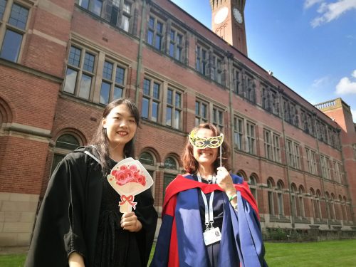
Like any hardcore scientist/science enthusiast, I enjoy gossiping about science. I just couldn’t help seeking out ways to spread my passion for it. So after finishing my BSc project in the Alicia Hidalgo Lab at the University of Birmingham, I stayed over the summer to help them with outreach. I designed a website for the lab, and produced 20 educational videos to show what neurobiologists do at the bench.
With Professor Alicia Hidalgo on Graduation Day
The videos were published on a YouTube channel Alicia set up, and got over 500 views in the first month. You can watch people in the lab explaining why study fruit flies, summarising what they’ve done in the latest research papers, and demonstrating how they work with DNA, proteins, cells, and lab animals. The lab website also has a strong emphasis on education ad science outreach. We have embedded videos, clickable 3D objects, and other interactive elements for interested people around the world to explore.
At first, I was as clueless as you probably are right now about how to make videos and websites. Over the two months, however, I taught myself the necessary skills. Now I can make animations, build a website with WordPress, film educational videos, and produce lay-people-friendly multimedia content.
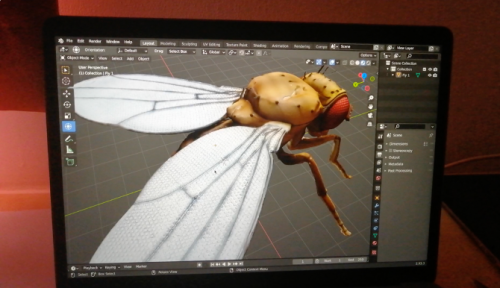
Towards the end of the project, I had another idea. A friend of mine created a digital 3D object using 2D photos of a fossil, as an assignment in class. So I asked her for advice and started learning how to do it myself. A week later, I created a clickable digital 3D model of the fruit fly Drosophila, using 600 photos taken in the lab. I published it on a 3D-object sharing platform and soon forgot about it. But a few months later, I was approached by a scientist in the field of fluid mechanics asking aerodynamics questions! It’s nice to know what I made have resonated with scientists in other fields.
Working on the digital fruit fly model
What I’ve Learned
Having just graduated, I was reflecting on my uni experience throughout the summer. Doing the outreach project helped me figure it out. Despite getting a First Class Honours degree, I could barely recall 20% of my class notes. Apparently, this happens to virtually everyone. So detailed knowledge isn’t the most important thing one gains from uni. Then what is? While doing this project, I realised that I’ve gained three key transferable skills and insight.
1) Insight: life in the lab.
During my four-year degree, I worked on three research projects and peeked at the workings of research labs. I learned about collaborations, group dynamics, and stories of people at various stages of their academic careers. This leads me to my current position, an MRes student in Experimental Neuroscience, and I’m confident to move on to a PhD and beyond.
2) How to teach myself something from scratch.
The ability to learn anything with the internet is a crucial skill that got me through uni and this project. With information overflow versus a limited lifetime, modern scientists need to quickly locate what we need and master the necessary skills for their own research. Being able to teach ourselves anything unleashes a lot of potentials. You will be surprised at the opportunities it brings.
3) How to communicate with different audiences.
Communication, be it verbal or non-verbal, online or face-to-face, is a transferable skill across all careers. Learning how to present and promote one’s work online is arguably the most important skill in the modern age. By learning to promote the lab’s work online, I realised that you don’t need to be a professional “influencer” to get your voice heard.
So I started gossiping about things I’m passionate about – neuroscience, productivity hacks – to people around me. During my summer project, I started blogging, made a personal website, and even started making podcasts with friends! Instead of passively interacting with my phone when I’m bored, I now initiate deep conversations with people and share what I’ve learned with people around the world.
How can this help you, a fellow scientist?
Well, for starters, no matter which career stage you’re in, face this reality – if your work isn’t online, it doesn’t exist. Digital journals have made it easier to share our work, but that is not enough. Successful scientists actively share their work online, by social media, or interviews, which are published as articles, videos, or podcasts.
Never before have there been so many brilliant ways to promote our work, but never before has it been so difficult to compete with other voices to make sure that ours get heard. Creating a lab website, a YouTube channel, or a social media account will be a good start.
What Next?
The insights and skills I gained this summer are invaluable. As I’m starting my Master’s degree, I want to continue blogging and make more outreach stuff for future labs. I’m also considering a career in academia with teaching and public engagement elements. Maybe I’ll become a professor, or a science writer, to inspire curious minds around the world.
Finally, I want to thank all the amazing people in the Hidalgo Lab – Alicia, Marta, Guiyi, Jun, Maria, Lizzie, Naser, Deepanshu, Mike, and Anna. They have been such an enthusiastic and supportive team for my thesis and summer project. I also want to thank the BSDB studentship for their support throughout my project. I would strongly recommend future students to apply to the studentship, not only for wet-lab projects but outreach projects as well.
Here’s a short introduction to the Hidalgo lab if you are interested:
In the latest episode of Genetics Unzipped, Kat Arney takes a trip to the zoo, to find out how studying tumours across the animal kingdom, from naked mole rats to elephants, can reveal insights into cancer in our own species.
In 2014, geneticists at the University of Kiel in Germany published a paper describing tumours in two different species of tiny freshwater Hydra. Little more than a tube with tentacles, Hydra comprise three distinct groups of stem cells. One of these groups, known as interstitial stem cells, turned out to be the source of the cancers, which severely impacted growth and fertility.
But while Hydra may be the simplest organisms currently known to develop cancer, they are far from the only example outside our own species. Kat explores how cancer has been found on virtually every branch of the tree of multicellular life, from the simplest to the most complex. And she tells the story of how a family trip to the zoo led to the University of Utah’s Professor Josh Schiffman discovering the biological secret that explains why elephants hardly ever get cancer.
If you enjoy the show, please do rate and review on Apple podcasts and help to spread the word on social media. And you can always send feedback and suggestions for future episodes and guests to podcast@geneticsunzipped.com Follow us on Twitter – @geneticsunzip
Mechanical regulation of cell division in developing tissues: Speed Vs Strength
During embryogenesis, dynamic mechanical forces act on developing tissues, inducing cellular mechano-responses. These changes in cellular behaviours such as cell division, adhesion, and motility are a vital aspect of tissue morphogenesis and homeostasis.
This summer, I was given the opportunity to work under Dr. Woolner at the University of Manchester’s Division of Cell Matrix Biology & Regenerative Medicine. Using a multidisciplinary approach, Dr. Woolner’s lab examines the cellular response of developing tissues to an applied mechanical force and seeks to identify the underlying molecular basis. This placement was an incredible and unique opportunity for me, as I was able to receive training and experience in a variety of new techniques used in biomechanics, mathematics, biomodelling and developmental biology.
Previous work by the Woolner lab demonstrated that the rate of cell division increases in epithelial cells following the application of a low-magnitude, uniaxial tensile force1. Work in other systems has shown that similar mechanically-induced increases in proliferation occur due to upregulation of the ERK1/2 pathway downstream of the stretch-activated calcium channel Piezo1, culminating in an upregulation of cyclin B2. Additionally, it is known that the orientation of cell division aligns with the axis of stretch1.
However, all current studies investigating the cellular response to tensile force involve rapid, instantaneous tissue stretching. Under physiological conditions, changes to mechanical tension in the developing embryo occur over a period of minutes to hours rather than seconds. In tumourigenesis, mechanical changes may take place over years. It is not currently known how cell division rate differs between fast and slow stretch regimes. Preliminary work suggests that slow-stretch regimes may not elicit the same division responses that are seen with instantaneous stretching.
My project aimed to help shed light on whether the speed or strength of an applied mechanical force is the major factor in altering cell division rate.
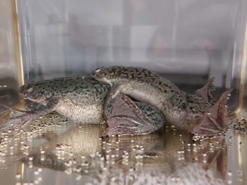
Using a tissue stretching apparatus, we applied an instantaneous, uniaxial stretch with reduced strength to tissues. For these experiments, Xenopus laevis embryonic tissue was used. Xenopus laevis embryos are a robust model organism for use in biomechanical research as they are large, develop externally and are easily visualised. I was very grateful for the opportunity to shadow members of the lab working with the Xenopus colony throughout the project. They are a unique model animal (I also have a few as pets!) and it was great to see how they are cared for and used responsibly in a research setting.
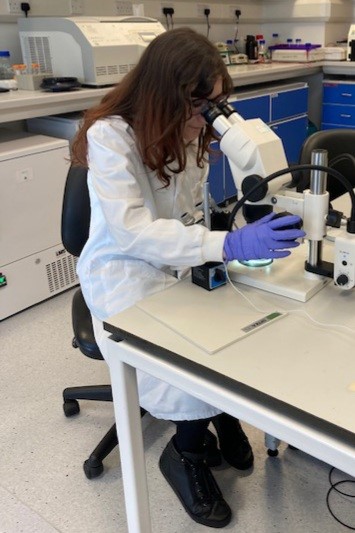
Fig 1. Selecting embryos at 2-cell stage for mRNA microinjection
In order to visualise the cell edge and nucleus, Xenopus embryos were injected at 2-cell stage with GFP-tubulin and Cherry-histone RNA. Straight away I was given the chance to jump in and get involved with the experiments, as I helped Gina (the Woolner Lab’s Research Assistant) with DNA miniprep and mRNA preparation. We proceeded with microinjection, which involved inserting a microscopic needle tip into each cell under an optical microscope. This was a very tricky procedure at first but by practicing alongside Gina, I was eventually able to go from struggling to inject 10 embryos in an hour to injecting over 50 in half the time!
Following overnight incubation, embryos were staged at early gastrula and the animal caps were dissected. Isolated animal cap explants are a versatile tissue able to survive and develop ex-vivo, making them ideal for live imaging. Dissecting the animal cap was done through an optical microscope using two sets of forceps. This was the most technically challenging aspect of my lab work, as it required a steady hand and patience but couldn’t be done too slowly or the embryos would become too developed. It was very rewarding to eventually get a perfect set of animal cap explants.
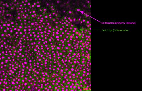
Following incubation on a fibronectin-coated silicone membrane, the animal caps were stretched and imaged. Shown in Figure 2 is a single frame from one of our live movies captured using confocal fluorescence microscopy. This was great experience, as imaging science was always of great interest to me but I had never previously had the chance to put my theoretical knowledge into practice. I also used image analysis software to calculate the mitotic index, as well as try cell population tracing. The Woolner lab uses tracing alongside vertex modelling3,4 to measure cell shape and infer mechanical stress across the tissue. The data collected during my project will be used to determine whether an increased cell division rate acts to relieve tensile stress across the tissue.
Fig 2. Fluorescent image of a Xenopus embryo animal cap explant experiencing a uniaxial stretch. Visualisation of the cell nucleus (magenta) and cell edge (green) allows image analysis techniques and cell population tracing to be performed. This was performed to calculate the mitotic index and biophysical properties of the tissue.
Alongside my core project work, I also successfully titrated the CDK-1 inhibitor RO-3306 to find the optimal concentration for cell division inhibition in Xenopus embryos. It is currently known that mechanical tension may increase cell division in fast-stretch regimes by promoting G1 to S phase transition5, which the Woolner lab will be investigating in slow-stretch regimes using a Fucci probe coupled with RO-3306 inhbition. Towards the end of my studentship, I was really grateful to have the opportunity to attend the 18th International Xenopus Conference. This was a great chance to discover the wide array of biomedical research using Xenopus currently being conducted worldwide and make valuable connections.
Fig 3. Xenopus laevis produce large, externally developing embryos which are easy to collect, visualise and manipulate. These properties make them particularly suitable for tissue stretch experiments.
I would like to thank everyone for all their support, guidance, patience and coffee & cake sessions throughout the internship. I am very grateful that I was able to receive the Gurdon/BSDB Summer Studentship and would recommend any student interested in developmental biology research to apply. Gaining first-hand lab experience in this field has given me invaluable skills and insight and has opened many doors for my future career.
References
Nestor-Bergmann A., Stooke-Vaughan G.A., Goddard G.K., Starborg T., Jensen O.E. and Woolner S. (2019) Decoupling the roles of cell shape and mechanical stress in orienting and cueing epithelial mitosis. Cell Reports26: 2088-2100
Gudipaty S.A., Lindblom J., Loftus P.D., Redd M.J., Edes K., Davey C.F., Krishnegowda V., Rosenblatt J. (2017) Mechanical stretch triggers rapid epithelial cell division through Piezo1. Nature543, 118-121.
Nestor-Bergmann A., Goddard G., Woolner S. and Jensen O.E. (2017) Relating cell shape and mechanical stress in a spatially disordered epithelium using a vertex-based model. Mathematical Medicine and Biology35 (Supplement 1): 1-27
Jensen O.E., Johns E. and Woolner S. (2020) Force networks, torque balance and Airy stress in the planar vertex model of a confluent epithelium. Proceedings of the Royal Society A476: 2237
Benham-Pyle B.W., Pruitt B.I and Nelson W.J. (2015) Mechanical strain induces E-cadherin-dependent Yap1 and β-catenin activation to drive cell cycle entry. Science 348(6238): 1024–1027.
Numerous efforts have been made to establish bona fide iPSCs from companion animals such dogs and cats. Generation of iPSCs from companion animals would provide useful unrestricted cell resources with a vast scientific potential. To name a few applications, they can be exploited as new models for regenerative medicine and as therapeutic veterinary tools to replace tissues; in veterinary pharmacology for drug development assays, and to elucidate function(s) of genetic variants that are associated with disease.
While protocols for producing human and mouse iPSCs are established, protocols for derivation of iPSCs from domestic animals are slowly developing due to difficulties encountered presumably in their reprogramming process. Only a few studies have indeed focused on the possibility of producing iPSCs from these companion species, and despite some describing their production, the burden of proof is largely lacking.
As an undergraduate student at the University of Edinburgh, this summer despite the current pandemic situation making it harder to find a lab-based studentship, I was lucky enough to have the opportunity to work in Drs. Schoenebeck’s and Burdon’s labs. The Gurdon/BSDB award allowed me to spend two months at Roslin Institute, a pioneering center for genetics and stem cell studies, collaborating with research groups with extensive experience in stem cell research (Dr. Tom Burdon’s) and canine genetics and genomics (Dr. Jeffrey Schoenebeck’s). During my time at Roslin I contributed to the research of an efficient protocol to derive canine iPSCs, supervised by the joint effort of these two excellent lab groups.
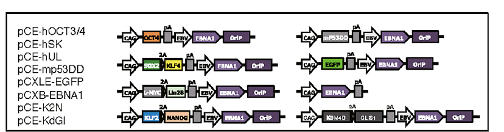
Based on the findings that iPSC generation is enhanced by P53 suppression and replacement of L-MYC with C-MYC(Okita et al., 2011) in the set of conventional reprogramming factors (OCT4, SOX2, KLF4, and C-MYC, collectively termed “OSKM” factors); recently, Yoshimatsu et al. (2021) have presented a study which provides insights on the possibility to facilitate canine cell reprogramming. They provided evidence of reprogramming somatic fibroblasts from a canine using an integration-free method. Their 8 episomal (Figure 1) vectors contain the OSKM factors including L-MYC, other pluripotency genes (LIN28 and NANOG), genes that have been shown to facilitate reprogramming (GLIS1and KDM4), and a dominant-negative form of the mouse TRP53 (mP53DD), which was shown to suppress endogenous P53 expression in human cells, and presumably should operate the same in canines.
Figure 1. Schematic of the plasmid mixture used for vector transfection. (Adapted from Yoshimatsu et al., 2021)
The episomal vectors contain OriP/EBNA1 sequences derived from Epstein-Barr virus (EBV), which ensure the stable extrachromosomal replication of the vectors, hence high expression of the reprogramming factors carried along, which facilitate the production of iPSC. However, the full applicability of this EBV-based system is still unclear as only two dogs were used to prove its actual functionality.
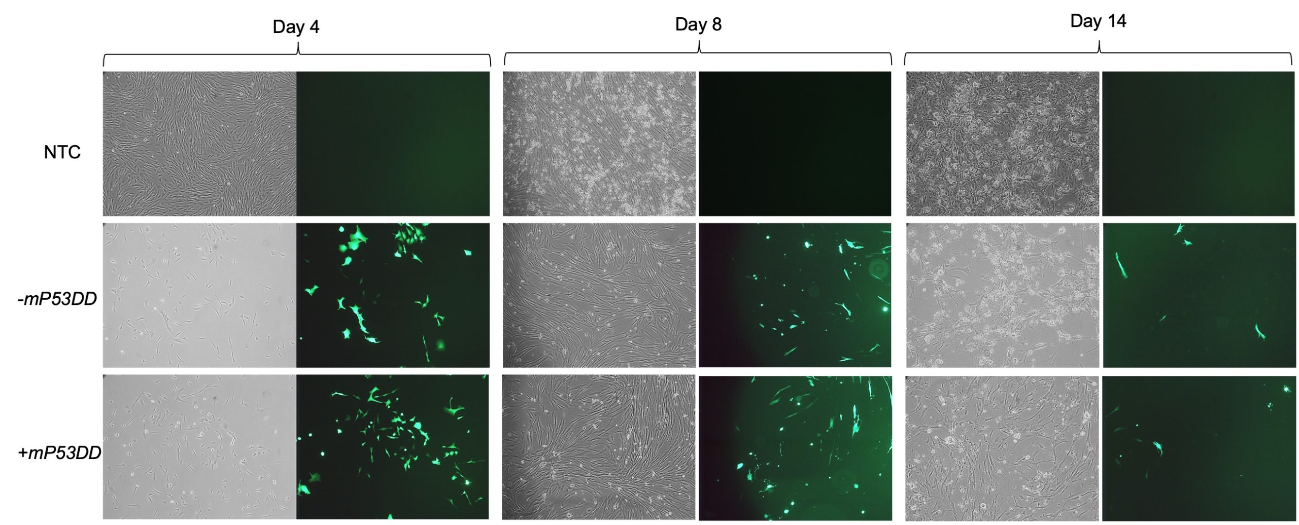
The aim of my project was to assess the ability of the aforementioned system to reprogram canine fibroblasts, testing the capability of facilitating reprogramming by the inclusion of dominant-negative P53. After being introduced to the fundamental cell culturing techniques and practiced such skills on mouse feeder cells, I expanded canine fibroblast from testis in feeder medium prior to transfection of the EBV-based vectors. I then electroporated such cells with 2 different mixtures of vectors, one consisting of the 8 plasmids including the dominant-negative P53 (+mP53DD), and the other without it, consisting of 7 plasmids (-mP3DD). Right after transfection the medium used to feed the cells was changed to M10. Since one of the transfected vectors carried EGFP, I took GFP imaging to directly assess if the transfection was successful, comparing the transfected fibroblast with the non-transfected control (NTC). Images (Figure 2) show a high extent of cell death following electroporation of the cells, while GFP expression in a high proportion of the survived cells indicate uptake of the vectors. Cell recovered and showed prolonged GFP expression until day 14.
Figure 2. Bright field (left) and GFP (right) images of canine fibroblast at days 4, 8, and 14 after transfection.
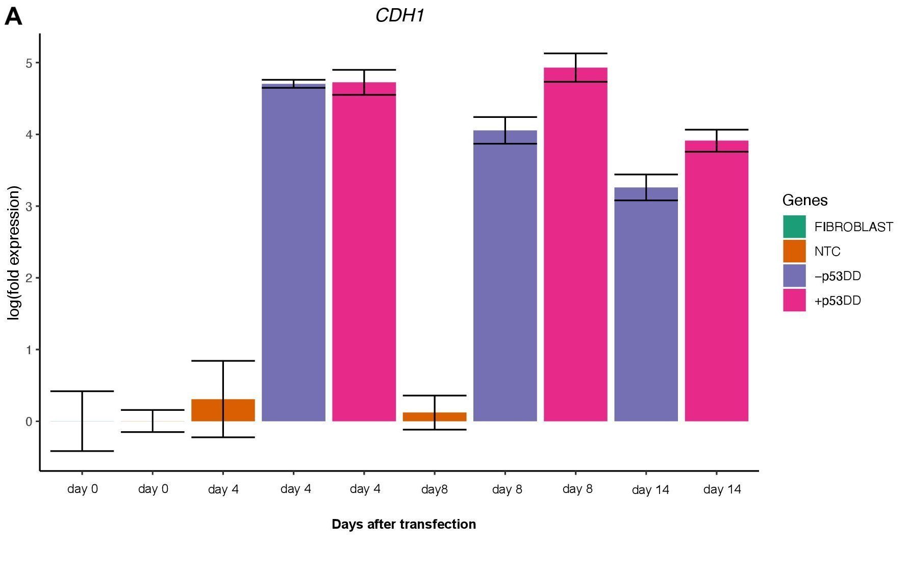
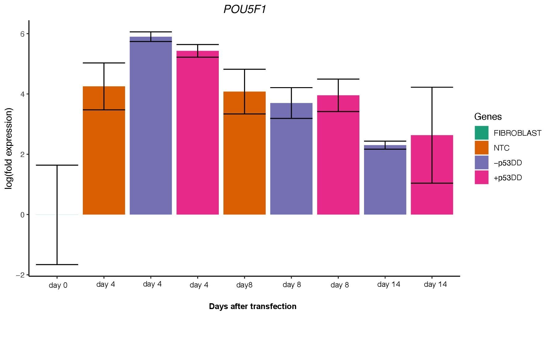
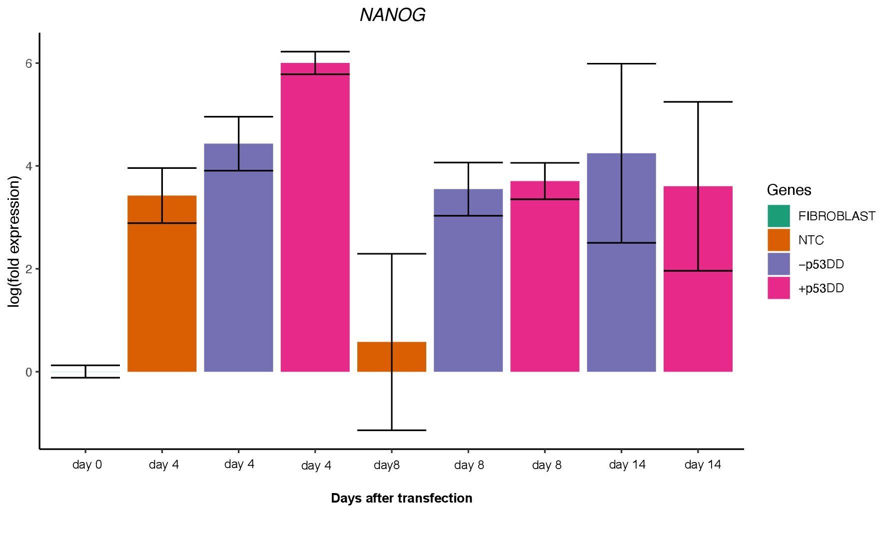
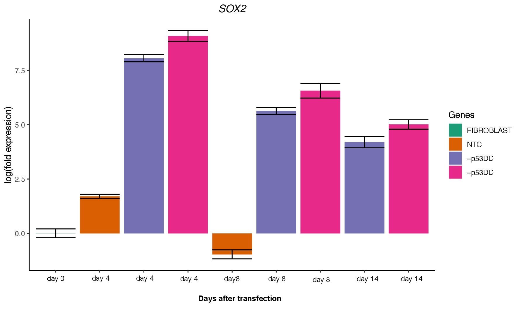
Following pre-expansion for 8 days after transfection, fibroblasts were transferred onto STO feeder cells and changed medium with NSM for induction of iPCS colonies. During reprogramming I sampled cells periodically to harvest their RNA (days 4, 8, and 14). From such RNA samples I obtained cDNAs that I used to perform subsequent RT-qPCR analyses. Using canine specific primers – some of which I personally designed and formerly validated – for endogenous expression of pluripotency markers (POU5F1, NANOG, and SOX2) and other genes of interest (CDH1, CD44, CDNK1A), I was able to assess the reprogramming status of the cells during the process.
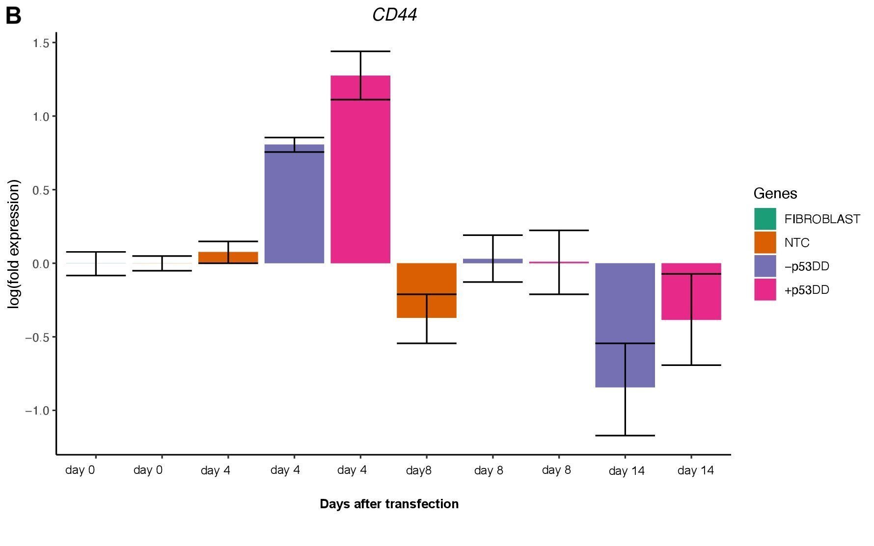
Changes in expression of two markers of reprogramming (CDH1 and CD44) was consistent with what shown in another study (O’Malley et al., 2013). CDH1 (E-cadherin) is upregulated (Figure 3A) indicating mesenchymal-to-epithelial transition which is a typical behavior of the cells entering reprogramming. Furthermore, consistently with O’Malley et al. (2013) CD44 was found to be upregulated at day 4 and progressively downregulated passing the time (Figure 3B), the final population of iPSC are expected to be indeed CD44–.
Unfortunately, I was not able to identify any iPSC colony by day 14, as showed in the Yoshimatsu et al. (2021) study, or later in time under either condition (+/- mP53DD). Upregulation of the core pluripotency markers POU5F1, NANOG, and SOX2 during the experiment, demonstrate the ability of the used EBV-based vector system to induce endogenous expression of pluripotency genes in canine cells; however, such expression dissipates throughout time (Figure 4). The reason I could not obtain any iPSC colony might be that this vector system was shown to not maintain sustained enough endogenous expression of the pluripotent genes to overcome the full barrier of reprogramming.
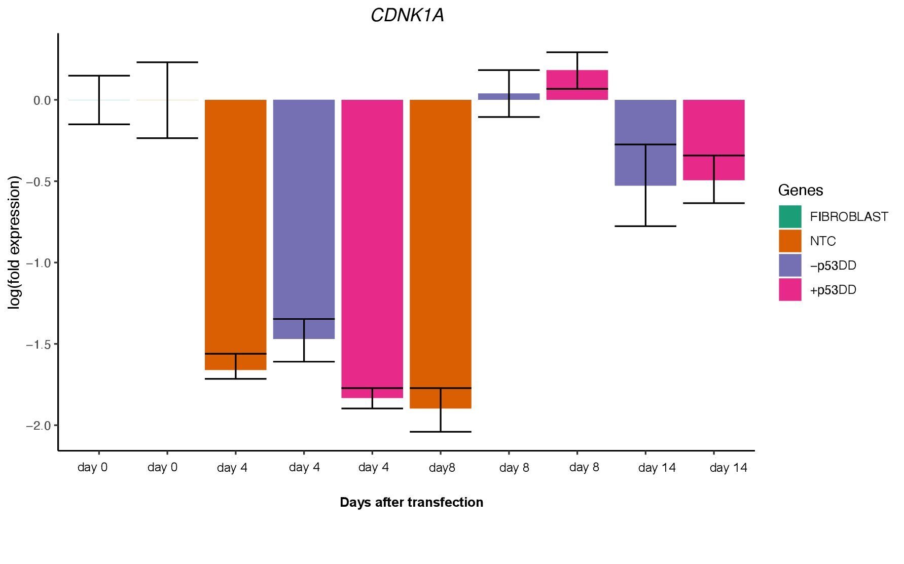
The two vector mixtures +/- mP53DD did not show distinguishable effects, since CDNK1A, direct target of P53showed no difference in expression levels between the two conditions (Figure 5). This suggest that either mP53DDwas expressed at not effective levels or not at all, or that this dominant-negative form of P53 do not interact with the canine form of P53.
Ultimately, this project surely helped to broaden my knowledge in stem cells and reprograming methods, as to learn numerous lab techniques fundamental to pursue hopefully my research career in the future.
Figure 3
Figure 4
Figure 5
References:
O’Malley, J. et al. (2013) High-resolution analysis with novel cell-surface markers identifies routes to iPS cells.
Okita, K. et al. (2011) A more efficient method to generate integration-free human iPS cells. Nature methods. [Online] 8 (5), 409–412.
Yoshimatsu, S. et al. (2021) Non-viral Induction of Transgene-free iPSCs from Somatic Fibroblasts of Multiple Mammalian Species. Stem cell reports. [Online] 16 (4), 754–770.
Maria Diaz de la Loza is an Andalusian researcher and scientific illustrator who has worked in genomic instability and development in both Spain and the UK. Her passion for science has been a huge artistic inspiration and has led to her illustrations and videos being used in scientific publications and events. She is currently working in London as a freelance illustrator and fly technician at King’s College London. Her work can be seen at http://be.net/MariaDiaz_delaLoza and you can follow her on Twitter at @Maria_Diaz_Loza
Maria Diaz de la Loza
Where are you originally from and what do you work on now?
I am originally from Chipiona, a little town by the seaside in the South of Spain. Thanks to the support of my family, I was able to move to the capital of my region to complete my degree in Biology followed by a PhD in Genetics, studying genomic instability in yeast, at the University of Seville. I was ready for a change for my first postdoctoral position, and I moved to a new field to work on development in the fruit fly, in the Andalusian Centre for Developmental Biology. That was a life changing decision. The fly community is remarkably creative, and it truly inspired me to further apply my artistic side to science. For my second and last postdoc, I moved to London, to the new Francis Crick Institute, where I continued working in development. These years as a scientist put me in contact with many different (and amazing) people, mentors and experiences, and they inspired me to pursue a career that combined my two sides: art and science. Now, I work as a freelance illustrator, collaborating with other scientists around the world to illustrate their work. I find it extremely satisfying. I did not want to completely leave academia, and I am lucky to maintain a direct contact with the research community by working as a fly technician at King’s College London, where my supervisor is delighted with my illustrator side and gives me the flexibility that I need to balance my two jobs.
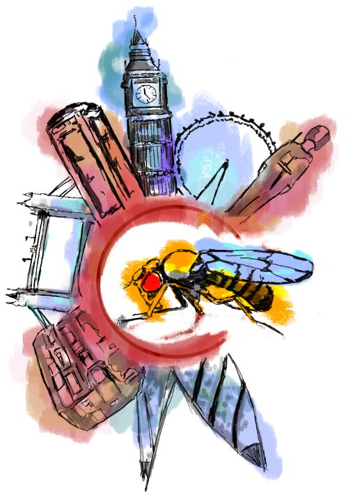
This image was one of my first official commissions; the logo for the 25th European Drosophila Research Conference in London 2017, organised by the European Drosophila Society. This year I designed the logo for the EDS!
Were you always going to be a scientist?
I always wanted to be a biologist (although, at nine years old, I also fancied the idea of being a vet or a hairdresser). Being in close contact with the sea all my life, I thought I would specialise in marine biology. However, after taking my first genetics class I was totally fascinated, and I started to take all the genetics subjects I could. It was after getting in closer contact with that community that I considered the possibility of starting a PhD, and I was lucky they gave me that opportunity in the Department of Genetics of the University of Seville. Even though I started my PhD without a clear idea of how an academic career works, I found that I liked the environment a lot and I have enjoyed many aspects of it since. Being a scientist is not only about science, it allows you to meet all kind of people, to see the world and to grow as a person, and of course you gain an incredible number of new skills, so I will say it is a path worth taking.
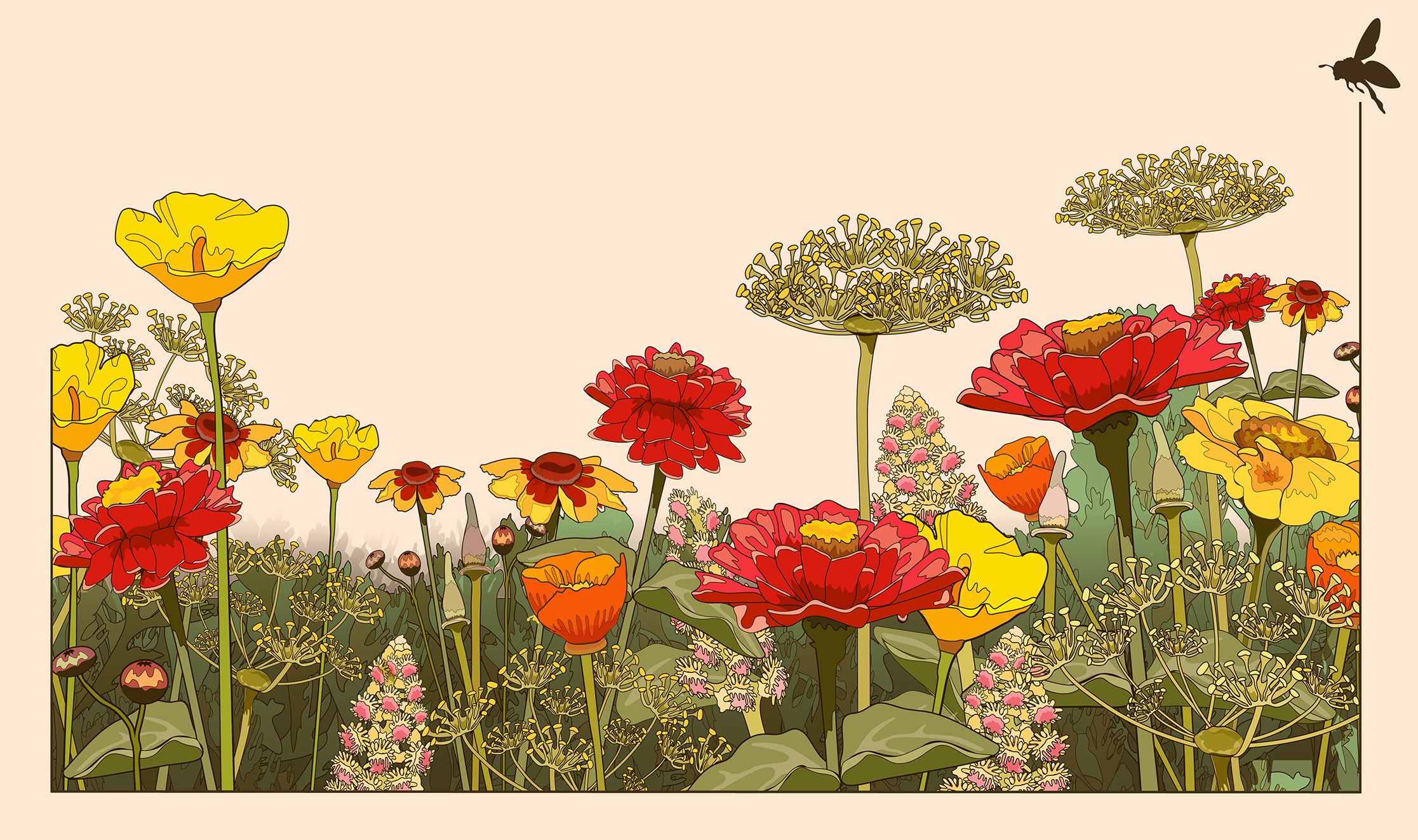
This was a lovely commission from Iris Salecker’s Laboratory in 2020. The lab works on the visual cortex in Drosophila and this artwork was part of their contribution for the Science Fair in Paris (Fête de la Science in the École Normale Supérieure). They wanted to create a flower meadow that they could animate to represent how we see colour and movement. I used Illustrator to recreate a movie that Iris had taken of a garden in full bloom, close to her lab in Paris.
And what about art – have you always enjoyed it?
Absolutely! I have been creating and drawing since I was a little. My mother was an avid reader who liked to copy illustrations from her favourite books, and she always encouraged me to do both. I continued drawing through school as a way to learn and embellish my homework, and I started to learn what I could about visual arts by myself. I even had a moment of doubt, just before starting University, when I played with the idea of enrolling into an Art Bachelor. In the end, biology won – I wanted to do that my entire life – but I never stopped painting and drawing. During my PhD, it was a pleasant surprise to discover how both science and art complement each other. Science is more easily understood with the help of an image, and illustrations are not only useful for communicating your work to others, but also an excellent tool to improve our insight into the process you are studying. I can say that through science, I continued learning and developing my artistic skills.
“During my PhD, it was a pleasant surprise to discover how both science and art complement each other.”
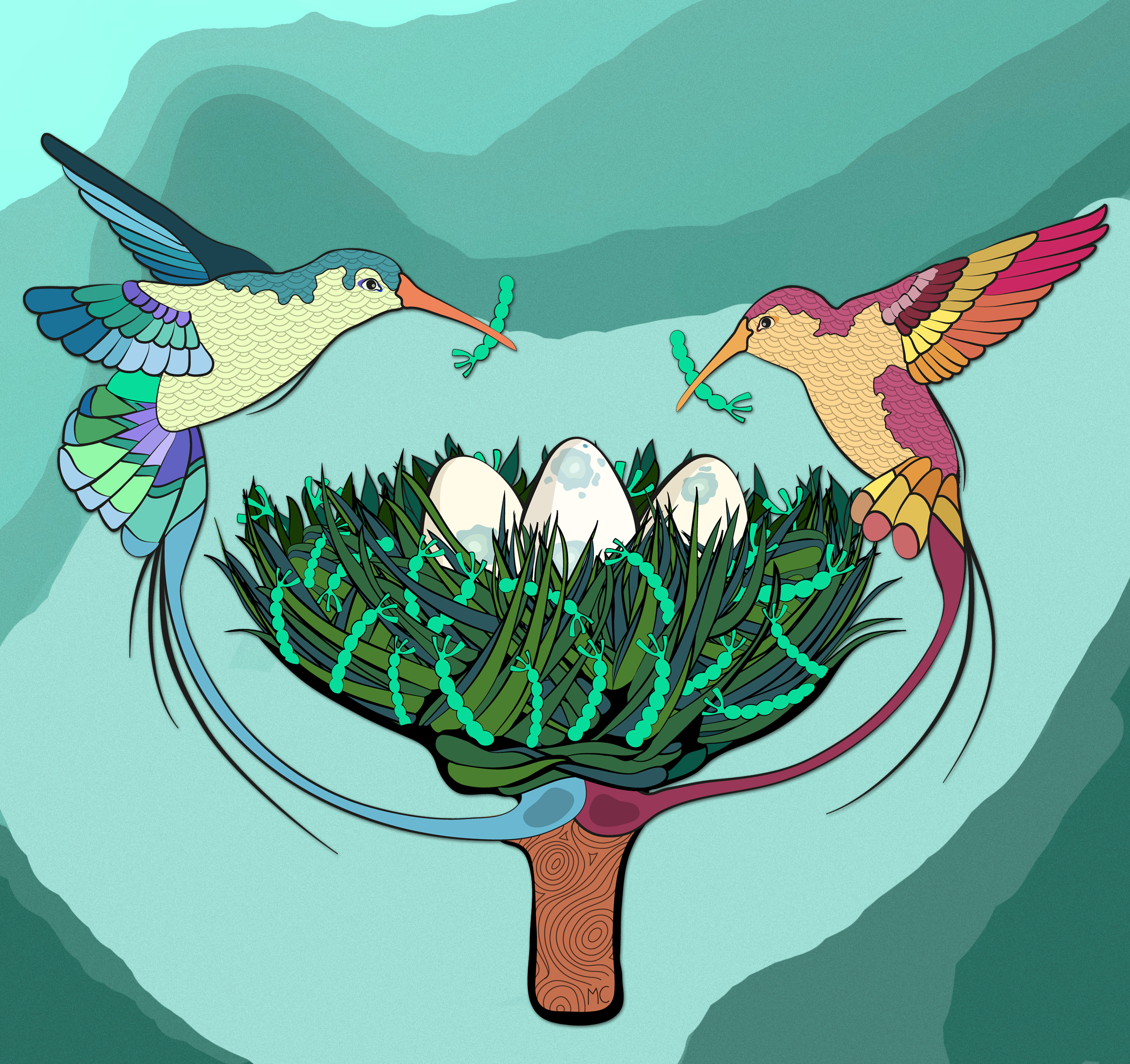
I started to work in developmental biology in my first postdoc, in Acaimo González-Reyes and Maria Dolores Martín-Bermudo labs in the Andalusian Center for Developmental Biology, and now I am thrilled to work with them as an illustrator. This image represents extracellular matrix deposition in the stem cell niche, by ‘hummingbird’ somatic cells, and it was Acaimo’s proposal as a cover for their last publication in Current Biology in 2021. Just a few months ago, we worked together in a video abstract for their publication in Development, showing in detail, biological processes during the cell cycle of germline stem cells.
What or who are your most important artistic influences?
Absolutely everything! I get obsessed with anything that catches my eye and learn from it. Sometimes it is about visual arts, mostly classic and modern painters, or some visually attractive movies. Another major influence is nature itself and certain urban landscapes; I am completely fascinated with the amazingly eclectic London architecture since I moved here. I usually take photographs that I can use for future projects, and I often navigate through them to come up with ideas. Same goes for science, as a developmental biologist I have always worked with microscopy images, and I learn a lot from other people’s work. But inspiration can be found everywhere, sometimes I spend hours watching crafts, makeup, or tattoo videos, and very often I have used everyday objects to come up with a design. I think you can pick up ideas from enjoying any kind of artistic manifestation, which later, can offer you exactly what you need to make a design.
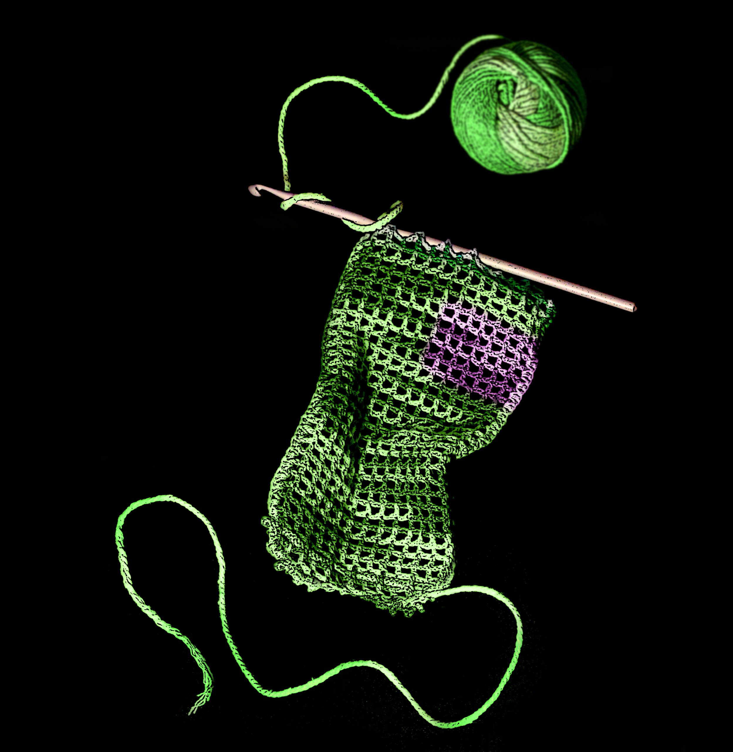
This is my first accepted cover, and it is one of my favourite techniques. I took several photographs of traditional knitting tools and arranged them in a composition with Adobe Photoshop to represent constant secretion and degradation of the extracellular matrix. We even included a small square in magenta to represent an area in which we switch from green fluorescence to magenta. We published it in Developmental Cell, as one of the latest works from Brian Stramer’s lab at King’s College London.
How do you make your art?
As a freelance illustrator, what really fulfil me is the process of illustrating someone else’s work and making it beautiful and precise. The process usually starts by having a chat with the authors to understand their work and the general idea of what they want (which is made easier by my scientific background). Then I start to think about alternative options. At some point, there it is the perfect one. The drawing starts then. First, I prepare some pen and paper sketches, to show my concept to the client, and once we have decided the major points, I move to digital platforms. One of the great aspects of working with other scientists is that they are used to being very clear and concise in what they want. This means my preliminary ideas are usually well received, and the first feedback from the authors helps improved them substantially. The process continues with some back and forth with the authors, to be sure that I am showing exactly what they want. The final product usually consists of digital diagrams and illustrations for publications, journal covers or events, but I also like to combine them with different media. One of my favourite techniques is to take real photographs of everyday objects and combine them to illustrate a biological process as an analogy. Recently I have had some fun working with several labs to create video abstracts, and I am eager to explore more in that direction.
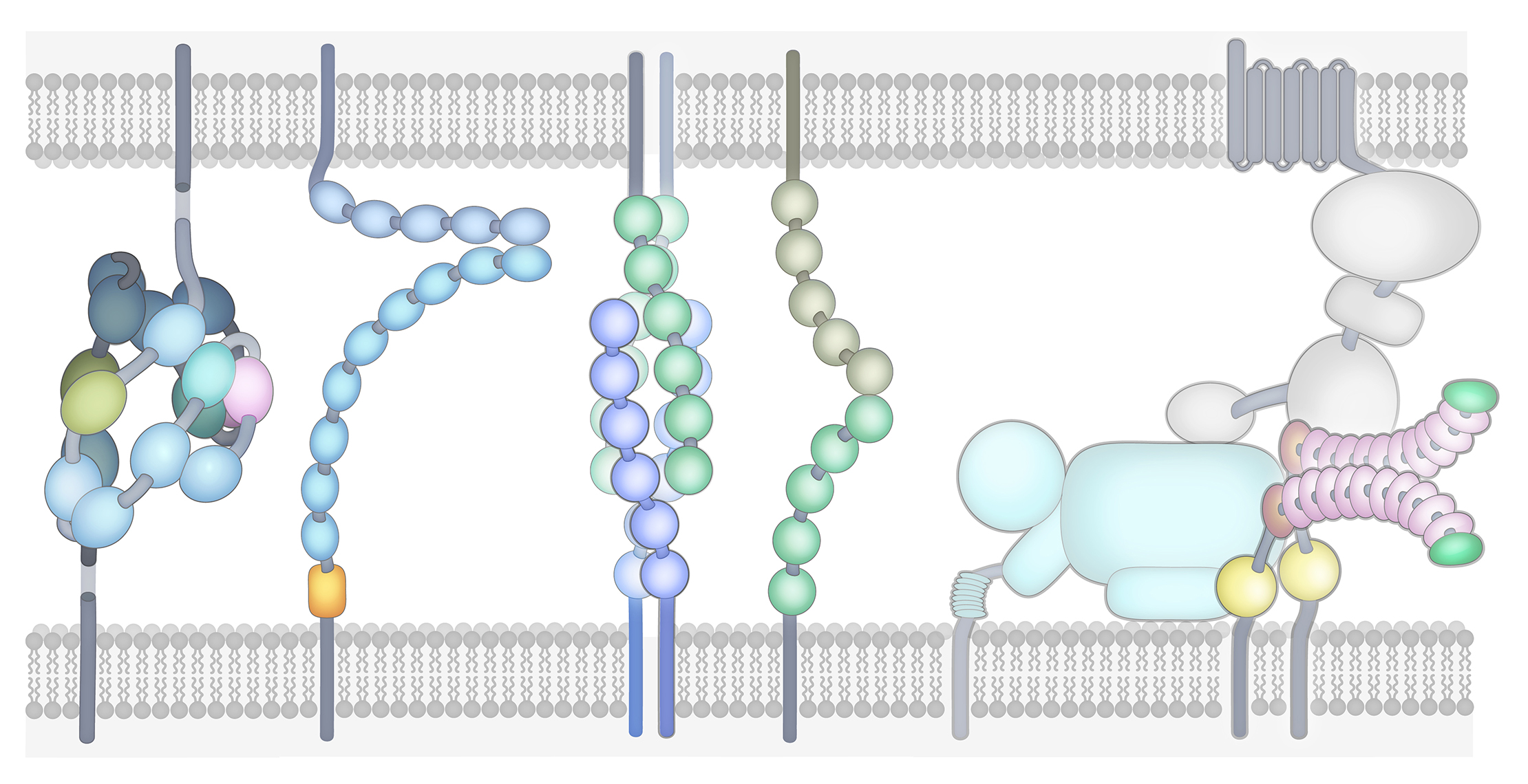
Larry Zipursky and Joshua Sanes (University of California and Harvard University, respectively) were the first scientists that chose me to work on a big project; a very detailed review in the assembly of neural circuits, combining findings in the fruit fly and mice. This work was published in Cell. I learnt a lot from them, both about their fields and on how to work on a big project. This is one of my favourite diagrams in the review, in which we summarised, in one image, the transmembrane proteins that help neurons to interact, making them pleasant to look at while also maintaining a great level of scientific detail.
Does your art influence your science at all, or are they separate worlds?
I think that what inspires me the most to create art, is to apply it to something practical. When I started in academia, art and science started to evolve together and now my artistic side is almost completely focussed on science. I chose to study development because it is strongly based on imaging (I was jealous of their microscopy images!), and I used my artistic side to improve the way I acquire and show my results. Also, I think designing diagrams was a deal-breaker for me. I started to create my own diagrams early on during my PhD, primarily to try to understand what I was doing, but also to help to explain my work to others, and with time I started to do the same for my colleagues. Now I barely attend any kind of talk without drawing their ideas on my notebook. Working with scientific images also helped me to better understand photography, specially of very tiny things, and my connection with other laboratories is very helpful to get specific material for my illustrations. The last step was to officially combine them both, and I am extremely grateful that I have been able to do this during the last two years.
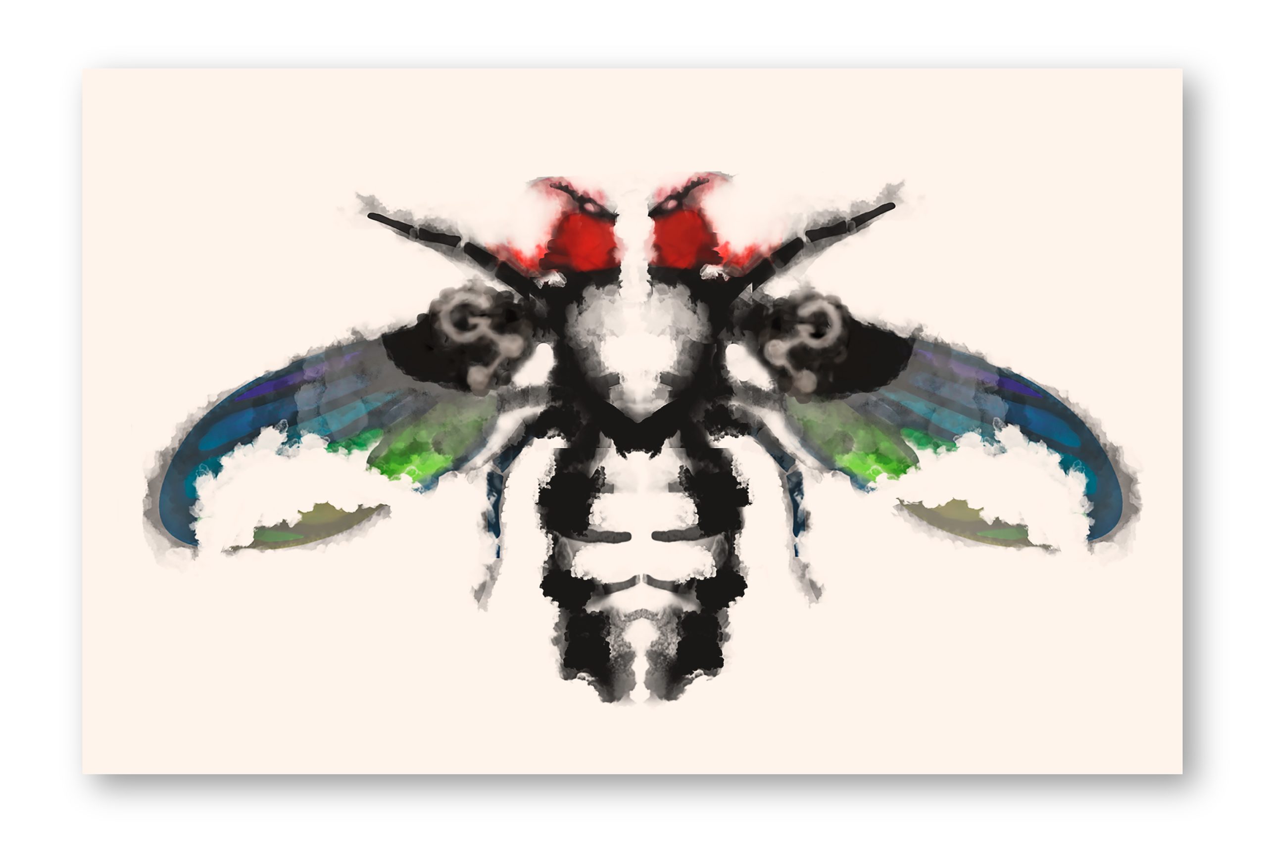
For the new addition to the Genesse Sci Fly Card Collection in 2021, I created a fly design inspired by the famous Rorschach Test cards (because Drosophilists see flies everywhere, right?), highlighting the two tissues more frequently used to study patterns, eyes and wings, including the company logo blended into the design.
What are you thinking of working on next?
I always wonder how it would be to work in illustration full-time; for a journal or a company. Being a freelancer allows you to interact with people around the world and learn about many different projects, which is exciting and it is something that I will miss for sure. However, I have learnt a lot from other illustrators during my freelance stage, and I think working in a department dedicated to science illustration could improve my training further. For now, I am thrilled of working in different projects as a freelancer, but who knows what the future has in store for me!
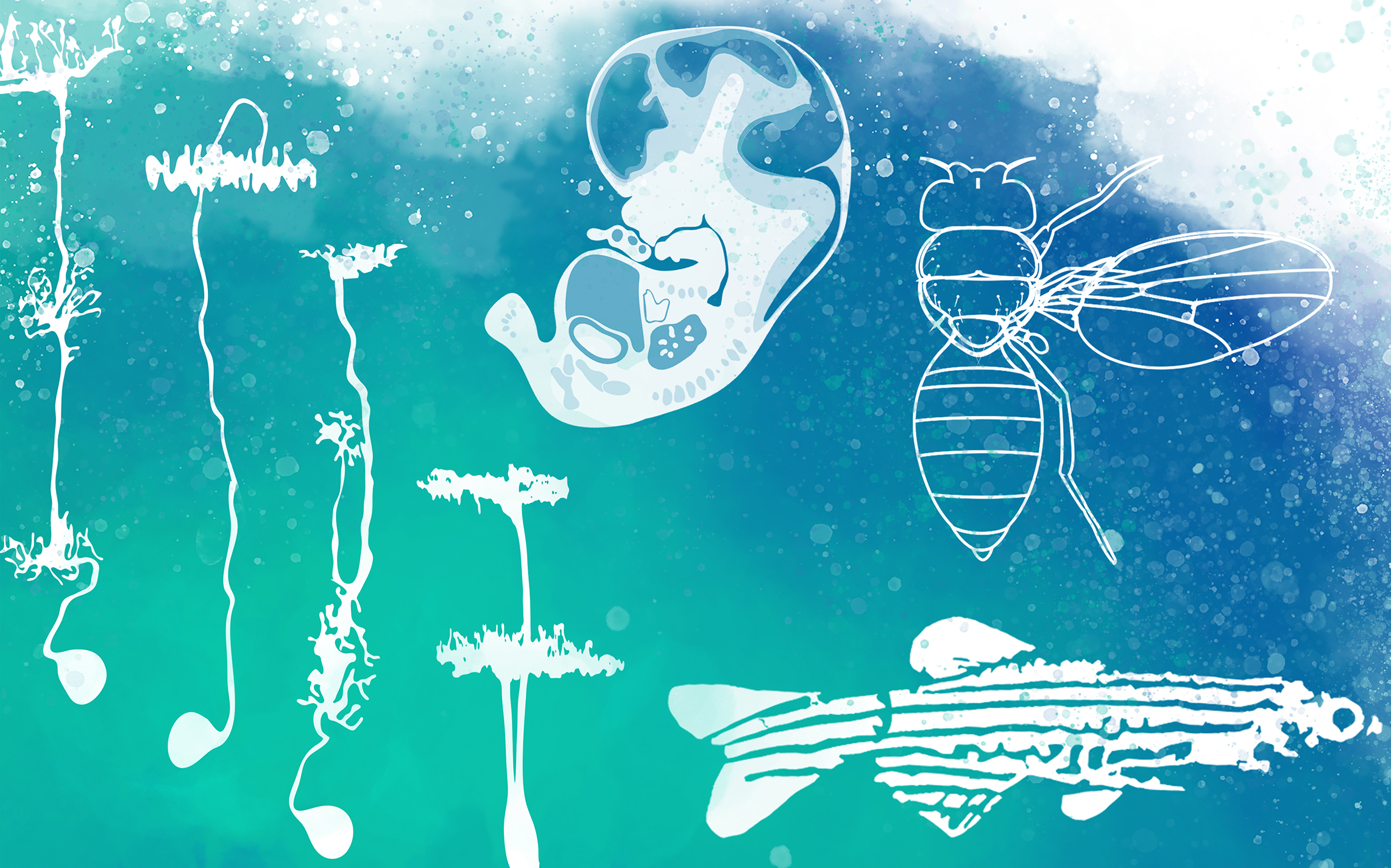
I created this image for myself, to be my banner in social media. It represents how my scientific career has evolved, studying different fields and model organisms, and the different styles I use to create my illustrations.. From single cells to human embryos, realistic shapes or schematics, I always try to be truthful to the scientific context and always, to make science beautiful.
Thanks to Maria and all the other SciArtists we have featured so far.We’re looking for new people to feature in this series – whatever kind of art you do, from sculpture to embroidery to music to drawing, if you want to share it with the community just email thenode@biologists.com (nominations are also welcome!)
 (No Ratings Yet)
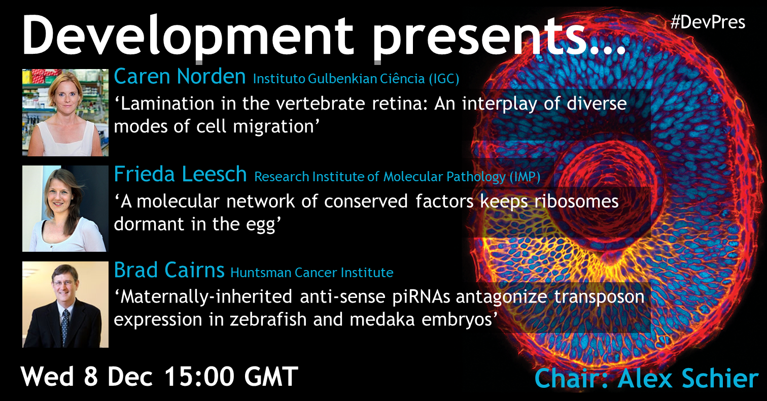
 (1 votes)
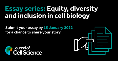
 (3 votes)