Can you hack your hormones to be happier or boost your height? Could a hormone supplement be the key to beating ageing? Are humans heading for a fertility cliff? And is the ‘manopause’ real?
Hormones: The Inside Story, the podcast from the Society for Endocrinology uncovering the facts about hormones and health with an expert-led, myth-busting and entertaining format, is back for a second series.
Hormones affect growth, sleep, body fat, fertility and almost every aspect of our daily lives and health. Sadly, the mainstream media is brimming with misinformation and potentially dangerous advice from a host of non-experts and dubious commercial enterprises.
Building on the huge success of last year’s debut series, the Society for Endocrinology and First Create The Media have just released series two, which continues examining the stories and the science behind hormones, cutting through the myths and misinformation, providing real facts and enabling you to make better decisions about your health.
With the help of presenter Georgia Mills, this series uncovers the truth about how hormones affect our growth, weight, mood, how we age and our declining fertility. Speaking with leading experts, she’ll be finding out about the controversies around the male menopause, fasting and weight loss, whether there really is a fertility crisis and if you can beat the aging process or boost your happiness by hacking your hormones.
Tune in to learn how monkey testicles could link to the fountain of youth, why Irish giants are not just the stuff of legend, the secrets of lengthy prairie vole ‘romance’ and how brushing your teeth could save your life.
Listen and subscribe now on Podbean, Spotify, Apple Podcasts or just search for ‘Hormones: The Inside Story’ wherever you get your podcasts.
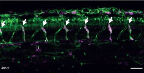
Axons are slender extensions of neurons which can be meter-long and form the biological cables that run through our nerves and brains to hardwire the nervous system. In humans, axons must survive for up to a century; we lose ~40% of axons towards high age and many more in neurodegeneration, but the causes are poorly understood.
To understand axon maintenance and pathologies, we are focussing on the bundles of microtubules (MTs) that run all along axons – be it in a tiny fly or in a human [Ref1]. These bundles determine axon structure and form the highways for life-sustaining axonal transport. Accordingly, MT bundle decay causes axon degeneration. But the mechanisms that maintain these bundles (and might fail in pathology!) are little understood [Refs2,3].
Here we will test the long-standing, but poorly proven hypothesis that axonal bundles are cross-linked by MT-binding proteins. For this, we will study potential architectural functions of MAP1B/Futsch proteins, known to be enriched in axons across the animal kingdom [1]. They show an intriguing evolutionary profile: N- and C-terminal regions are potentially MT-associating and evolutionary well conserved, whereas middle regions show enormous sequence and length differences [FigS1 in Ref1]. MAP1B/Futsch proteins may therefore act as flexible spacers, with their length differences explaining the variations in MT spacing observed in different species [1].
To study MAP1B/Futsch, you will use inter-disciplinary experimental approaches that equip you with a wide range of skills relevant for the biomedical sciences and evolution biology. (a) To determine the precise sub-cellular localisation of MAP1B/Futsch you will use CRISPR/Cas9-mediated protein tagging and apply expansion/electron microscopy; molecular mechanisms will be determined via biochemical and in vitro assays. (b) To study MAP1B/Futsch family evolution you will use computational bioinformatics retrieving and analysing Futsch sequences from multiple species. (c) To determine the functional consequences of evolutionary variability you will generate hybrid proteins and assess their impacts on axon architecture.
The project will be supervised by experts in the field. Andreas Prokop has studied the Drosophila nervous system for 30 years, has long-standing experience with electron microscopy, and is also an expert in science communication (https://poppi62.wordpress.com/publications). Viki Allan studies MT-based transport with expertise in biochemical and in vitro assays to dissect functions of MT-associating proteins [Ref4]. Matthew Ronshaugen specialises on evolutionary biology and his lab is equipped to perform the computational analyses and generate CRISR/Cas9 variants [Ref5].
Entry Requirements
Applicants must have obtained or be about to obtain a First or Upper Second class UK honours degree, or the equivalent qualifications gained outside the UK, in an appropriate area of science, engineering or technology.
Applicants interested in this project should make direct contact with the Primary Supervisor to arrange to discuss the project further as soon as possible.
Equality, Diversity and Inclusion
Equality, diversity and inclusion is fundamental to the success of The University of Manchester and is at the heart of all of our activities. The full Equality, diversity and inclusion statement can be found on the website.
Funding Notes
Funding will cover UK tuition fee and stipend only. The University of Manchester aims to support the most outstanding applicants from outside the UK. We are able to offer a limited number of scholarships that will enable full studentships to be awarded to international applicants. These full studentships will only be awarded to exceptional quality candidates, due to the competitive nature of this scheme.
Background: We discovered that integrins and Rac1, form a pathway essential for axon growth during normal nervous system development. Axon growth establishes connections between neurons across the nervous system, essential for normal cognitive function. Accordingly, integrin mutations cause intellectual disability in mouse and humans [1,2], and we discovered that germline RAC1 mutations cause a human genetic disorder likewise characterised by intellectual disability [3].
Aims: In this project the student will identify further components of the integrin-Rac1 pathway involved in axonal growth, establish their potential disease links and test therapeutic strategies.
Methods:
(a) The student will identify further axon growth-relevant components of the integrin-Rac1 pathway using mass-spectrometry experiments and their bioinformatic analysis.
(b) Candidate proteins will be experimentally assessed in neuronal cell culture and in vivo to assess whether and how they function within the integrin-Rac1 pathway; this will involve extracellular matrix extraction, primary neuron cultures, brain dissections, Drosophila genetics and advanced imaging.
(c) The student will identify disease-relevant mutations in integrin-Rac1 pathway components through interrogating large human genomic datasets via computational bioinformatics analysis and clinical correlation.
(d) Through in vivo studies, the student will establish whether/how candidate proteins impact on axon growth leading to miswiring as a cause for intellectual disability.
(e) The student will have the opportunity to test approved drugs to assess potential ameliorating effects on aberrant axon growth caused by these mutations.
Training: The experimental approaches used are highly inter-disciplinary and will equip the student with a wide range of skills relevant for the biomedical sciences. To guarantee high quality training and optimal progress, the project will be supervised by experts in the field. Martin Humphries is an expert on integrins and the mass-spectrometry analysis of integrin complexes [4]. Andreas Prokop has 30 years experience of applying genetics, cell biology and imaging approaches to study the Drosophila nervous system, integrins and the cytoskeleton [5], and is also an expert in science communication (https://poppi62.wordpress.com/publications); he has established the cellular model used here. Siddharth Banka is an expert in human genetics, analysis of the human genome and has discovered more than 20 novel human disorders [3]. Tom Millard is highly experienced with Drosophila genetics, molecular cloning and imaging and has established a set of disease-relevant Rac1 mutations available for this project [3].
Entry Requirements
Applicants must have obtained or be about to obtain a First or Upper Second class UK honours degree, or the equivalent qualifications gained outside the UK, in an appropriate area of science, engineering or technology.
Funding will cover UK tuition fee and stipend only. The University of Manchester aims to support the most outstanding applicants from outside the UK. We are able to offer a limited number of scholarships that will enable full studentships to be awarded to international applicants. These full studentships will only be awarded to exceptional quality candidates, due to the competitive nature of this scheme.
Thanks to the #DevBio community for sharing their thoughts, especially on twitter. If you have some news that you think we should share on our blog, please get in touch at thenode@biologists.com. If you are interested in getting involved with writing preLights you can find out more here.
Doing great science depends on teamwork, whether this is within the lab or in collaboration with other labs. However, sometimes the resources that support our work can be overlooked. In our new series, we aim to shine a light on these unsung heroes of the science world. The first article in the series is by Lindsay Henderson (NARF Academic Liaison and Post-Doctoral Scientist at Roslin Institute) who describes the work of the National Avian Research Facility, UK.
What is the NARF and where is it based?
The National Avian Research Facility (NARF) was founded in 2013 and is based at The Roslin Institute, on the University of Edinburgh’s Easter Bush campus, UK. The NARF produces and maintains poultry lines for use in scientific research and consists of two units, a conventional facility and a facility that has specified pathogen free (SPF) status.
The Greenwood building conventional facility, an example of our egg incubator (setter) and a Roslin Green (eGFP) chick on the right and wild-type chick on left under blue light1.
What services does the NARF offer for developmental biologists?
The NARF supplies fertile eggs from our range of chicken lines that have a broad utility in developmental biology. These include, commercial layer and broiler chicken lines, and Japanese quail.
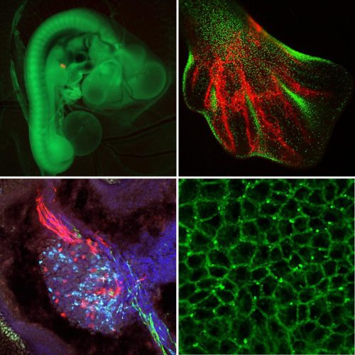
Researchers within the Roslin Institute Chicken Embryology (RICE) group have generated a number of genetically-altered (GA) fluorescent reporter chicken lines that are valuable tools for developmental biology2. These lines carry fluorescent reporter transgenes that are expressed ubiquitously or are inducible in cells of the developing embryo and are used for fate mapping, cell lineage tracing and tissue grafting. At this time the NARF has three ubiquitous reporter chicken lines; the Roslin Green (eGFP)3, Flamingo (TdTomato)4 and Membrane GFP5, and an inducible reporter chicken line, the Chameleon (Cytbow)2.
The NARF also maintains fluorescent reporter chicken lines that are gene specific. This includes a number of immune cell reporter lines, such as the CSF1R-reporter lines6, that can be used to visualise immune cells, including macrophages and dendritic cells.
From top left clockwise; 1) GFP chicken embryo with a ‘red’ graft placed into the limb bud with micro-surgery. The graft is about 50-100uM (Davey Group). 2) HH32 stage limb bud from the CSF1R-eGFP embryo. Macrophages (green) are concentrated in areas of cell death, blood vessels (red) are labelled with SNA lectin (Balic Group). 3) Fluorescent image of the dorsal skin (periderm) from membrane GFP chick embryo (Headon Group). 4) Dorsal Root Ganglion of the nervous system of a Chameleon transgenic chicken embryo. Nerves going into the dorsal root ganglion are red, and nerves coming out are green (Davey Group).
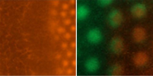
From left to right; The spreading wave of feather formation in a TdTomato chick embryo and the feather buds formed in the skin of a GFP/TdTomato chimeric embryo (both approx. HH32). Images from the Headon Group.
The NARF also holds a spontaneous mutation line, the talpid3line that is a classical recessive embryonic lethal chicken mutant with abnormal limb patterning and malformations7–9. The talpid3line has provided direct insights into clinical genetics and specifically the genes responsible for limb abnormalities and ciliopathies in humans9,10.
The NARF maintains one of the only SPF inbred lines, Line 0, that is free of Avian Sarcoma-Leukosis Virus (ASLV) ev loci. Embryos from this line may be particularly suitable for research using RCAS retrovirus vectors11.
Who are the developmental biologists based at the Roslin Institute that use the NARF?
RICE is a collective of research groups based at the Roslin Institute that use the chicken embryo to study vertebrate embryonic growth and development. RICE includes; the Balic Group that has produced the first transgenic chicken lines which allow specific sub-population of chicken immune cells to be visualised. The Davey Group examines the causative alterations of gene expression which lead to variations in phenotype using comparative anatomy, genomics and embryonic manipulation of avian species, with a particular interest in the molecular anatomy of the developing limb bud. The Headon Group focuses on the development, maintenance and repair of the skin and its appendages. The Rainger Group’s research is focused on the fusion of tissues in the developing retina using the chicken embryo as an experimental model system. The McGrew Group has made major advances in and continues to explore gene editing of avian germ cells and biobanking. The Clinton Group investigates the molecular control of sex determination and gonadal development, and the mechanisms underlying sexual dimorphisms in birds. The Sang Group has a broad focus on the development and application of technologies for genome engineering of the chicken.
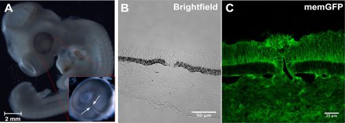
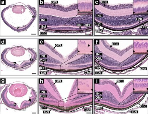
Membrane GFP (memGFP) is used to reveal optic fissure fusion dynamics in the chick eye. A) Whole mounted image of memGFP chicken embryo (HH26); inset – dissected eye showing optic fissure margin and the ventral region (arrows). B) Confocal optical section brightfield image of chick optic fissure at point depicted by arrows in A. C) Fluorescent image of same memGFP chick optic fissure used to distinguish fusion point. Figure provided by the Rainger Group.
What are the strengths of the chick model system in today’s developmental biology?
Recent advances in genome modification in the chicken led by Mike McGrew using the NARF facilities, have made possible the rapid and cost-effective production of GA chicken lines. In addition to new methods to create precise and targeted gene modifications, like CRISPR/Cas9 technologies, this greatly advances the potential of gene editing in chickens for developmental biologists. These advances in gene editing and transgenesis in conjunction with the experimental accessibility of chick embryos, and the ease of live culture and imaging, make the chick a powerful model for future developmental biology research.
Gene editing in chickens can be used to create targeted mutations, to knock-out genes of interest and enable inducible ablation of cell types that contain specific genes12. Recently, the NARF’s gene editing technologies have made possible research investigating avian sex determination using targeted mutations in the DMRT1 gene13. Gene editing in the chicken will soon be used to explore the genes responsible for defects of the optic fissure closure in the eye that cause ocular coloboma14.
How can researchers get involved with the NARF?
The NARF can supply fertile eggs for research and can supply a quote for eggs for inclusion in grant applications. Please find the full list of the lines we provide here. To order eggs from any of the poultry lines or to obtain costings for grant applications, contact narf@roslin.ed.ac.uk.
We continue to develop new GA chicken lines to aid in research under both conventional and SPF conditions. We welcome new collaborations with developmental biologists based at other research institutes. If you have any questions or are interested in a collaboration please contact Lindsay Henderson the NARF Academic Liaison.
The NARF is supported by the University of Edinburgh and the UKRI-Biotechnology and Biological Sciences Research Council.
Publications
Macdonald, J. et al. Efficient genetic modification and germ-line transmission of primordial germ cells using piggyBac and Tol2 transposons. Proc. Natl. Acad. Sci. U. S. A.109, 8803 (2012).
Davey, M. G., Balic, A., Rainger, J., Sang, H. M. & McGrew, M. J. Illuminating the chicken model through genetic modification. Int. J. Dev. Biol.62, 257–264 (2018).
McGrew, M. J. et al. Localised axial progenitor cell populations in the avian tail bud are not committed to a posterior Hox identity. Development135, 2289–2299 (2008).
Ho, W. K. W. et al. Feather arrays are patterned by interacting signalling and cell density waves. PLoS Biol.17, (2019).
Rozbicki, E. et al. Myosin-II-mediated cell shape changes and cell intercalation contribute to primitive streak formation. Nat. Cell Biol.17, 397–408 (2015).
Balic, A. et al. Visualisation of chicken macrophages using transgenic reporter genes: Insights into the development of the avian macrophage lineage. Development141, 3255–3265 (2014).
Ede, D. A. & Kelly, W. A. Developmental abnormalities in the head region of the talpid3 mutant of the fowl. J. Embryol. Exp. Morphol.12, 161–182 (1964).
Ede, D. A. & Kelly, W. A. Developmental abnormalities in the trunk and limbs of the talpid3 mutant of the fowl. J. Embryol. Exp. Morphol.12, 339–356 (1964).
Fraser, A. M. & Davey, M. G. TALPID3 in Joubert syndrome and related ciliopathy disorders. Current Opinion in Genetics and Development56, 41–48 (2019).
Davey, M. G., Towers, M., Vargesson, N. & Tickle, C. The chick limb: Embryology, genetics and teratology. Int. J. Dev. Biol.62, 253–263 (2018).
McNally, M. M., Wahlin, K. J. & Canto-Soler, M. V. Endogenous expression of ASLV viral proteins in specific pathogen free chicken embryos: Relevance for the developmental biology research field. BMC Dev. Biol.10, 106 (2010).
Ballantyne, M. et al. Direct allele introgression into pure chicken breeds using Sire Dam Surrogate (SDS) mating. Nat. Commun.12, 659 (2021).
Ioannidis, J. et al. Primary sex determination in birds depends on DMRT1 dosage, but gonadal sex does not determine adult secondary sex characteristics. Proc. Natl. Acad. Sci.118, e2020909118 (2021).
Rainger, J. Novel approaches to define tissue fusion mechanisms in embryonic development. Funder: UK Research and Innovation Available at: https://gtr.ukri.org/projects?ref=MR%2FS033165%2F1.
A press release from Universität Zürich on Lienkamp lab paper, published in Development
Using cutting-edge genetic engineering, UZH researchers have developed a model to study hereditary kidney disease with the help of tropical frogs. The method allows them to collect large amounts of data on anomalies, which can then be analyzed using artificial intelligence. The research opens up new opportunities in the search for new treatment approaches for the hitherto incurable disease.
Frogs’ anatomy and organ function are strikingly similar to those of humans. An international team led by Soeren Lienkamp, professor at the Institute of Anatomy at UZH, has now exploited this similarity by using a tiny tropical frog called Xenopus tropicalis to model human genetic diseases. The researchers focused on polycystic kidney disease, a congenital and currently incurable form of progressive kidney deterioration, and replicated it in frogs.
Observing disease processes in real time
Using CRISPR/Cas9, a methodology for turning off gene function, the scientists targeted genes known to play a role in cystic kidney disease. “Our novel frog models develop cysts in the kidneys within only a few days, allowing us to observe these disease processes in real time for the first time,” says lead author Thomas Naert. While most genetic studies are performed on mice, frogs have features that make them well-suited for larger scale studies. “One frog couple can produce hundreds or even thousands of eggs,” says Naert. “That’s why you see such large numbers of tadpoles in the Swiss lakes in springtime.” Similarly, in the lab large numbers of Xenopus tropicalis tadpoles can be manipulated to develop cystic kidney diseases.
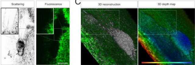
AI analyzes data from light-sheet microscopy
To analyze the data from such a large number of animals, the team employed a technique called light-sheet microscopy, which produced a 3D reconstruction of the entire tadpole and all its organs. Much like magnetic resonance imaging, light-sheet techniques make it possible to see through tissues in tadpoles to find disease-affected organs. The collected data was then processed using artificial intelligence to allow rapid, automated assessment of disease. “While it would normally take my team several days or even weeks to analyze data from hundreds of tadpoles, artificial intelligence can now do this task in a matter of hours,” says Lienkamp.
The findings from frog models analyzed in this way provide new insights into the early processes of polycystic kidney disease. These insights will form the basis for developing new treatment approaches for affected patients.
The study was funded by the Swiss National Science Foundation (SNSF), NCCR Kidney.ch, and ERC Horizon2020 (Starting Grant and Marie Skłodowska-Curie Program).
Many internal organs fill and empty periodically while carrying out their normal physiological function. Associated peripheral neurons act as specialised mechanoreceptors to detect changes in organ volume, and relay this information to the brain, where it is processed, and used to evoke appropriate physiological and behavioural response(s). For example, during a meal the stomach expands to accommodate ingested food, triggering neural circuits to inhibit feeding behaviour, promote digestion, and evoke feelings of satiety, fullness, nausea or pain, depending on the size of the meal. Despite being central to normal physiological function many basic questions remain about the mechanisms of organ volume sensing.
In this project you will explore how mechanisms of gut volume sensing control physiology and life cycle in a classic insect model—the blood-sucking bug Rhodnius prolixus. You will use state-of-the-art techniques in genomics, imaging, genetic manipulation, and gene/protein expression analysis to identify the molecular and cellular mechanisms of mechanotransduction in the Rhodnius gut and define the relevant neural circuits that act to control profound changes in this creature’s physiology and development in response to feeding.
This project is a curiosity-driven exploration into a fundamental question relating to how animals sense and respond to their internal world. The molecular mechanisms of organ volume sensing are not well understood for any animal, including humans, but are likely to be conserved. Therefore, the project is likely to provide insight into physiological processes that are key for maintaining health in humans, illuminating areas relating to appetite, overeating and disorders connected to visceral pain.
Specific details about the project:Rhodnius prolixus is a blood-sucking bug of immoderate feeding habits. It can take from its host a volume of blood sufficient to increase its own weight by about tenfold. The food-swollen gut distends the abdomen and sets in train a series of endocrinological processes that culminate in profound changes to the physiology and life cycle of the animal. These include (i) a rapid diuretic response, enabling the animal to jettison excess salts and water, returning the animal to a more comfortable state, and (ii) stimulation of body growth and maturation that precede the transition to the next life phase (an older nymph or metamorphosis to adult form).
A major goal of the current project is to determine how abdominal distension is sensed and transduced to the brain to elicit such dramatic changes.
For more details please contact: Barry.Denholm@ed.ac.uk
Completed application form along with your supporting documents should be sent to our PGR student team at sbms-postgraduate@ed.ac.uk by 16 December 2021. References: Please send the reference request form to two referees. Completed references for this project should also be returned to sbms-postgraduate@ed.ac.uk by the closing date: 16 December 2021.
It is your responsibility to ensure that references are provided by the specified deadline.
Promoter repression and 3D-restructuring resolves divergent developmental gene expression in TADs Alessa R. Ringel, Quentin Szabo, Andrea M. Chiariello, Konrad Chudzik, Robert Schöpflin, Patricia Rothe, Alexandra L. Mattei, Tobias Zehnder, Dermot Harnett, Verena Laupert, Simona Bianco, Sara Hetzel, Mai Phan, Magdalena Schindler, Daniel Ibrahim, Christina Paliou, Andrea Esposito, Cesar A. Prada-Medina, Stefan Haas, Peter Giere, Martin Vingron, Lars Wittler, Alexander Meissner, Mario Nicodemi, Giacomo Cavalli, Frédéric Bantignies, Stefan Mundlos, Michael I. Robson
The p53-p21 axis plays a central role in lymphatic homeostasis and disease Rohan Mylavarapu, Molly R. Kulikauskas, Cathrin Dierkes, Nema Sobhani, Michelle Mangette, Jeffrey Finlon, Wanida Stevens, Farinaz Arbab, Neil F. Box, Mark Lovell, Ajit Muley, Carrie J. Shawber, Beth Tamburini, Friedemann Kiefer, Tamara Terzian
A kinase-dead Csf1r mutation associated with adult-onset leukoencephalopathy has a dominant-negative impact on CSF1R signaling Jennifer Stables, Emma K. Green, Anuj Sehgal, Omkar Patkar, Sahar Keshvari, Isis Taylor, Maisie E. Ashcroft, Kathleen Grabert, Evi Wollscheid-Lengeling, Stefan Szymkowiak, Barry W. McColl, Antony Adamson, Neil E. Humphreys, Werner Mueller, Hana Starobova, Irina Vetter, Sepideh Kiani Shabestari, Matthew M. Blurton-Jones, Kim M. Summers, Katharine M. Irvine, Clare Pridans, David A. Hume
ACTN2 missense variant causes proteopathy in human iPSC-derived cardiomyocytes Antonia T. L. Zech, Maksymilian Prondzynski, Sonia R. Singh, Ellen Orthey, Erda Alizoti, Josefine Busch, Alexandra Madsen, Charlotta S. Behrens, Giulia Mearini, Marc D. Lemoine, Elisabeth Krämer, Diogo Mosqueira, Sanamjeet Virdi, Daniela Indenbirken, Maren Depke, Manuela Gesell Salazar, Uwe Völker, Ingke Braren, William T. Pu, Thomas Eschenhagen, Elke Hammer, Saskia Schlossarek, Lucie Carrier
In the latest episode of Genetics Unzipped, presenter Kat Arney is getting in harmony with the science of music. Is there a music gene? Does musical talent really run in families? And how does the inability to perceive music impact on daily life?
Music is a deeply human characteristic – whether it’s clapping, tapping, singing or playing, most of us love to listen – and maybe move – to good tune or a funky beat, and there are plenty of music makers in the world, from schoolkids playing the recorder or making beats on a laptop to virtuoso concert pianist and global pop stars. But where does our musical urge come from? And is it in our genes?
Kat chats with Reyna Gordon. associate professor and director of the Music Cognition Lab in the Department of Otolaryngology and the Genetics Institute at Vanderbilt university in Tennessee. She’s the recipient of a prestigious NIH Director’s New Innovator Award for her work looking at the underlying biology of why rhythm means so much to us.
Our second guest is Jasmin Pfeifer, from Heinrich-Heine University in Dusseldorf, Germany. A linguist by training, Jasmin has found herself involved in the world of genetics through her studies of a condition called congenital amusia, or hereditary tone-deafness.
If you enjoy the show, please do rate and review on Apple podcasts and help to spread the word on social media. And you can always send feedback and suggestions for future episodes and guests to podcast@geneticsunzipped.com Follow us on Twitter – @geneticsunzip
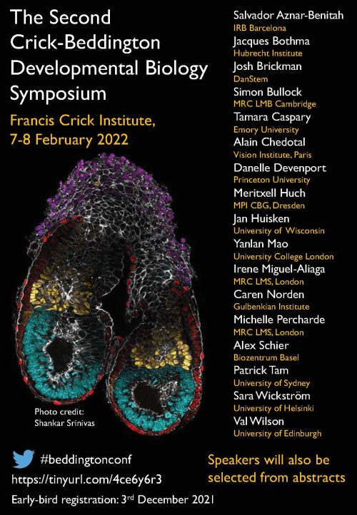
This meeting will showcase state-of-the-art developmental biology across the lifecourse. The multidisciplinary programme will highlight the latest innovations in live imaging of developing systems, organoid approaches to unravel developmental mechanisms, and the mathematical modelling of morphogenesis and organ homeostasis. Featuring 17 internationally renowned plenary speakers, alongside short and flash talks selected from the submitted abstracts of early career researchers.
Discounted early-bird registration until 3rd December
Abstract submission deadline: 16th December
Registration, abstract submission and speaker biogs here:

 (No Ratings Yet)