One of the central questions in developmental biology is how different cell fates are generated from a single founding cell. Although great strides have been made in our understanding of this problem in animals, the evolutionary origins of this process are not understood. It is known that many unicellular organisms progress through different cell stages during their life cycle, known as temporal cell differentiation, and it has been hypothesized that spatial cell differentiation (as seen in animals) evolved from this more ancient differentiation-mode. A full understanding of how this occurred has been hampered by a lack of information on the basic principles underlying temporal cell differentiation in the closest relatives of animals, the unicellular holozoans.
In recent years, several studies have revealed that many of the genes and pathways directly related to development and cell fate in animals were already present in their unicellular ancestors. Moreover, many examples have shown the formation of specialized cell types in response to specific environmental ques and transient multicellular structures have been reported in many unicellular holozoan lineages. Therefore, recent discoveries strongly point towards an earlier origin of several developmental processes, including cell differentiation, than was previously thought and make a strong case that understanding the mechanisms underpinning “development” in these unicellular lineages will be key to understand the emergence of definitive animal cell differentiation and development.
The Workshop will consist of sessions of talks and discussions centred around various aspects of development to unicellular holozoans. Each session will contain a mixture of researchers working on unicellular holozoans and those working on other eukaryotic systems who will provide alternative insights. Through these sessions the Workshop will build knowledge aiming to produce a white-paper document outlining the emerging conceptual framework in the field, the major outstanding questions as well as seeding collaborative efforts to address these questions.
Organisers & speakers
Elena Casacuberta Institute for Evolutionary Biology, Spain James Gahan University of Galway, Ireland
Detlev Arendt EMBL, Germany David Booth University of California, San Francisco, USA Thibaut Brunet Institut Pasteur, France Pawel Burkhardt University of Bergen, Norway Susana Coelho Max Planck Institute for Biology Tübingen, Germany Omaya Dudin University of Geneva, Switzerland Nicole King University of California, Berkeley, USA Lucie Laplane CNRS, Université Paris, France Eric Libby Umeå University, Sweden Aurora Mihaela Nedelcu University of New Brunswick, Canada Àlex de Mendoza Queen Mary University of London, United Kingdom Iñaki Ruiz-Trillo The Institute of Evolutionary Biology, Spain Florentine Rutaganira Stanford University, USA Arnau Sebé-Padrós Centre for Genomic Regulation, Spain Hiroshi Suga Prefectural University of Hiroshima, Japan Katrina Velle UMass Dartford, USA Renske Vroomans University of Cambridge, United Kingdom
We offer 10 funded places for early-career researchers (PhD, postdocs and PIs in the first three years of their first appointment) to attend our Workshops along with the 20 invited speakers. We just ask that you pay for your own travel costs. If you would like to attend please complete the online application form and include a one page CV and a letter of support from your supervisor. If your supervisor would prefer to send the letter directly to us please ask them to email it to workshops@biologists.com
All attendees are expected to actively contribute to the Workshops by asking questions at presentation sessions and taking part in discussions, as well as giving a short talk on their research.
The early-career research deadline is on Friday 12 June 2026. For more information, visit the Company’s Workshops page.
This meeting was particularly meaningful to me, as it marked my first engagement with the developmental biology community in India since beginning my academic training abroad four years ago in 2021. I joined the laboratory of Smadar Ben-Tabou de-Leon at the University of Haifa, Israel, for my master’s research and subsequently continued in the same group for my doctoral studies. My research focuses on the regulation of sea urchin larval skeletogenesis, with a particular emphasis on the role of cytoskeletal remodelling proteins in controlling morphogenesis. Against this backdrop, the meeting provided a valuable opportunity for me to reconnect with the broader research community and situate my work within the expanding landscape of developmental biology.
The conference brought together a diverse group of established and early-career scientists, including principal investigators, graduate students, and postdoctoral researchers from across India and internationally. It highlighted the rapidly evolving nature of the field, where classical embryology is increasingly integrated with stem cell-based models, quantitative approaches, and systems-level perspectives. Personally, one of the best parts of this conference was that there were no parallel sessions – so, we didn’t have to choose between different talks (or seminar halls!).
Understanding gastrulation, human gastruloids and metabolism in context of stem cell-based models
One of my favourite talks was by Alfonso Martínez Arias (Universitat Pompeu Fabra, Spain), a renowned developmental biologist and one of the authors of the exceptional book: Wolpert’s Principles of Development (Wolpert’s Principles of Development – Paperback – Lewis Wolpert; Cheryll Tickle; Alfonso Martinez Arias; Marysia Placzek – Oxford University Press). He presented the gastrulation from the perspective of pluripotent stem cells and gastruloids, highlighting how self-organizing stem cells aggregate when exposed to defined signalling and can generate post-gastrulation-like body plans.
Extending the gastruloid paradigm to human biology, Maneesha Inamdar(Institute for Stem Cell Science and Regenerative Medicine, India) presented a robust, accessible human gastruloid-based platform for studying early cell fate decisions and teratogenicity. Metabolism plays an instructive role during early development – was the major takeaway from Maneesha’s talk. Her work addressed a longstanding challenge in developmental biology and medicine: the lack of ethically permissible and human-relevant models for post-implantation development. Towards the end of her talk, she also introduced the Centre for Research, Application and Training in Embryology (CReATE) – a research institute within theInStem, that aims to conduct innovative research to advance understanding of early human development.
The discussion on the role of metabolism in development was continued by Sally Dunwoodie (Victor Chang Cardiac Research Institute, Australia), who focused on the role of nicotinamide adenine dinucleotide (NAD) metabolism in embryogenesis. She presented that during early development, NAD deficiency can lead to a plethora of congenital abnormalities, since metabolism is a key regulatory layer in embryogenesis.
Organogenesis, spatial patterning, and regeneration
The meeting also included a dedicated session on the themes of organogenesis and regeneration across organisms and developmental systems. Peter Currie (Monash University, Australia) discussed comparative insights into muscle stem cell systems across vertebrates, highlighting how distinct stem cell populations are regulated during growth, maintenance, and regeneration. James Briscoe (Francis Crick Institute, UK) gave the Jean Brachet Memorial Lecture, highlighting the importance of temporal dynamics and spatial patterning during spinal cord development. His talk emphasised how cells can interpret morphogen signals in a time-dependent fashion to precise neuronal identities. He famously presents the “6 Ps of Developmental Biology”—position, pattern, proportions, precision, pace, and purpose. Adding on to this theme, Raj Ladher(National Centre of Biological Sciences, India) presented findings on cochlear morphogenesis, showcasing how tightly regulated cell differentiation and tissue remodelling generate the complex architecture of the inner ear. Complementing these studies on organ morphogenesis, Richard Behringer (MD Anderson Cancer Centre, USA) talked about tissue remodelling during sex differentiation, highlighting how anti-Müllerian hormone–dependent epithelial-mesenchymal interactions drive Müllerian duct regression during male development.
Physical forces and morphogenesis work hand in hand
Several talks highlighted the emerging importance of mechano-genetic regulation of the developmental program. Speakers Tamal Das (Tata Institute of Fundamental Research, India) and Priti Agarwal (National Centre of Biological Sciences, India) discussed how cytoskeletal dynamics, actomyosin contractility, and mechanical feedback influence cell behaviour and organ morphogenesis. Their work demonstrated that morphogenesis commences from a close interaction between biochemical signalling and physical constraints. On par with this perspective, Shankar Shrinivas’s (University of Oxford, UK) work demonstrated patterned heterogeneities in tissue mechanics during early mammalian development, showing how spatial differences in mechanical properties guide cell migration and anterior patterning. All together, these talks emphasized the importance of mechanical forces’ role as an active regulatory layer across different developmental scales. Richa Rikhy (Indian Institute for Sciences, Education and Research, India) delivered the Anne McLaren Award Lecture. Her work using Drosophila as a model system emphasized that mitochondria are not merely metabolic organelles but function as active regulators of developmental processes. She highlighted how mitochondrial morphology—regulated by coordinated fission and fusion events—modulates cellular metabolism, redox state, and signalling pathways, ultimately influencing cell fate decisions.
Translational impact
Bringing a developmental disease perspective, Loydie Majewska (McGill University Health Centre Research Institute, Canada) showed that TMED2-dependent protein trafficking is essential for craniofacial and heart morphogenesis. Using conditional mouse models, she linked defects in secretory pathway function to congenital malformations, underscoring how embryological mechanisms inform human disease. Extending the translational scope beyondcongenital disorders, Tina Mukherjee (Institute for Stem Cell Science and Regenerative Medicine, India) discussed how developmental programs governing sensory perception shape mosquito vector competence and disease transmission. She presented how developmental programs, which govern sensory systems, shape host-seeking behaviour and disease transmission. An intellectually stimulating talk came from Kavita Babu (Centre for Neuroscience, Indian Institute of Life Sciences, India), who demonstrated that long-term associative memory in Caenorhabditis elegans can be transmitted between individuals via extracellular vesicles. Her work extended the concept of biological information transfer beyond genetic inheritance and intracellular memory.
Legacy of P. Babu, the man who brought C. elegans to India
In addition to the scientific sessions, the Society for Developmental Biology (SDB) recognised the historical contributions of Padmanabhan Babu and honoured him with the society’s inaugural Campus Award. The Campus Award, as described by Richard Behringer (MD Anderson Cancer Center), recognizes research discoveries that serve as guiding milestones for the field and have paved the way for major conceptual breakthroughs. Babu’s early work in C. elegans genetic screens led to the isolation of the e912 mutant, an allele foundational to the discovery of the first microRNA. This mutation, obtained by Babu during his post-doctoral research in Sydney Brenner’s laboratory in the early 1970s, later enabled pivotal studies that revealed the role of lin-4 in post-transcriptional regulation, a discovery honoured by the 2024 Nobel Prize in Physiology or Medicine for microRNA research. The SDB highlighted his work as a landmark contribution that helped shape the modern field of developmental biology and inform subsequent research on gene regulatory mechanisms (Society for Developmental Biology | Resource).
Concluding remarks
Altogether, the 2025 Joint Meeting of InSDB and ISD was a great success in bringing together researchers across the globe with expertise in classical embryology, stem-cell models, metabolic regulation, organelle dynamics, mechanical control and regenerative biology. The final day of the conference concluded with a gala dinner at the beautiful Mayfair Resort, Orissa, India, on the coast of the Bay of Bengal. The evening culminated in an impromptu dance session by many attendees, which beautifully captured the vibrant and diverse cultural spirit of India.
It is my pleasure to announce that a new evo-devo textbook will soon be available. It will even be more than evo-devo, as its name suggests: Eco-Evo-Devo: The Environmental Regulation of Development, Evolution, and Health. This book is a radical metamorphic molt of the Gilbert and Epel Ecological Developmental Biology. It shows how developmental biology, evolutionary biology, and ecology each form the context for studying the others. My new co-author is David Pfennig, a card-carrying evolutionary ecologist whose expertise is the evolution of plasticity. The book contends that the field of evolution must include a developmental framework which integrates population genetics with alternative inheritance systems, symbiosis, and plasticity.
Both the format and the individual chapters have been updated. Indeed, there are eight new chapters in the book. The initial chapters are mostly new and are introductions to the principles of development, evolution, and ecology. These should allow each student, no matter in which discipline they were originally trained, to take part in subsequent discussions. Plasticity and symbiotic relations during development are highlighted in these chapters and especially in the new introductory chapter. If species are united vertically by evolution and horizontally by ecology, developmental biology provides a third axis permeating them both.
The second portion of the book integrates these concepts to emphasize
•the organism as an ecosystem (holobiont theory)
• heredity as the transmission of genes, epigenetic patterns, cultures, and even particular environments
• evolution through developmental regulatory genes and symbiosis
• phenotypic plasticity and evolution
• the origins of complexity.
The third section of the book concerns the “downsides” of having such entangled systems of development: teratogenesis, endocrine disruptors, and the developmental origins of adult disease. The book ends with an assessment of what eco-evo-devo science can do to alleviate the biodiversity crisis.
Over 50 years ago, Leigh van Valen wrote, “A plausible argument could be made that evolution is the control of development by ecology.” This well-illustrated volume provides evidence for that argument. We think that Eco-Evo-Devo will show undergraduate and graduate students how developmental biology helps form a new evolutionary framework for the origins and maintenance of biodiversity.
An overview, description, table of contents (and incredibly beautiful cover) can be seen at the Oxford University Press website.
Although Marianne and I first met at the Aspen Center for Physics, USA, in the summer of 2022, our scientific careers had, in a sense, been pre-slated to converge.
Marianne began her research life as a statistical physicist, studying the dynamics of ultracold gases (about as far from developmental biology as you can get). But she followed her curiosity steadily toward living systems. This path eventually landed her in Bill Bialek’s group at Princeton University, USA, one of the world’s great incubators for quantitative approaches to biology. I never overlapped with her there, but I spent my own PhD and postdoc years in Princeton’s Molecular Biology Department, orbiting the same weekly biophysics seminar, the kind of room that trains you to ask the most fundamental version of your question and hold out for a real answer. We started our independent labs at similar times, I at UC Santa Barbara, USA, and Marianne, a few years later, at TU Delft, The Netherlands. Whether by attraction to the same scientific community or simply by the shared foundation of our training, I suspect it was only a matter of time before we found each other.
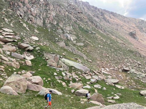
But it took a mountain, or nearly.
Twining Peak, Colorado. Elevation 13,711 feet.
One of the best-kept secrets of the Aspen Center for Physics is its mandatory downtime. Every couple of days, the entire group stops working and goes hiking. No laptops, no slides. Just altitude, views of the Rockies, and several hours of unstructured conversation. I am convinced that some combination of thin air and long trails produces a particular quality of scientific thinking that is very hard to replicate in a seminar room or at a desk.
Max and Rico Rojas (professor at NYU) walking down Twining Peak discussing the biophysics of cell shape. Photo by Marianne.
My lab had just engineered a new suite of optogenetic tools that allowed precise, programmable control of developmental signaling pathways in human embryonic stem cells, including, critically, the Wnt pathway, which governs cell fate decisions and patterning in early development and adult tissues. Borrowed from neuroscience and adapted for developmental biology, optogenetics allows you to use light to activate specific signals in cells with millisecond precision. For the first time, we could systematically interrogate how cells respond to signals delivered at different rhythms and timescales. I was looking for theorists who wanted to think about these questions seriously.
I found one on a mountain.
Twining Peak sits directly on the Continental Divide, where precipitation from the same slopes drains west toward the Roaring Fork River and east toward Arkansas. On this mountain, the same water flows in two different directions at once. The trail is not a casual outing, and the terrain is unforgiving at altitude. Somewhere on that mountain, probably on the way back down, we sketched the outlines of several project ideas and agreed to stay in touch. It felt inevitable.
“If I didn’t know this was biology, I’d almost say you’ve hit it at a resonance or something.”
What happened next was mostly emails and Zoom calls, which is to say, the unglamorous reality of international collaboration. Marianne is in Delft; I am in Santa Barbara. The overlap in our waking hours was narrow, so most of our early meetings required one of us to be awake either very early or very late. Reading back through our correspondence from late 2022, I am struck by how many messages were essentially calendar negotiations: a wedding, a conference, a colloquium that ran long, nine emails to lock in a single Zoom meeting. When we did finally connect, our calls would often be interrupted by a Dutch overhead announcement that rang through Marianne’s building at 5 or 6 PM, a recorded voice cheerfully informing everyone that the working day was over and it was time to go home. It became a kind of running joke.
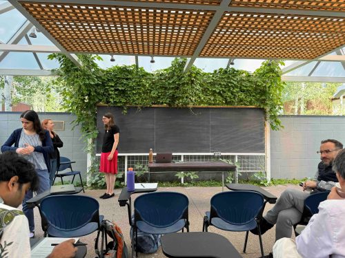
Marianne standing in front of a clean blackboard before a chalk talk session at the Aspen Center for Physics
But those early conversations were productive and fun. Marianne took on a graduate student, Olivier Witteveen, to work on the theory side. I began sending data. And it was during one of these data-sharing exchanges, in March 2023, that the concept at the heart of our paper was first named.
I had shared a set of experiments tracking how beta-catenin, the central transcription factor of Wnt signaling, responded to optogenetic inputs of varying durations and intensities into the upstream receptor system. The traces were very complex. Peaks where you expected them, but also puzzling dips, and an intriguing non-monotonic response to signals delivered at certain intervals. Marianne, looking at the data with fresh eyes and a physicist’s intuition, wrote back, “If I didn’t know this was biology, I’d almost say you’ve hit it at a resonance or something.”
I replied: “I was thinking resonance as well!”
As it turned out, the cells were doing the opposite. Anti-resonance is not where the response peaks but where it vanishes. We had identified the right concept and the wrong sign, a class of error that is actually quite common in physics.
Resonance and anti-resonance are well-developed concepts in physics and engineering. Resonance is when a system responds especially strongly to inputs delivered at a particular frequency. It’s why bridges can shake apart in the wind, why wine glasses shatter at the right musical note. Anti-resonance is the counterpart. A frequency at which the system’s response drops out almost entirely, even as neighboring frequencies drive a strong reaction.
These ideas apply to any physical system that receives inputs that vary in time, and yet they have rarely been applied to biology. The reason, until recently, was that you could not watch the signaling dynamics in a living developmental system over the timescales needed, and you certainly couldn’t deliver precisely timed, reproducible signals to probe them. The convergence of low-phototoxicity long-term imaging and cellular optogenetic tools changed both of these things at around the same time. So with these new tools, we could treat a developing tissue in the same way engineers have studied circuits for almost a century: put in a signal, vary its frequency, and measure how the system responds.
What we found in the Wnt pathway is that cells do not respond uniformly across frequencies. At certain input rhythms, the transcriptional response (the downstream readout of whether a cell “heard” the signal) falls to near zero, even as nearby slower or faster signals drive robust activation. The system has a blind spot. That is anti-resonance.
The deeper question, why, is one we speculate about but do not claim to have answered. Is anti-resonance a developmental gatekeeper that helps cells avoid dangerous intermediate identities by filtering out certain signal dynamics? Is it an anti-cancer mechanism that blocks runaway activation by particular signaling patterns? Or is it a spandrel in the Gould-and-Lewontin sense, an architectural byproduct with no function of its own, present simply because of how the underlying circuit is built? We genuinely don’t know. What we do know is that we are very early in our understanding of developmental signaling as a temporal phenomenon, and that most of what we currently call a signaling pathway is really a static snapshot of something that is fundamentally dynamic.
I visited Marianne in Delft in the spring of 2024; she came to present at our departmental seminar at UCSB that fall. In between, we had monthly meetings, almost always early morning Pacific time, which is early evening in the Netherlands, punctuated by that cheerful Dutch announcement that the working day was officially over.
We were both Assistant Professors, both trying to build our labs, find funding, attract students, figure out how to operate independently, and both aware that the tenure clock does not pause for transatlantic collaborations. But the calls were genuinely fun, which I had not counted on. We never had to convince each other that the questions were interesting, which turns out to be more than half the battle. And when an experiment did not match what the theory predicted, or a theoretical prediction seemed to have nothing to do with what the cells were actually doing, we could say so without it becoming a diplomatic incident. That back-and-forth, a physicist poking holes in the biology and a biologist pushing back on the theory, is where most of the real ideas in this paper came from.
The most satisfying moment of this project had nothing to do with the science directly. At some point in the past year, I tried to schedule a meeting with my graduate student Sam Rosen, who is co-first author on the eLife paper. He couldn’t make it. He was on a scheduled call with Olivier Witteveen, Marianne’s student and his co-first author, a call they had set up themselves, independently, without prompting from either of us.
They had built their own version of what Marianne and I had built. We are still figuring out why cells ignore certain signals. Apparently, our students were paying attention to different signals entirely.
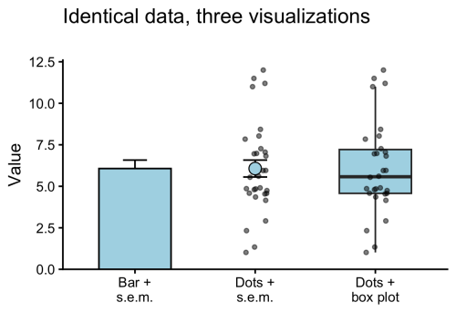
Thanks to community campaigns (#barbarplots) and opinionated papers (Drummond & Vowler, 2011; Weisgerber et al 2015) the dynamite plunger plot (a bar plot together with an error bar) has been abandoned as the default graph. The main reason to reject bar plots is that they display only an abstraction of the actual data and therefore oversimplify it. For full transparency and interpretability, the all data should be displayed. This can be achieved effectively by displaying the data as dots (or other symbols).
Figure 1: Identical data, visualized in three different ways. The bar plot conceals the data and is an oversimplification. Showing all observations as dots improves transparency. The data-points can be accompanied by statistics, e.g. mean and standard error of the mean (s.e.m.) or a box plot.
The dot plots are often accompanied by a graphical statistical summary. Common statistics are the mean or median. A more comprehensive statistical summary is provided by the box plot. The box plot was first proposed by Mary Eleanor Spear in her book “Charting Statistics” and publicised by the work of John W. Tukey. Open source tools, such as the user-friendly web tool BoxPlotR, have contributed to a wider adoption of box plots in publications. The box plot is characterised by 5 values, the median, the two borders of the box that indicate the IQR, and two whiskers. The whiskers can reflect multiple things, but most commonly indicate the most extreme data-point that is maximally at 1.5 x IQR from the border of the box (Krzywinski & Altman, 2014).
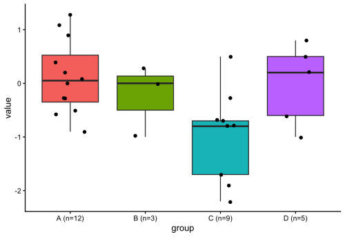
Figure 2: Four conditions with varying numbers of observations. When n is 5 or smaller (conditions B & D), the box plot does not add any information.
Since a box plot summarises the data distribution with 5 values, it does not add any information when the data consists of only 5 or less points. This can also be seen in figure 2 for conditions B & D. Adding a box plot to a condition that has only 5 datapoints would be similar to adding the mean for only 1 datapoint. Since some datasets have variable numbers of observations per condition, it would be ideal to only display the box plot when sufficient observations (n>5) are present. To do this in R with {ggplot2}, I considered defining a new geom (if you are interested in that, I recommend this tutorial), but then I realized that it can be done by filtering the data within the box plot function (inspired by the work of June Choe on {ggtrace}, see also this video: https://youtu.be/dUBnitXf5mk).
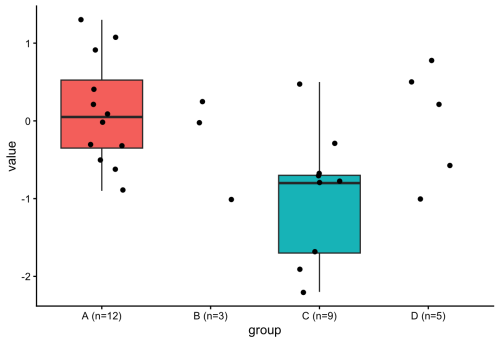
The trick is to use a filter() function within the geom_boxplot() definition to keep only the conditions for which n>5 (aggregated for each condition by group_by(group)). Here’s the R code:
#Filtered box plot, only drawing a box for conditions that have n > 5
ggplot(demo_data, aes(x = group, y = value, fill = group)) +
  geom_boxplot(
    data = ~ .x %>% group_by(group) %>% filter(n() > 5)
  ) +
  geom_jitter(width = 0.2, size = 1.5) +
  theme_classic() +
  theme(legend.position = "none")
The resulting plot only shows a box plot when n>5:
Figure 3: Similar to figure 2, but the box plot is only shown for conditions where n>5.
The use of a filter() function within the definition of a geom is an elegant method for getting rid of the box plot when the number of observations is too low. In general, this approach is very powerful and gives more control over plotting with ggplot2. There’s probably a ton of other applications, and one that comes to mind is to filter data based on some criterion and changing the color, e.g. for outliers. And, fun fact, Figure 1 was also created using the filter() function. Check out the R code (for all plots) here: https://github.com/JoachimGoedhart/Unboxing-data
by Irene Amblard, Thamarailingam Athilingam, Alejandra Guzman-Herrera,Dimitra Mouzourou, and Andrew Plygawko
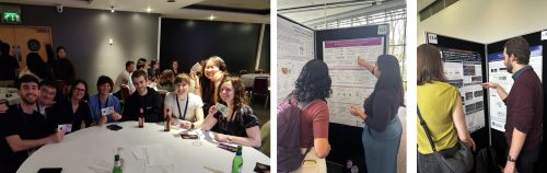
As members of the British Society for Developmental Biology, its annual meetings are always a highlight. This year’s ‘from Molecules to Morphogenesis’ meeting was held at University of Warwick in March, bringing together an inspiring range of talks spanning classical and modern developmental biology as well as a wide range of model systems.
Day 1: Seven Decades of Developmental Biology
On the first day we were all welcomed by a heartwarming introduction from Marysia Placzek (BSDB chair) talking about the history of the BSDB and the pioneer work of researchers that have shaped this society since the beginning, such as Philip Ingham who was present in the audience. The first session thus commemorated Seven Decades of Developmental Biology with a great line up of invited speakers highlighting the power of revisiting classical developmental questions with new technologies.
Starting with Denis Duboule remarkably showing in vitro embryo models retaining axis elongation and A-P polarity despite removing Hox gene function but showing defects in endodermal and mesodermal fate. Ruth Lehmann talked about the role of Nanos in early specification and maintenance of the Primordial Germ Cell (PGC) program in Drosophila embryos. Cliff Tabin added an evolutionary perspective on duck syrinx formation and morphological asymmetry through different types of Shh gradients. Yun Xia gave a fascinating talk on using ‘assembloids’ to study the interplay of cell repertoires in kidney development and disease progression. After a short coffee break, Alicia Hidalgo spoke about Toll signalling in brain plasticity and degeneration in flies. A standout talk for me was Norbert Perrimon’s on a decade long effort (also in flies!) to lay a framework of metabolic crosstalk between growing tumours and the host tissues that result in cachexia, a complex wasting syndrome. Andy McMahon described the underlying principles to direct iPSCs and generate human kidney organoids.
The last talk of the day was Valentina Lorenzi from the Wellcome Sanger Institute, who received the Beddington Medal for her impressive PhD work characterising spatiotemporal developmental trajectories of the human reproductive tract using cutting-edge technologies, but also for her work advancing and communicating women’s health as the former president of the Cambridge Femtech Society and through a collaborative zine called ‘Pelvic Matters’.
The Annual General Meeting followed, where all BSDB members get to hear from the committee what the society has been doing for the past year. All members also vote on changes to the constitution and on new committee members. The day ended with welcome drinks and dinner to encourage networking across all attendees.
Day 2: Cell Identity & Gene Regulation, Patterning & Morphogenesis
The first session of day two on Cell Identity and Gene Regulation, chaired by Vicki Metzis, began with Edith Heard on the dynamics of X-chromosome inactivation (XCI), followed by two short talks from Amruta Vasudevan, examining a Wnt/Nodal/Notch temporal module underlying symmetry breaking in mouse gastruloids, and Oliver Davis, highlighting the role of FoxG1 in regulating chromatin accessibility and neural fate in cerebral organoids. Joshua Brickman talked about using in vitro models to study chromatin remodelling and Sox2 activity. Following a short break and snacks, the session continued with Hilary Ashe, discussing ribosomal pausing and protein synthesis control in Drosophila embryos, and two more short talks. Christos Kalaitzis on the Hox code mediating regional diversity of vagal neural crest cells, and Daniel Goszczynski on dual developmental origins of mammalian PGCs.
There were also two rounds of flash talks from poster presenters, spanning varied topics including nuclear mechanosensing, transgenic avian lines, cell size scaling, cell morphology and more. During lunch time, the poster session provided a fantastic opportunity for delegates to dive deeper into the science, spark new conversations, and discover exciting work being done across the community. With packed rooms, lively discussions, and an exceptionally high standard of posters, the poster sessions were a highlight of the meeting.
The afternoon session covered Patterning & Morphogenesis under the lens of model organism diversity, chaired by Shankar Srinivas. Ranging from Drosophila to Arabidopsis, this was a unique opportunity to learn about how very distinct systems develop and shape their body plan or organs (e.g. mouse heart, fly blastoderm and epithelium, chick and shark telencephalon, mayfly eye, marsupial trunk, plant roots!). Several talks also highlighted the question of developmental timing. Kenzo Ivanovitch showed how early versus late primitive streak progenitors make distinct parts of the heart. Sergio Menchero from the Crick Institute gave the Dennis Summerbell Award lecture, showcasing his study on temporal diversity in marsupials’ developmental programs and how they prioritise differentiation of necessary structures for their survival. Dana Fakhreddine presented her work identifying heterochronic differences in molecular events shaping the distinct telencephalon identities. Erik Clark refined the classical morphogen-gradient models by adding a temporal lock mechanism to explain how striped expression is appearing in the fly embryo. Lastly, Bert De Rybel presented work from his lab on Arabidopsis root meristem, introducing the idea of developmental timers during development and aging.
After dinner two medal winners were announced. First, the Wolpert Medal was awarded to Prof. Neil Vargesson from U. of Aberdeen to recognise his outstanding contributions to our understanding of chemically induced birth differences for the past 20 years. Neil’s work has led to changes in health policies and help for affected families in the UK and abroad. His commitment to public engagement through numerous outreach activities has raised awareness of developmental biology and medicine safety. Next, the Waddington Medal lecture by this year’s awardee, James Briscoe from the Crick Institute. James’ fundamental discoveries have shaped our understanding of how morphogens work. He has also promoted developmental biology through numerous roles, including as Director and then Editor-in-Chief of the journal ‘Development’ from the Company of Biologists.
To end the evening, we had a Student/Postdoc ECR social event that lived up to expectations in every way. A science-themed pub quiz drew 120 attendees into teams that brought their competitive best. The questions ranged from easy to tricky and delightfully niche, a favourite was ‘when dealing with chick development, what does YSL stand for?’ and no, the answer is not Yves Saint Laurent but Yolk Syncytial Layer. Spirits were high and competition was tough, but one team came on top: ‘Shut up nurds!’ (a nod to Joshua Brickman’s talk). Perhaps the most creative team name came second, ‘Bad Scientists Doing Biology’ or, of course, B.S.D.B., and in third place we had the ‘Mighty-Chondria’ team. A great way to end day two!
Day 3: Cell Communication & Fate, Organogenesis & Regeneration, Conference Dinner
Wednesday morning started with the Cell Communication & Fate session, chaired by David Turner. This series of talks opened with Sally Lowell summarising elegant work from her lab generating new tools to track the neighbourhood of a cell and discussed how these tools can be applied to diverse model systems. Following this, a series of fascinating talks covered distinct systems where cell fate is controlled by intrinsic mechanisms, such as post-transcriptional modifications shaping neural crest fate as presented by Lara Busby, and extracellular cues provided by the environment, such as mechanical cues generated by neighbouring cells. On this topic, Stefan Harmansa presented his work on how mechanical forces shape epithelium architecture in the drosophila wing disk by combining experimental and modelling approaches. Vikas Trivedi shared recent work from his lab investigating the role of temperature on the shape of the zebrafish epithelial layer during gastrulation. Nine flash-talks concluded this session by covering a diversity of questions, ranging from the impact of metabolic alterations to the role of cellular geometry.
After lunch and the second poster session of this meeting, we returned for the afternoon session on Organogenesis & Regeneration, chaired by Timothy Saunders. This session underscored how the study of diverse systems furthers our general understanding of morphological and regenerative processes. Beautiful work from Emily Noël focused on the importance of ECM asymmetry in shaping the developing zebrafish heart. Cristina Newnes on muscle development in Drosophila embryos, showed the power of established model systems. Meanwhile, the comparative analysis of muscle development in zebrafish versus sharks from Peter Currie’s lab, the study of head scale formation in diverse reptiles by Rory Cooper, and the in vitro generation of hindlimb progenitors from hPSCs by Sude Uyulgan, all made clear how much there is to learn from studying novel or less conventional systems.
The Marie Johansson Prize was introduced for the first time by Corinne Houart, in memory of an outstanding Postdoc in her lab to recognise leading ECR researchers contributing to developmental biology. The first recipient of this prize is Giulia Boezio from the Crick Institute for her exceptional work on spinal cord patterning and establishing new techniques in complex embryonic tissues in vivo, as well as her contributions to public engagement, mentoring, and network. The Tickle Medal was then awarded to Cynthia Andoniadou from KCL for her incredible work as a developmental endocrinologist on the pituitary gland, building the human pituitary atlas, and championing female scientists in the UK and internationally.
Ending the last full day, we had the long-awaited Conference Dinner, during which Marysia Placzek acknowledged and thanked the meeting organisers with a special gift for making this meeting happen. She then announced the poster prize runner ups and winners: Gareth Moore (£150) and Luke Simpson (£300) in the postdoc category, Achira Karunaratna (£150) and Noura Maziak in the student category. Noura won the big prize of an all-expenses-paid trip to attend the Society for Developmental Biology meeting in the US! The new members of the BSDB committee were also announced, congratulations and welcome to Paula Alexandre, Gi Fay (Geoffrey) Mok, and Teresa Rayon. Finally, it wouldn’t be a BSDB meeting without lots of scientists cheering and dancing the night away!
Day 4: Human Development & Disease
Our final session of the conference mercifully started at 9.30, giving us extra time to recover after the night before. This session was all about Human Development & Disease, chaired by Anahi Binagui-Casas. Sarah Teichmann showed the incredible efforts of the Human Developmental Cell Atlas, which has mapped >450 cell types across pre- and postnatal development, an invaluable resource for the community. This work also highlighted an underlying theme across the conference that many progenitor populations and lineage commitments emerge earlier in development than previously anticipated. Presentations from Lila Allou, Teresa P. Silva, and Lorenz Studer also made clear, in a sometimes-poignant manner, how garnering a deeper understanding of developmental processes such as organogenesis and axis formation can help us to better understand how such processes change in the context of congenital diseases. Kathy Niakan shared the molecular mechanisms regulating the first cell fate decisions in human embryos but also made a noteworthy remark about supporting model organisms, highlighting that “we wouldn’t be able to know what to look for [in humans] if it wasn’t for work in other organisms”.
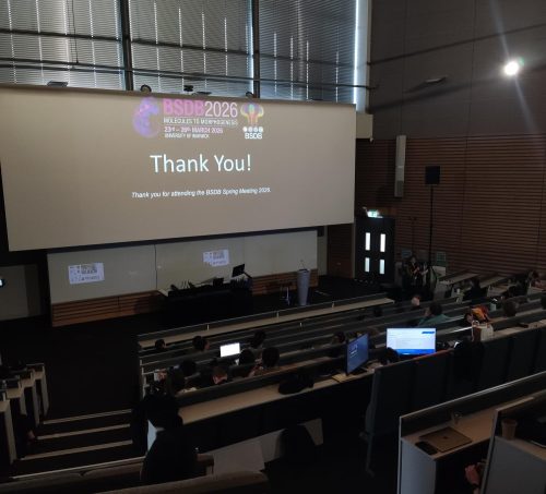
Overall, this was an enjoyable and memorable meeting thanks to all the attendees, speakers, organisers, and everyone in the community coming together to share their passion for developmental biology. Looking forward to next year’s BSDB meeting in Edinburgh!
P.S. Check the programme to learn more about the speakers, the BSDB website for more information on the awards and to become a member, and the BSDB social media to relive the highlights of the 2026 meeting! (@bsdb.socials on Instagram and TikTok)
A press release from the University of Colorado Anschutz on a study published in Development
Written by David Kelly
Researchers at the University of Colorado Anschutz may have identified why many cancer patients say food suddenly tastes unpleasant during treatment.
The study, published today in Development, found that a class of targeted cancer drugs known as tyrosine kinase inhibitors (TKIs) can change how taste buds are maintained—reducing the ability to taste sweet foods and altering flavor perception overall. While the study was conducted in animal models, researchers believe similar changes likely occur in humans.
The findings offer the clearest explanation to date for a common but often overlooked side effect of cancer treatment.
What researchers found
Using mouse models and lab-grown taste tissue, scientists studied the cancer drug cabozantinib and discovered:
Taste buds as a whole were not damaged or reduced in number by drug treatment
The composition of cells inside taste buds shifted
Drug treatment reduced the number of cells that detect sweet tastes
Drug treatment increased the number of cells that detect bitter and savory (umami) tastes
Mice lost their preference for sweet-tasting solutions
Researchers identified an unexpected cause: a protein called KIT.
While TKIs are used to block cancer growth pathways, they also unintentionally block KIT—an important regulator of taste cell development.
When KIT is blocked:
Sweet-sensing cells fail to develop properly
Bitter/umami-sensing taste cells take their place
The proportion of sweet and bitter taste bud cells is very tightly controlled. When this proportion is altered, taste perception may drastically change.
“If you lose the sweet component of everything you eat, your entire sense of taste becomes distorted,” said senior author Linda Barlow, PhD, professor of cell and developmental biology at CU Anschutz.
Why it matters
TKIs are important anti-cancer drugs that significantly extend survival in several types of advanced cancers. However, an estimated 10% to 50% of patients taking these drugs experience taste changes, known as dysgeusia.
Though often considered minor, the impact of dysgeusia can be significant:
Loss of appetite
Weight loss
Poor nutrition
Social withdrawal and reduced quality of life
“It’s difficult for them to enjoy a meal with their family and friends,” Barlow said. “Nothing tastes good to them so they withdraw and become isolated. Isolation leads to depression.”
Study co-author Elaine Lam, MD, professor of medicine and medical oncology at the CU Anschutz Cancer Center, said the drugs are meant to block blood vessels developing in tumors, effectively starving them. Unfortunately, they also cause unintended consequences.
“People don’t eat and they lose weight. This sometimes leads us to reduce or interrupt the dose of their drugs,” said Lam, a kidney cancer specialist. “This research is important because it identifies the underlying mechanisms that affect taste. Now we have to figure out the best way to treat this.”
Lam said possible solutions include designing cancer drugs that avoid blocking KIT or developing treatments to protect taste function.
What’s next
Future research will focus on confirming these findings in patients and identifying ways to prevent or reduce taste changes.
Bottom line
Targeted cancer drugs called tyrosine kinase inhibitors may not destroy taste buds—but they can change their cellular makeup, shifting the balance away from sweet-sensing cells and potentially changing how food tastes.
The study in Development is titled “Tyrosine kinase inhibitors affect sweet taste and dysregulate fate selection of specific taste bud cell subtypes via KIT inhibition”. The lead author is Christina M. Piarowski, PhD. Additional authors are Jennifer K. Scott, Courtney E. Wilson, PhD, Heber I. Lara, PhD, Ernesto Salcedo, PhD, Andrew S. Han, Peter J. Dempsey, PhD and Jakob von Moltke, PhD. The study is available here.
This press release was originally published on the University of Colorado Anschutz news page.
We are pleased to announce that the Physics of Living Matter conference is back in Cambridge for its 19th edition!
This will be on the 24th and 25th of September 2026, at the Centre for Mathematical Sciences (Wilberforce Rd, Cambridge CB3 0WA, UK).
As per tradition, the conference will showcase a diverse set of biological problems that are tackled through the lens of the physical sciences and in addition to an exciting programme presented by renowned international speakers, oral presentations will be selected from the submitted abstracts.
The PLM series started 19 years ago from an interest in promoting the interface between the Life and Physical Sciences in Cambridge. Over the years, PLM has grown from a local meeting to a popular international event that attracts interdisciplinary scientists from all around the world.
Confirmed speakers for this edition are:
Nancy Kleckner (Harvard University, USA) – Bragg Lecture
Rosana Collepardo (University of Cambridge, UK)
William Durham (University of Sheffield, UK)
Zena Hadjivasiliou (Francis Crick Institute, UK)
Pulin Li (Whitehead Institute – MIT, USA)
Jean-Leon Maitre (Institute Curie, FR)
Jeremie Pallaci (Institute of Science and Technology Austria, AT)
Marco Polin (IMEDEA UIB-CSIC, ES)
Victror Sourjik (Max Planck Institute for Terrestrial Microbiology, DE)
Peter Swain (University of Edinburgh, UK)
Berta Verd (University of Oxford, UK)
Andrea Weisse (University of Edinburgh, UK)
The call for abstracts is open now! You can submit an abstract for a talk or a poster presentation using this form. The deadline for abstracts is 12 of June.
We will be opening the egistrations in the next few days; if you are interested, please fill in this form for updates.
For further information, you may check our website, or contact Maria Bargués-Ribera at admin@physbiol.cam.ac.uk.
We are looking forward to welcoming you to PLM19!
Best regards,
The organising committee
James Locke (Sainsbury Lab, University of Cambridge, UK) Teuta Pilizota (Department of Physics, University of Cambridge, UK) Ben Steventon (Department of Genetics, University of Cambridge, UK)
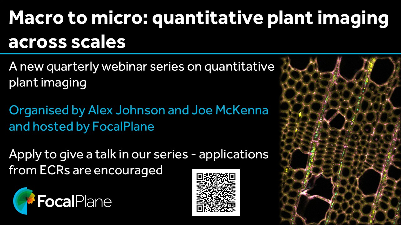
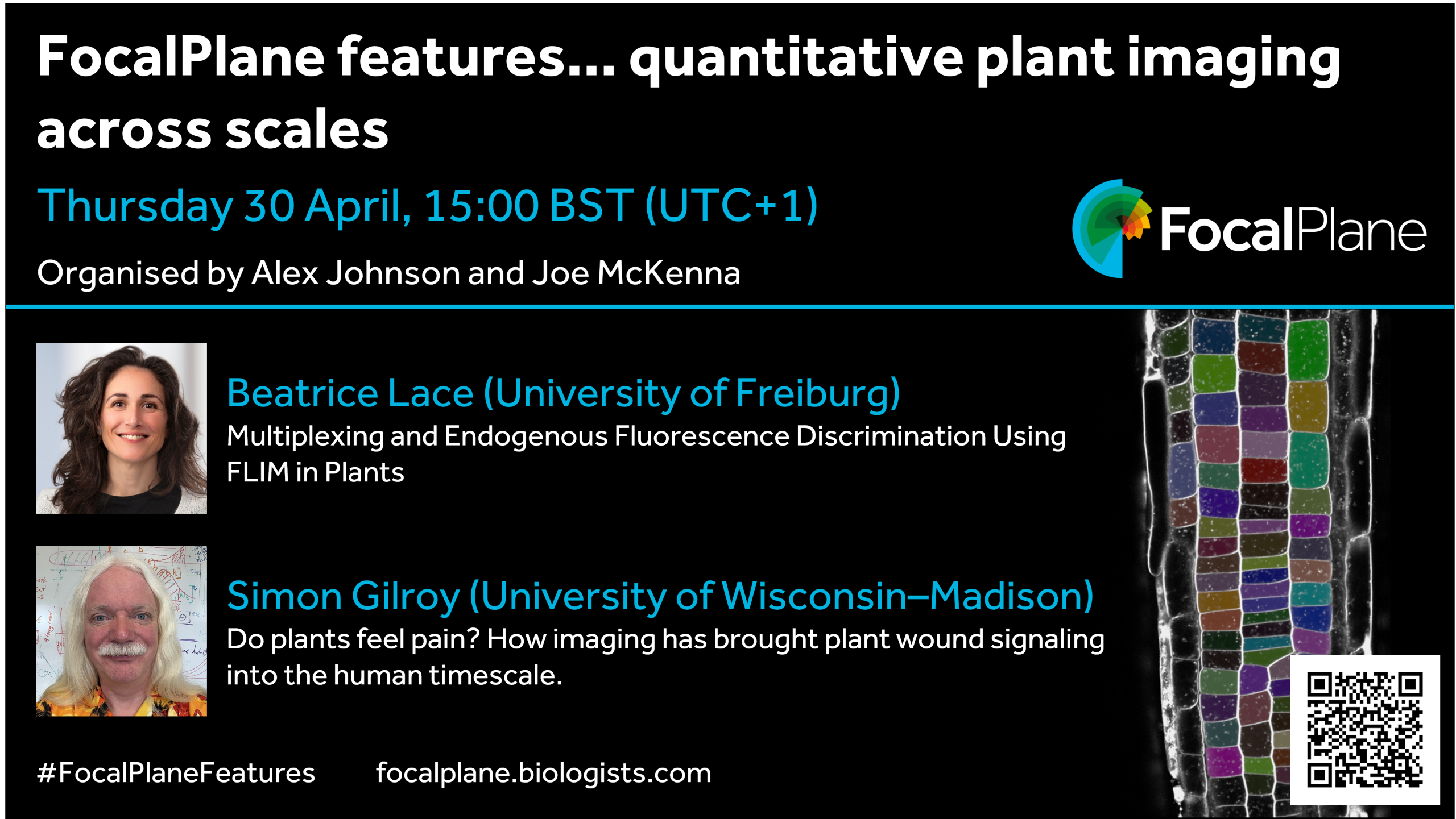
We are delighted to launch our new webinar series, Macro to micro: quantitative plant imaging across scales, with Alex Johnson and Joe McKenna. In this series, we’ll be highlighting the latest research using imaging to investigate questions in plant biology. We’ll also hear talks from imaging experts from outside the plant biology field.
You can find more information on the webinar series, including how to register for event notifications and to sign up to give a talk, on our dedicated webpage.
Our first webinar is on Thursday 30 April at 15:00 BST and will feature talks from Beatrice Lace and Simon Gilroy. Beatrice will present, ‘Multiplexing and Endogenous Fluorescence Discrimination Using FLIM in Plants’ and Simon will ask, ‘Do plants feel pain? How imaging has brought plant wound signaling into the human timescale’.
We are happy to announce the forthcoming workshop entitled “Roadmap for EvoDevoMec”, Nov. 2nd – 5th, 2026, Université Paris Cité.
The link with evolutionary history of an organism is key to understand embryonic development. Much of the focus has been so far on genetic circuitry. However, it has become increasingly clear that physical constraints are an essential aspect at the basis of morphogenetic processes. We have now reached an exciting stage where it becomes possible to integrate mechanical considerations in our view of how development has been shaped during evolution: EvoDevMec.
This new approach has recently raised great interest, but biological questions still need to be reformulated with precision. In addition, due to its integrative nature, it poses a scientific and technical challenge: What type of experimental systems could be used? How could we define experimental proof of concepts? How can theoretical biophysical models help integrate various questions and approaches?
We aim at discussing these questions in an informal context, i.e. with chalk talks. The workshop is limited to 30 persons, from PhD students to senior scientists from various countries. We will welcome biologists using a breadth of models (from unicellular organisms to animal and plant models), physicists (theoreticians, numericians, experimentalists), and mathematicians.
 (No Ratings Yet)
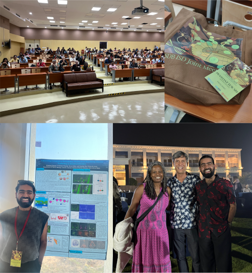
 (3 votes)