Today’s my last day working at the Node and Development. I started in June 2016, which really feels like a different world looking back now – at the first conference I went to, the SDB in Boston, the TV screens in the hotel lobby flipped between speeches from the two recently nominated Presidential candidates; at my second conference, also in Massachusetts but down in Southbridge, the first debate played in the bar to an eerie silence. I came here from the bench – I had done a PhD in Brighton and a postdoc in Cambridge but had always known that, as much as I love science, research (and/or being a PI) was not what I wanted to do with my life. The Node job allowed me to stay in touch with science and scientists. I’ve really come to appreciate the global community of developmental biologists.
Leaving has led me to wonder: what makes developmental biology such a rewarding field to be a part of? Of course, there’s the embryos, the time-lapses, the magic of it all. Those vertiginous shifts in scale – experiments that go from a misplaced nucleic acid to a funky protein structure to a misdirected cell to a novel tissue structure to a confused embryo – which you can then contextualise in the scope of evolution, ecology, physiology. It feels like we can have it all, not many disciplines can beat that (although try speaking to a misty-eyed cosmologist like my dad). Then there’s the field’s history, from bespoke experimental embryology in nineteenth century marine labs to the same embryos lit up by lasers and deconstructed by single cell sequencing; the golden ages keep coming, the old ways are repurposed. I’ve also always liked the mix between basic and applied research, a bit of a false dichotomy of course since one is not separate from the other; better to look at it as leveraging the rich body of developmental biology research to help understand and cure terrible diseases and make more food for the world – what’s not to love about that?
At the bottom of it though, I think it’s all about the people. On that maiden conference in Boston I got to do my first interviews for Development: Doug Melton, Dave McClay and the late Kathryn Anderson. Three totally different personalities, distinct career trajectories, but what tied them all together was a reverence for the embryo. Just as rewarding were the conversations later on in the poster hall, ten dollar beer in one hand and slice of pizza in another, with graduate students and postdocs, getting energised by their excitement. I wanted to showcase researchers young and old(er) in ‘The People Behind the Papers’, an interview series which started on the Node and has since moved to Development. Satisfyingly, my hundredth interview just came out, with tunicate researchers Izumi Oda-Ishii and Yutaka Satou (my last, number 101, will come out in the next few weeks). One of the hardest things about the pandemic for many of us has been the loss of personal contact without a screen in the middle, those chance encounters in conference bars…the people make the science, and it seems to me the people of developmental biology are a particularly good bunch.
It’s been gratifying to develop the Node, help a community journal promote the work of its authors, and work with a fantastic team in our not-for-profit publisher The Company of Biologists. Whatever you think about academic publishing, I’ll insist that we are one of the good guys. I’d encourage everyone to:
join the Node Network, our global directory of developmental biologists
Oh, and write for the Node – it’s free and easy, and could be anything from a job ad to an event notification to a behind the paper story to a polemical diatribe against funding inequity (honestly, we love it all). Why not look at our new topic pages for inspiration, up in the Archive dropdown
I’ll stop before I start excessively rambling. After this, I’m staying in science (communication), going to the Sanger Institute to be a science writer, combining my two favourite things. And you’ll still find me on Twitter, looking out for the next embryo time-lapse.
Developmental biology is a global science, but Europe and the USA get a lot of the airtime, and it can be hard for those outside these regions to get support and recognition for their work. Added to this, conducting research during the pandemic has been a considerable challenge, especially so for new PIs. But the pandemic has also brought new opportunities and support networks – just check out New PIs in Cell and Developmental Biology, a platform for e-seminars, collaboration and peer support (for more information read our 2020 interview with organiser Salah Elias). One of these new PIs is Rio Sugimura, who established his lab in the University of Hong Kong in 2020. We asked Rio to share some of his experiences of this current time as well as his life and research in four different countries.
Why I got into science
I first dedicated myself to science when I lost my mother to liver cancer. Before this, I had gradually come to be interested in science as I was a first-year medical student in Japan. In my final years of medical school, I rotated in hospital wards. I chose the auto-immune diseases ward first, hoping that cutting-edge immunology had been applied to patients. However, it was quite disappointing: diseases were classified according to rough symptoms, with very heterogeneous groups of patients all classified as having the same disease. Steroids were still being used with no understanding of why they work in some patients whilst make symptoms worse in others. I remember encountering one patient at the terminal phase of systemic sclerosis. We had no clues to cure them, could only watch this progressive illness till the end. At that time, I lost my mother to liver cancer: she had received a blood transfusion during the caesarean of my birth, and contacted the hepatitis C virus that cost her her life. So at that time, I was quite disappointed by modern medicine, and decided to dedicate myself to medical science.
From Osaka to Kansas City to Boston
I did my Ph.D. at the Stowers Institute in Kansas City in the USA. It was a big change in my life. I had just got my MD from my medical school, and all but me became residents: I was the only person to choose the path to M.D./Ph.D., with the Ph.D. part trained in the USA. I moved from one of the busiest cities in the world (Osaka) to a nicely remote and vast landscape. I enjoyed the Kansas life, the BBQ and the camping, and at the time the Stowers Institute was new and growing. Its science was top-notch in each field, and I particularly engaged in stem cell biology. I worked day and night, just the same as my classmates in their residencies. We were on the opposite sides of the globe, but I tried to keep up with the energy and motivation.
I then moved to Harvard Medical School as a postdoc. Boston was the opposite end from Kansas. Scientifically, it was very crowded, and competition and even aggression were encountered daily. I think I was competitive enough in science. New ideas and cutting-edge technologies were everywhere and I felt I could do anything. There you can talk to researchers, editors, and patient advocates, and access all the networks you need for science. I usually went to seminars in random departments of Harvard every day to scavenge sandwiches and pizza (I never cooked at home). The knowledge and directions I heard during the talks have been still navigating my research programs – you can grab collaborators, not only sandwiches!
Home but not home
After my time at Harvard, I moved to Japan and joined the Parthenon of iPSC research, the CiRA in Kyoto. It was a glorious place: every student was optimizing the recipe for factory-size production of iPSCs and their cell therapy products. I found that what we did at Harvard was 5 years behind the recipes there… I sought PI positions inside Japan, but soon realized that the academic system was too foreign to me. The internal pedigree of researchers, although obviously important in other countries, was a critical factor there. I had been told that I lacked strong enough Japanese roots to be a PI at several places I interviewed. A PI who had known me for nearly 10 years kindly advised me that Japan has no place for me. These experiences were enough to convince me that Japan was no longer my country although I was born there.
To Hong Kong via Italy
I flew to Italy during the pandemic with my family, including a 1-year old baby. I was going to establish my lab in Italy during the pandemic. The northern Lombardy region I stayed in was hardest hit by COVID19. I still found that the small town of Pavia was home to our family – people were very supportive. I very much appreciate my director who drove us to the weekly grocery so we could thrive during the lockdown. There I set up my research program based on CAR-T cell technology, which is the basis of our lab now.
So how did I end up in Hong Kong? It was quite simple – I was asked to join the newly established stem cell center at the University of Hong Kong. I joined a zoom chatting with PIs there and was amazed by the conversation, finding shared interests and direction. I saw that I could find my feet there, but it was a tough decision. I was building a life in Italy, but after endless discussions with my family we realized that not only the research but also the international city life and educational opportunities for our children were attractive to us. So, we decided to move.
And here we are in HK, where my lab works on precision medicine for cancer immunotherapy. We want to know: Can we predict the response to immunotherapy in cancer patients? How can we target solid tumors by CAR-T cells? What makes some immune cells target cancers, but others fail to? These are old questions, but we employ cutting-edge technologies to give insights. We work at the intersection of stem cell technology and cell engineering. Our lab has built strong ties with physician-scientists in HK’s biggest teaching hospital nearby our campus. HK has a nearly 8% prevalence of the hepatitis B virus in elderly generations, and liver cancer is one of the deadliest cancers here. Since my experience in losing my mother to liver cancer, I have always dreamed that my research contributes to conquer this disease, and now we can.
Starting a lab during the pandemic
When the pandemic began, I was already in transition to Italy, and had to wait for a few months until the border of the EU opened. The access to the lab in Japan had been limited due to pandemic, and the same happened in Italy. Fortunately, HK did not have such limitations because the pandemic was under control. My move to HK was smooth, as the country did a great job in controlling the pandemic. I do not know if the pandemic affected the laboratory setup that much really, aside from some item ordering was getting slow though, still enough manageable. Luckily, we did not receive any dead cats! I am very lucky to have funding, and our lab is expanding. Most of our students are from mainland China, followed by local HK students. They are really motivated and well-trained.
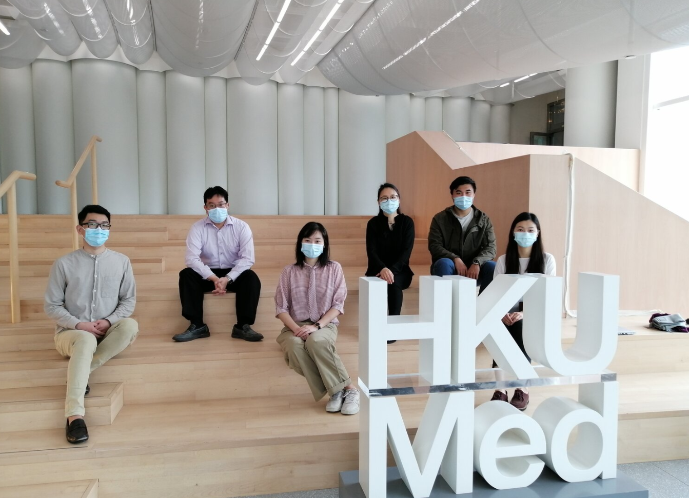
Rio’s Blood Engineering Lab in Hong Kong University.
Although international students and postdocs seem to be elusive, I feel that we have a good pool of candidates actually because of the pandemic. These students would usually go to the USA or Europe, but this pandemic may have forced them to change course. Indeed, we are seeing candidates for students, postdocs, and even PIs, whose CV could land in the USA safely if there was no pandemic, who are trying to come back to HK. I believe that the key to enhancing the strong research environment in HK is to secure those returnees. Our university is recruiting 140 faculty up until 2027, so I believe that it might be a positive impact of the pandemic.
Another big change for us is Zoom. We are not in the USA, not in Europe, and so access to top-notch seminars, conferences, collaborators, and chances to meet with editors had been quite limited. This inequity has big consequences for where you publish, how much funding you get, you recognition and invitations, and where you land as a PI. After leaving Boston, I had always felt outside of the scientific circle, but since the pandemic, Zoom brings us everywhere. Communications regarding collaboration and joint grants overseas have been much easier. Most importantly, while we are being so remote to the ‘center of science’ aforementioned, we can gain much exposure and recognition digitally. It is a big advantage for PIs not in the USA nor Europe. I do hope that societies will keep the Zoom option after the pandemic is over.
In term of the standing of developmental and stem cell biology in HK, we are in transition, increasing a new generation of scientists and mostly recruiting from overseas. I believe that the next 5 years will be crucial to determine the activity and the level of this field in HK. The key is to increase the exposure and recognition of new PIs of HK internationally. The one thing I would request of the global community to better help HK: please think of HK scientists when inviting for key-note talks, seminars and journal editorial boards! I think we deserve some more recognition.
On this note, you can find Rio’s profile in the Node Network, our global directory of developmental biologists, which you can sort by country if you are hoping to geographically diversify the pool of researchers, speakers or reviewers! Find out more here: https://thenode.biologists.com/networkinfo/
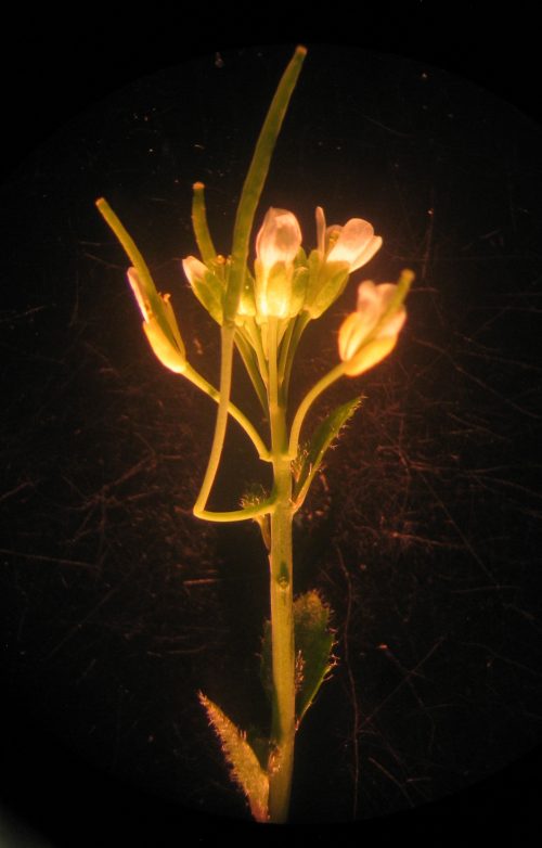
A previously unreported anatomical structure named the ‘cantil’ has been described in the popular plant model, Arabidopsis thaliana. Scientists from The Pennsylvania State University, USA, reveal that the cantil forms between the stem and flower-bearing stalk when flowering is delayed. Published in the journal Development, this study highlights that there are still discoveries to be made, even in some of the most meticulously-studied species, and provides new clues for understanding conditional growth in plants.
A FLOWERING LOCUS T mutant strain (ft-10) flowering under a long-day length. One long and two short cantils are visible. Credit: Timothy Gookin
For many, the Thale cress (Arabidopsis thaliana) is little more than a roadside weed, but this plant has a long history with scientists trying to understand how plants grow and develop. Arabidopsis was first scientifically described as early as the 16th century and the first genetic mutant was identified in the 1800s. Since the 1940s, Arabidopsis has increased in popularity within the scientific community, which continues to use it as a model system to explore plant genetics, development and physiology to this day.
One might expect that after decades of scientific scrutiny the structure of Arabidopsis had been fully documented, but a new study from scientists from The Pennsylvania State University, USA, has revealed that this humble plant still has some surprises. The researchers describe a previously unreported structure called the ‘cantil’, which connects to the stem at one end and hangs in the air to hold up the flower-bearing stalk, similar to the function of a cantilever in structural engineering.
“I first observed the cantils in 2008,” said Dr Timothy Gookin, a postdoctoral researcher working in the group of Professor Sarah Assmann. “I initially didn’t trust any of the results; I thought it must be an artefact of genetic contamination, perhaps combined with environmental contamination of the water, soil, fertilizer or even the building air supply.”
How have cantils eluded scientists for so long? First, cantils are rare; they only develop under certain conditions that cause the plant to delay flowering, such as short day lengths, and cantils only form at the precise point at which the plant begins to flower. In addition, as Dr Gookin discovered, some popular Arabidopsis strains have genetic mutations that make them incapable of producing cantils at all.
Nonetheless, Dr Gookin set about the gargantuan task of proving that cantils are a naturally occurring structure and not an artefact of mutation or contamination – an effort that took more than a decade. “It took over 12 years of experimentation to really get a grasp on what we were seeing and to understand how cantils were regulated. This study required the growth of 3,782 plants to full maturity and the manual inspection of over 20,000 flower-bearing stalks in 34 unique plant lines,” explained Dr Gookin. “I finally deemed the cantils a natural phenomenon after identifying them in wild-type (non-mutant) plants from different sources, which were growing in independent locations and diverse conditions.”
During his extensive research, Dr Gookin identified a number of mutant plants in which cantils appear more frequently, revealing some of the genetic factors that control cantil development. The discovery of cantils is not only a lesson in the virtues of perseverance, but their development also provides important clues for understanding the conditional growth of plant structures in response to their environment. “One speculative interpretation is that the cantil represents a highly repressed ancestral linkage between different types of flowering plant architectures; the multiple layers of genetic and environmental factors that regulate cantil development are certainly quite striking,” said Dr Gookin.
On Wednesday 9 June Development welcomed three more researchers with interests in chromatin and epigenetics to our ninth Development presents… webinar.
Below you’ll find each of the talks, plus a Q&A chaired by Development Editor Maria-Elena Torres-Padilla. Be sure to subscribe to our mailing list for updates for information on future events.
Jessica Zuin (Friedrich Miescher Institute for Biomedical Research) – ‘Nonlinear control of transcription through enhancer-promoter interactions’
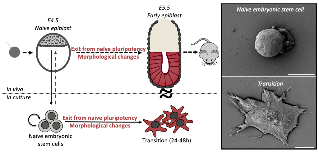
Mouse Embryonic Stem Cells (ES cells) have the capacity to generate any tissue in the organism; this remarkable ability is called naïve pluripotency. Intriguingly, when ES cells start to differentiate they undergo a striking shape change.
Figure 1: Shape and fate change are concomitant during early differentiation in mouse embryonic stem cells. Schematic and scanning electron microscopy images depicting the change of shape, from round to spread, of differentiating ES cells.
During my interview with Ewa (Paluch), my PhD supervisor, we were discussing potential projects for me, when she showed me some preliminary scanning electron microscopy pictures of differentiating ES cells. I was immediately struck by the idea of fate changes coupled to cell shape changes. ES cells are round and smooth, whilst differentiating cells are spread with many membrane folds. As cell shape is dictated by cell mechanics, this shape change suggested a profound mechanical change during differentiation. We wondered if the shape change was a consequence of the fate transition, or was it a cause? This sounded like a great question to investigate for a PhD, at the interface between two cool labs (Ewa Paluch and Kevin Chalut) and so a few months later I joined, ready to dive into mechanics of cell fate transitions.
When observing ES cell fate transition in more detail, we noticed cells blebbing (blebs are pressure-driven spherical protrusions) intensely before spreading. This was pretty interesting because such blebbing is often a sign of changes in the organisation of the actin cytoskeleton, and notably membrane to cortex attachment. We measured this attachment during early differentiation (using microscopy & Western Blots) and found striking differences between naïve and differentiated cells. This was an important result for us as we knew from the literature that membrane to cortex attachment is the main regulator of effective membrane tension, which has been shown to play an important role in regulating cell shape and function.
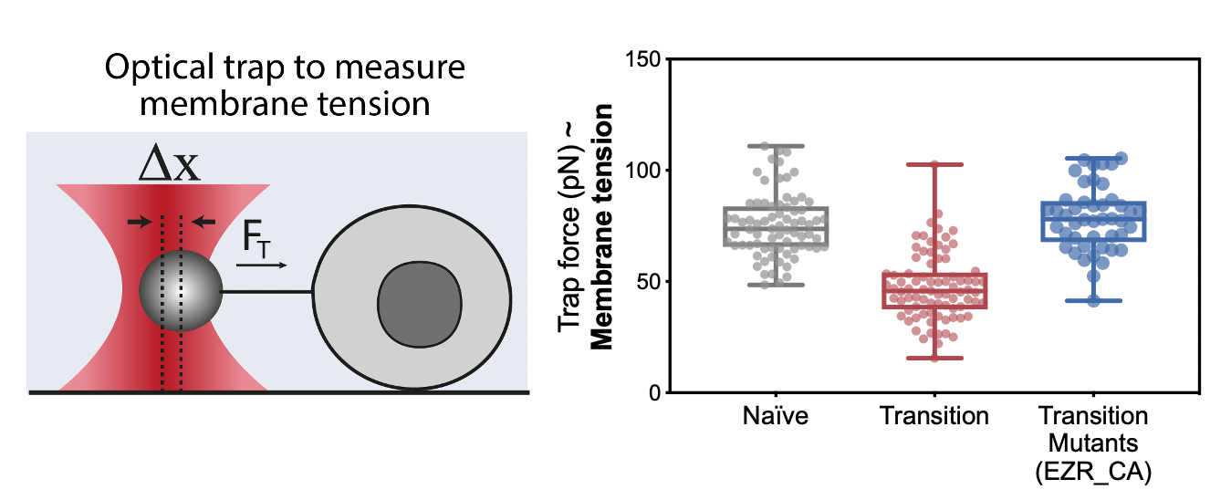
Figure 2: Using a tether pulling assay, we measured membrane tension in ES fate transition. We found that ES cells undergo a drop in membrane tension as they spread during early fate transition. We could prevent this drop using mutants that maintain high membrane tension during this transition
The next step was to measure membrane tension during early differentiation; however, we didn’t have the setup ready for it just yet. In this frustrating interregnum, we decided to push ahead anyway and to try mutants and drugs that should generate defects in membrane tension. These preliminary experiments turned out to be a key milestone of this project: drugs and constructs increasing membrane tension led to major differentiation defects.
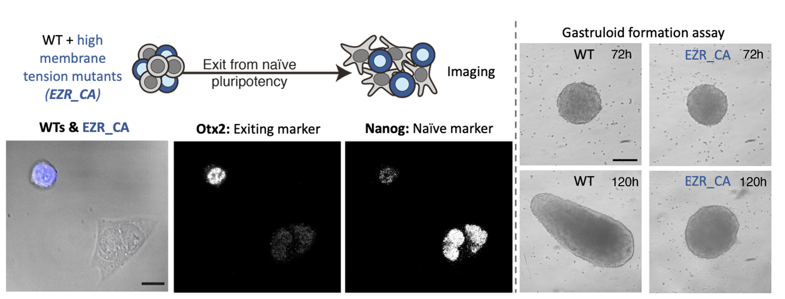
By the time we got these results, we had set up our system to measure membrane tension: a tether pulling assay using an optical trap. This technique is great because it enables precise membrane tension measurements without disturbing cell shape. As we hypothesized, we found that as cells differentiate, they undergo a dramatic drop in membrane tension. Furthermore, we were able to verify our membrane tension mutants, and establish that maintaining high membrane tension during fate transition (using drugs or mutants) leads to significant early differentiation defects. We also used our mutant cell line to show that maintaining high membrane tension resulted in developmental defects in gastruloids and cultured embryos.
Figure 3: Preventing the decrease in membrane tension results in early developmental defects. On the left is depicted an immunofluorescence assay in which we mix WT and mutant cells (which maintain high membrane tension during differentiation). On the right are pictures of our gastruloids formation assay, which shows that maintaining high membrane tension results in major morphological defects.
The next goal was to identify a mechanism by which changes in membrane tension could regulate differentiation. We tried to perturb various mechanosensors to no avail until one day Kevin had the idea of looking at endocytosis. Endocytosis is negatively regulated by membrane tension and is also a major regulator of signalling. We measured endocytosis in ES cells and found that it increases sharply as cells decrease their membrane tension and change shape. We next figured out a way to increase endocytosis levels in cells with high membrane tension (mutants) and found that this was sufficient to rescue their differentiation defects. At that stage we knew endocytosis was downstream of membrane tension but what was downstream of endocytosis? In other words, what was the link between increase in endocytosis and ES fate transition?
We decided to look at ERK activation, because ERK activation is required for early differentiation and recent work by the Scita lab beautifully showed that ERK is, in part, activated in the early endosome (Palamidessi et al., 2019). The Scita lab kindly provided us with a FRET sensor which allow to measure ERK activity in early endosome. We found that ERK activity sharply increased in cells as soon as they spread, which correspond to their drop in membrane tension and increase in endocytosis.
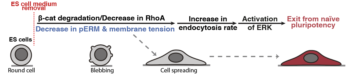
When we put the story together for preprint, we agreed that the main missing bit of the story was that we didn’t know what was triggering the observed change in membrane tension. After a few exploratory experiments we found out that this drop in membrane tension was triggered by Beta-catenin degradation. This degradation is coupled to a significant drop in RhoA activity, which is a known regulator of cell mechanics and notably membrane tension. This result was pretty incredible because it tied together two key pluripotency pathways, beta-catenin and ERK, via an intrinsic change in cell mechanics (membrane tension).
Figure 4: Membrane tension gates early differentiation. (Belly et al., 2021)
One aspect I really like about this paper is that it highlights the role of intrinsic cell mechanics in regulating cell mechanics. Before working on this, I always considered the role of mechanics from the environment point of view, for example how different substrate stiffnesses could regulate fate. But here we could show that cells can intrinsically modulate their mechanical properties, independently of their environment, and in turn regulate signalling. This change is clearly tied to changes in cell state, and speaks to another level on which cells tune their own receptiveness to signalling.
A mutation from a human patient with a rare metabolic disorder has been replicated in the Japanese rice fish. Researchers from the Centre for Organismal Studies Heidelberg, Germany, have developed a fish model to study disorders caused by a deficiency in the process of adding sugar molecules to proteins. These findings, published in the journal Development, provide a system to study the causes of complex metabolic disorders in humans.
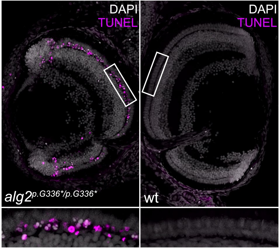
In Alg mutant embryos, rod cells are initially born but not maintained and undergo programmed cell death indicated in magenta (TUNEL staining). Credit: Clara Becker.
Human cells are kept healthy by the activity of millions of proteins. These proteins are modified in different ways, such as by adding sugar molecules to them, which can be crucial for them to function properly. Given this importance, defects in the sugar-adding process are often lethal at the very early stages of development. In rare cases, however, patients can develop sugar-adding deficiencies that result in a range of metabolic diseases, known collectively as ‘congenital disorders of glycosylation’ (CDG). These disorders are caused by defects in the enzymes involved in the sugar-adding process. For example, ALG2-CDG (or CDG-Ii) is a disorder caused by mutations in the ALG2 enzyme, which combines sugar molecules together. ALG2-CDG patients appear unaffected at birth, but later develop problems in different organs, such as the eyes, brain and muscles.
The rarity, variety and complexity of these disorders has made them difficult to study, especially in the context of the whole body. Now, scientists have developed the Japanese rice fish (also known as the medaka) as a model system for studying such disorders. “Fish are particularly good models for these disorders because they develop outside the mother, making them very suitable for studying early embryonic defects,” said Professor Joachim Wittbrodt from the Centre for Organismal Studies, who led the study with Dr Thomas Thumberger. “The medaka is particularly well suited to this type of research, because we can edit the genome with high efficiency and we can utilise genetically identical lines.”
The team used CRISPR/Cas9 genome editing to introduce mutations in the medaka’s alg2 gene, in the same region where mutations had been found in a patient with ALG2-CDG. The scientists found that many of the symptoms of the patient, such as neuronal problems, were replicated in the fish. “We basically discovered a large fraction of the symptoms that had been described in the patient. Unlike studies of cells in a dish, the fish model provides the full spectrum of different cell types in an organism, which produced some unexpected results. For example, even though all the cells lack ALG2 enzymatic activity, only some cells respond, while their neighbours do not. In the fish eye, the cone (colour-sensing) cells are unaffected, whereas rod cells (which are required for vision in low light) form initially, but are then eventually lost. This defect, known as retinitis pigmentosa, is a symptom of many patients with congenital disorders of glycosylation,” explained Professor Wittbrodt. “We want to identify the proteins that require ALG2 in rod cells to understand their involvement in maintaining rod-cell function,” he added.
Importantly, these defects could be prevented by supplying fully-functional Alg2 to the fish eggs. Moving forward, the researchers plan to use this animal model to study the effects of human ALG2 mutations further. Professor Wittbrodt said, “the fact that this disorder can be efficiently rescued opens the door for understanding how different mutations in ALG2 affect its function. We especially want to study the cell type-specific responses in the context of a whole organism.”
The Marín-Juez laboratory, at the CHU Sainte-Justine Research Center, is recruiting a PhD student and a postdoctoral fellow (4-year fully funded positions). Our laboratory is interested in the cellular and molecular mechanisms regulating cardiac regeneration. The successful applicant will join the Marín-Juez laboratory at the CHU Sainte-Justine Research Center, where s/he will have access to state-of-the-art facilities and technology platforms including Advanced imaging platform (light-sheet, spinning-disc confocal, multiphoton, STED super-resolution, etc.), genomics (DropSeq, 10x, Illumina Novaseq) and bioinformatics platforms. CHU Sainte-Justine Research Center provides a thriving scientific environment where the successful applicant will have the opportunity to work with multidisciplinary scientific teams and to collaborate with talented clinicians and researchers.
Research project description
For this project, we are particularly interested in understanding how the cardiac endothelium regulates different aspects of cardiac regeneration and how alterations in the coronary network formation impact the ability of coronary vessels to support tissue replenishment. We have recently found early coronary regeneration as a key determinant of heart regeneration (Marín-Juez et al., PNAS 2016), and identified mechanisms regulating coronary network replenishment to form a vascular scaffold that supports cardiomyocyte regeneration (Marín-Juez et al., Dev Cell 2019). We now seek to define how the different components of the cardiac endothelium regulate tissue replenishment and identify the different mechanisms involved in their regulation of CM proliferation and migration.
Required training and profile
Ph.D. student position: Applicants should have training in vascular biology, molecular biology, cell biology, or related fields. Suitable candidates should be enthusiastic about regenerative and vascular biology. Previous research experience with zebrafish and/or heart regeneration is desired.
Postdoctoral position: We are looking for candidates with a Ph.D. in the biological sciences and laboratory experience in tissue repair/regeneration, cellular, molecular biology, or genetics. Previous experience working with zebrafish, imaging and histology are highly valued but not essential.
Both positions: Candidates with experience in confocal/light-sheet imaging and/or genome engineering are strongly encouraged to apply. Preference will be given to applicants with excellent collaborative and communication skills. The Marín-Juez lab and the CHU Sainte-Justine Research Center subscribe to the principle of equal access to opportunities and encourage women, members of visible and ethnic minorities, persons with disabilities and Indigenous people to apply.
Submit your application
Candidates must send the required documents before 07/31/2021 to Rubén Marín Juez at ruben.marin.juez.hsj@ssss.gouv.qc.ca
Please provide: Curriculum vitæ, Cover letter and References (2 or 3).
Royal Society Publishing has recently published a special issue of Interface Focus entitled Interdisciplinary approaches to dynamics in biology organized by Rubén Pérez-Carrasco and Berta Verd, and featuring lots of content relevant to developmental biology.
We are happy to announce the upcoming EMBO workshop “The Evolution of Animal Genomes”.
The event will take place virtually, from 13-17 September 2021 (Registration and abstract deadline: 12th July 2021)
The development of novel tools to analyze and reconstruct entire genomes open exciting possibilities to understand the appearance of phenotypical traits. This workshop will bring together internationally recognized scientists with distinct, but complementary, expertise in interpreting the effects of genomic variability. The combination of such aspects allows a comprehensive overview that goes from fundamental principles encoded in genomes to their ultimate biological significance on the formation of living, evolving organisms.
An exciting line-up of speakers (keynote lecture by Mike Levine) will cover the following topics:
– Principles of genomic adaptation – Evolutionary impact of regulatory variation – From linear to spatial – The 3D genome – Transposable elements as drivers of evolution – Evolution at single-cell resolution
There will be short talks selected from abstracts, as well as ample time for networking.
In the latest episode of Genetics Unzipped, we discover how researchers have used genetic engineering to turn genes into lifesaving drugs such as insulin for people with diabetes, and monoclonal antibodies that are used to treat autoimmune conditions, cancer and infectious diseases like COVID-19.
If you enjoy the show, please do rate and review on Apple podcasts and help to spread the word on social media. And you can always send feedback and suggestions for future episodes and guests to podcast@geneticsunzipped.com Follow us on Twitter – @geneticsunzip
 (6 votes)
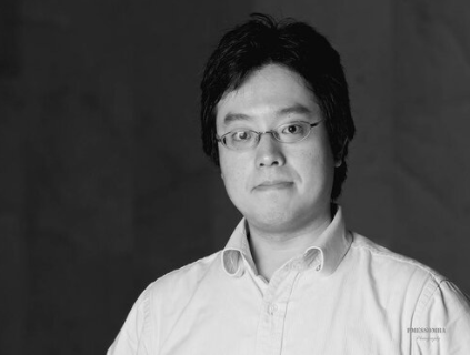
 (8 votes)