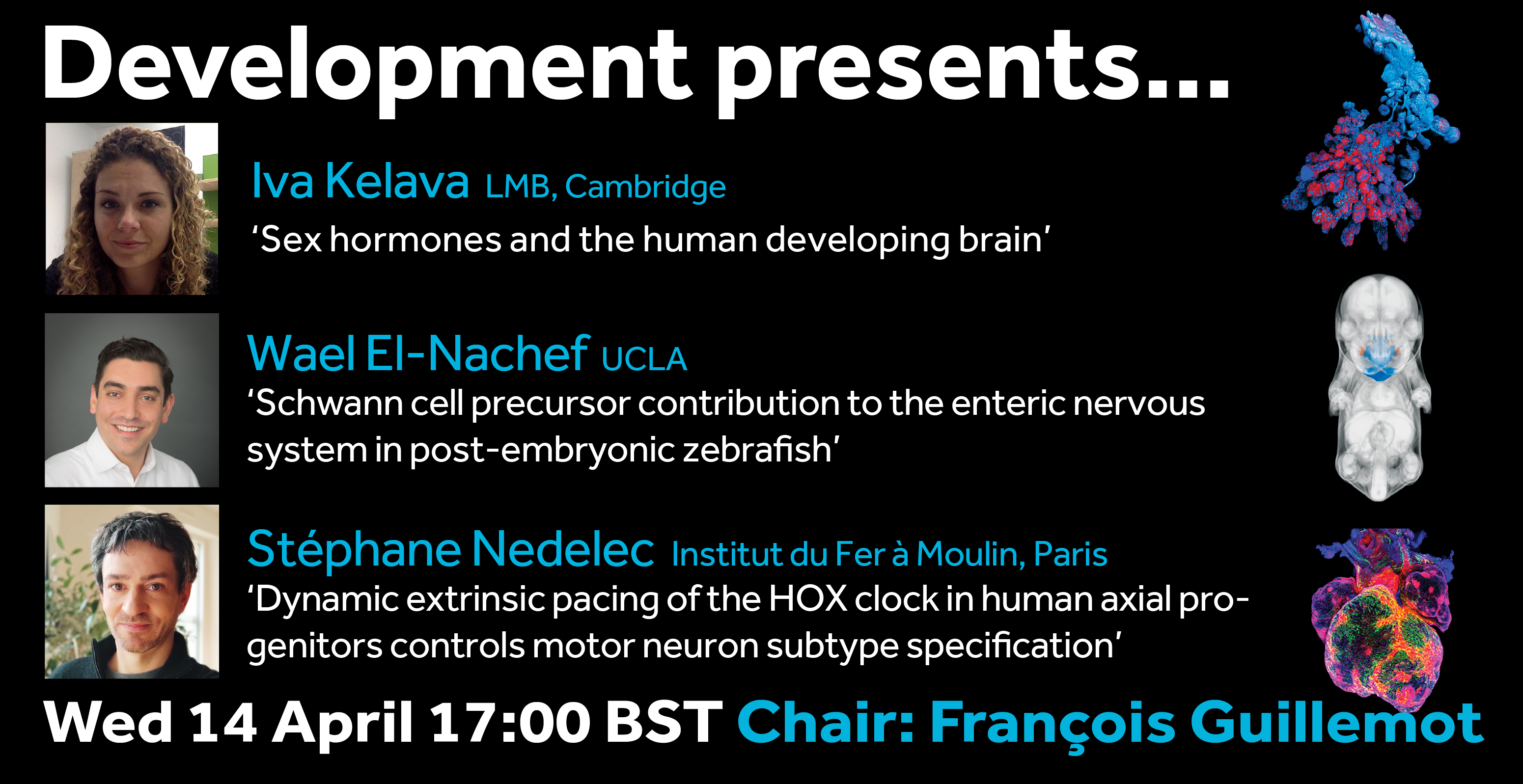
The seventh webinar in our Development presents… series will be chaired by Development Editor, François Guillemot (The Francis Crick Institute), who has brought together three exciting talks on the development of the nervous system.
Wednesday 14 April 2021 – 17:00 BST (GMT+1)
Iva Kelava (Postdoc in Madeline Lancaster’s lab at the Laboratory of Molecular Biology) ‘Sex hormones and the human developing brain’
Stéphane Nedelec (from the Institut du Fer à moulin) ‘Dynamic extrinsic pacing of the HOX clock in human axial progenitors control motor neuron subtype specification’
The webinar will be held in Remo, our browser-based conferencing platform – after the talks you’ll have the chance to meet the speakers and other participants at virtual conference tables. If you can’t make it on the day, talks will be available to watch for a couple of weeks after the event; details will be posted on the Node or you can sign up to our mailing list for email alerts.
Feel free to share this poster with your colleagues:
In mucociliary epithelia, such as the mammalian airway epithelium or the embryonic epidermis of Xenopus tadpoles, the correct balance between multiciliated cells (MCCs) and secretory cells provides the functional basis for removal of particles and pathogens to prevent infections and to maintain organismal oxygenation (Walentek and Quigley, 2017).
While the rest of the Walentek lab is working on how mucociliary epithelia are established during development, I am the “black sheep” of the lab, because I wanted to know how this tissue is remodeled to become a non-ciliated epithelium during metamorphosis. Indeed, mucociliary epithelial remodeling and MCC loss are observed in human chronic lung disease as well as during metamorphosis of the Xenopus epidermis. However, it remained unresolved how and why MCCs were lost in Xenopus, and how the process compares to observations made in mouse models of airway inflammation and human cells from chronic airway disease patients. By addressing this question, we hoped to find the underlying molecular mechanism for MCC loss in Xenopus, and to establish a new model to study mucociliary remodeling in the vertebrates.
Thus, I started my PhD on the loss of MCCs during Xenopus tadpole development. I found it especially interesting that everyone in the field was aware of this loss, but no one really looked at it in detail before to see how and when this was precisely happening. The first paper that I found on this topic dated from 1988 (Smith et al, 1988), where the authors describe a loss of MCCs from areas around the developing lateral line. Additionally, another group described MCCs with reduced ciliation that were positive for mucus staining in advanced tadpole stages (Nishikawa et al., 1992). But then, I could not find further work that would explain these phenomena. It remained unclear how the complete loss of MCCs from the tadpole epidermis was accomplished and why MCCs were lost in the first place. Therefore, I got interested in the case and I was hoping to resolve this mystery like a detective.
At the start of my investigations, I first established the time line of MCC loss during Xenopuslaevis tadpole development by immunofluorescence confocal- and scanning electron-microscopy.Being new to the field of Xenopus epidermis biology, my first discovery was to see how amazing normal MCCs look, with their hundreds of motile cilia and their dense apical F-actin network. Then, I started to observe how their morphology changed over time of epidermal remodeling. During these studies, I found that MCCs were lost during a first “local” phase in areas where lateral line neuromasts (NMs) would emerge. A bit later, MCCs were also lost everywhere else in the epidermis. This suggested to me that there could be two distinct mechanisms for MCC loss, depending on the location and the timing. So, I set out to investigate both processes in more detail.
MCCs undergo lateral line-induced apoptosis
Investigating the relationship between MCC loss and neuromast (NM) development seemed like a good start, because it confirmed previous findings by Smith et al. in other frog species and demonstrated a conservation of this phenomenon.
Figure 1 : MCCs are lost locally around the neuromasts of the lateral line. A)MCC stained for acetylated-α-tubulin and actin. MCC express low level of p27::GFP. B) Neuromast of the lateral line labeled by p27::GFP and stained for acetylated-α-tubulin and actin.
To understand the temporalrelationship between the migration of the lateral line primordium, NM deposition and the loss of MCCs, I started to transplant fluorescently labeled lateral line primordium cells into non-fluorescent hosts, and to use a transgenic reporter line (p27::GFP) (Rubbini et al., 2015) which expresses GFP in the lateral line primordium and NMs. Interestingly, I found that MCCs are still present while the primordium is migrating, but are lost when NMs emerge through intercalation in the epithelium (Figure 1). In parallel, I conducted immunofluorescent staining, confocal microscopy and analyzed scanning EM images, which showed that MCCs could be shed from the epithelium, suggesting removal through apoptosis. Therefore, I stained tadpoles with an anti-cleaved Caspase 3 antibody and performed TUNEL assays that showed signals exclusively in MCCs. This confirmed that MCCs over the lateral line were lost via apoptosis. Thus, we hypothesized that emergence of neuromasts induces loss of MCCs via shedding-apoptosis.
As we like to do in the lab, I first performed an easy and fast experiment to provide a proof-of-concept for our hypothesis that NM emergence is really the cause of local MCC loss. For that, I simply ablated the anterior part of the embryo where the lateral line primordium originates from and from where primordial cells migrate out in various directions to populate head, trunk and tail with NMs. This experiment confirmed that MCCs were not locally lost in absence of NM deposition.
But how did NMs induce this loss of MCCs? Looking into the literature, we realized that NMs are signaling centers that express Notch ligands. During specification, MCCs are inhibited by Notch signaling, and mature MCCs retain some level of Notch receptor expression, which means that they could also respond to Notch signaling changes. This led us to hypothesize that high Notch signaling from NMs could signal to MCCs and induce apoptosis. It did not take too long to find out which ligands are expressed in NMs, because fellow graduate student Magdalena Brislinger in the lab is working on Notch signaling and has analyzed the expression of all Notch ligands and receptors throughout early Xenopus development. On her beautiful images of sectioned tadpoles stained for Notch ligand expression, we found that the lateral line primordium and NMs express jag1 at high levels and induce hes1 expression in the overlying epithelial cells. This validated that NMs are Notch signaling centers that communicate with epidermal cells. By incubating the embryos in DAPT, which inhibits Notch signaling, and by performing Caspase 3 and TUNEL assays, I could show that MCC apoptosis and loss over the lateral line were suppressed in absence of Notch activation, confirming that Notch signaling is required for MCC loss via apoptosis.
The majority of MCCs coordinately trans-differentiates into Goblet secretory cells
Figure 2: MCCs trans-differentiate into a mucous-secretory goblet cells. Left: Normal MCC stained for acetylated-α-tubulin (grey), PNA (magenta) and actin (green). Right: Trans-differentiating MCC stained for acetylated-α-tubulin (grey), PNA (magenta) and actin (green) shows reduce ciliation and acetylation as well as mucin production and apical actin remodeling.
But my investigations were not finished yet! Broad epidermal TUNEL staining was missing from areas farther away from the lateral line, which made us think that an alternative mode of MCC removal was used there. To find out what was going on, I stained tadpoles throughout the time of global MCC loss to visualize MCC cilia, to identify secretory cell types via mucus staining, and for F-actin to outline cell borders and to assess cell morphology. Interestingly, confocal microscopy on these samples revealed altered apical F-actin morphology in a subset of MCCs, which also stained positive for mucus. I will always remember the moment when I found those cells and, still new to the Xenopus field, I ask Peter naively if it was normal to see some MCCs with mucus, and he got all excited about the finding (Figure 2).Quantification of this dataset showed that while the overall number of MCCs decreased over time, the proportions of mucus-positive MCCs increased. This suggested MCC to goblet cell trans-differentiation as an additional mechanism for MCC removal in the Xenopus epidermis.Subsequently, we also found that mesoderm-derived intermediate Notch signaling levels cause MCC to goblet cell trans-differentiation, but only when thyroid hormone was produced, which elevated Jak/STAT signaling that has an anti-apoptotic effect and is required to allow MCCs to undergo this transition- probably by making them more resistant against stress. Based on these findings, a key aspect of the paper became the dual role of Notch in MCC apoptosis and cell fate change. We (and the reviewers of our paper) thought that a genetic manipulation of Notch signaling, which could induce both behaviors in young MCCs, would strongly support our statement. Thus, I wanted to use a Notch gain-of-function approach to manipulate MCCs specifically. So, I generated a construct that expresses constitutive active Notch intracellular domain NICD fused to GFP under the control of a MCC-specific promoter. The cloning seemed easy but not if you consider the unexpected magic of cloning. After struggling for weeks to have this construct ready and perform my last experiments for this paper, I finally succeeded to generate the construct and open a bottle of Champaign to celebrate my success. After injecting the construct, I could see nuclear GFP in MCCs, but importantly, a significant proportion of GFP-positive cells showed goblet cell morphology, demonstrating that Notch signaling activation in MCCs can trigger fate change. Additionally, TUNEL assays showed the induction of apoptosis in early stage tadpoles. Together, these experiments provide evidence that ectopic Notch signaling can induce apoptosis as well as cell fate conversion in MCCs.
MCCs retract cilia and loose basal body components
Figure 3: Trans-differentiating MCCs remodel basal body distribution and composition. Confocal micrograph of a normal MCC and a trans-differentating MCC reveals disorganized basal bodies (Centrin4-CFP, grey), cilia de-acetylation (Ac.-α-tubulin, green), F-actin remodeling (Actin, green) and reduce levels of basal body distal appendages proteins (mCherry-Cep164, magenta), actin interactors (FAK-RFP, magenta), and rootlet components (Clamp-RFP, magenta).
We also found that trans-differentiation is initiated through loss of ciliary gene expression, including foxj1 (a master transcription factor for motile cilia maintenance) and pcm1 (a protein that protects cilia and basal bodies from degradation). At the cellular level, we could observe altered proteostasis, cilia retraction, basal body elimination (Figure 3, Figure 4G) as well as initiation of mucus production and secretion. Some of these changes resembled processes observed during primary cilia retraction, which is initiated in cycling cells when they re-enter the cell cycle to divide. So, I wondered if MCCs that trans-differentiate and become goblet cells could they also re-enter the cell cycle and divide again? I found that trans-differentiating MCCs also lost expression of the cell cycle inhibitor p27, supporting the idea that MCCs could re-enter the cell cycle, and presence of a hybrid cilium (Liu et al, 2020) could suggest that MCCs retrain a parental centriole that could serve as a base for mitotic division (Figure 4E). Therefore, I tried to follow individual cells using live-cell imaging and various techniques to label individual MCCs before trans-differentiation, including photo-convertible proteins, MCC-specific fluorescent labeling, etc. However, either the constructs turned out to be toxic to the cells, or the labeling could not be restricted to individual MCCs, thus, I could not exclude the possibility that I followed co-converted goblet cells, or I lost the cells during imaging. So, setting up this experiment properly would require a transgenic line, which takes a long time to establish in the Xenopus laevis system. Sadly, due to these technical limitations, we were not able to provide genetic tracing data in this paper. But we are looking forward to find out more about the cellular behaviors of MCCs during and after the trans-differentiation process in the future. This will fill an extremely important gap in our understanding of tissue remodeling and MCC loss!
Figure 4: Cilia and basal body structure visualised by electron microscopy. A) Transversal section and transmission electron microscopy of a motile cilium and its associated basal body. Motile cilia of MCCs are composed microtubules in a 9+2 configuration, a transition zone and a basal body. B-F) Parallel sections and transmission electron microscopy of a cilium (B), basal bodies with one (D) or two (E) basal feet and rootlet (F). G) Trans-differentiating MCCs are enriched in electron-dense structures corresponding to lysosomes.
So, in summary, our work describes two modes for MCC loss during vertebrate development, the signaling regulation of these processes, and demonstrates that even cells with extreme differentiation features can undergo direct fate conversion (Tasca et al.,2021). In addition to our scientific findings, this project was an amazing experience for me personally. It is a fantastic feeling to know that I could unravel, in large parts, a decades-long mystery, and to generate insights into the molecular processes of mucociliary tissue remodeling. I also enjoyed the scientific investigation, the collaboration with group members as well as with the Mitchell lab, and with Martin Helmstädter from the group of Gerd Walz in our department, who provided the beautiful electron microscopy images for the paper.
References
Liu, Z., et al., Super-Resolution Microscopy and FIB-SEM Imaging Reveal Parental Centriole-Derived, Hybrid Cilium in Mammalian Multiciliated Cells. Dev Cell, 2020. 55(2): p. 224-236 e6. DOI: 10.1016/j.devcel.2020.09.016
Nishikawa, S., J. Hirata, and F. Sasaki, Fate of ciliated epidermal cells during early development of Xenopus laevis using whole-mount immunostaining with an antibody against chondroitin 6-sulfate proteoglycan and anti-tubulin: transdifferentiation or metaplasia of amphibian epidermis. Histochemistry, 1992. 98(6): p. 355-8. DOI: 10.1007/BF00271070
Rubbini, D., et al., Retinoic Acid Signaling Mediates Hair Cell Regeneration by Repressing p27kip and sox2 in Supporting Cells. J Neurosci, 2015. 35(47): p. 15752-66. DOI: 10.1523/JNEUROSCI.1099-15.2015
Smith, S.C., M.J. Lannoo, and J.B. Armstrong, Lateral-line neuromast development in Ambystoma mexicanum and a comparison with Rana pipiens. J Morphol, 1988. 198(3): p. 367-379. DOI: 10.1002/jmor.1051980310
Walentek, P. and I.K. Quigley, What we can learn from a tadpole about ciliopathies and airway diseases: Using systems biology in Xenopus to study cilia and mucociliary epithelia. Genesis, 2017. 55(1-2). DOI: 10.1002/dvg.23001
Tasca, A., et al., Notch signaling induces either apoptosis or cell fate change in multiciliated cells during mucociliary tissue remodeling. Dev Cell, 2021. 56(4): p. 525-539 e6. DOI: 10.1016/j.devcel.2020.12.005
Dr Cagney Coomer received her PhD with Ann Morris at the University of Kentucky, where she studied zebrafish retinal development and regeneration, and is currently a postdoctoral researcher in Marnie Halpern’s lab in the Geisel School of Medicine at Dartmouth College. In 2020, she was awarded the Society of Developmental Biology’s inaugural Trainee Science Communication Award for her work with NERD SQUAD Inc, the non-profit STEM outreach organization she founded that is dedicated to inspiring the next great minds by bringing science to life. Over a virtual chat, we discussed her experiences in the lab, the classroom and the community centre, and why she thinks outreach and role models are vital to science.
When did you first become interested in science?
I wanted to be a scientist since I was 8 years old. I used to have this dream I was going to be a marine biologist and move to Alaska to study great white shark mating in the ring of death. But I grew up in Kentucky, which is a landlocked state, and I actually never saw an ocean until I was in college. I realised it’s nothing like a bathtub: it’s big and scary, and you can’t see the bottom! It was only when I was actually in the water that I appreciated my place would be at the bottom of the food chain – the human body is not made for the ocean. So I went back to campus and asked if I could change my major.
How did you first become involved in research?
Once I’d finished my undergraduate programme, when I was interviewing for jobs I’d get responses like ‘Well, yeah, you’ve done some things in the lab, but you’re really just a glorified dishwasher!’. I knew how to pour plates, clean dishes and stuff like that, but I didn’t have a lot of really technical skills, so I went to the Bluegrass Community and Technical College in Lexington to enrol in their biotech programme specifically to get those skills. In the process, my PI and I wrote an EPSCoR grant to start an undergraduate research programme at my local community college, where we developed a technique for growing aloe vera in a petri dish, and that’s how my actual research career started.
You ended up doing a PhD with Anne Morris at the University of Kentucky – what were you working on?
First of all, I picked Ann’s lab because she had the best reputation amongst the graduate students, and I really liked my project – I actually was meant to rotate with some other labs but ended up sticking with her from the start. I first started working on calpain 5, mutations in which are associated with this really devastating autosomal retinal disease in humans. It was interesting because even though it’s expressed in lots of different tissues, the mutation (which upregulates expression of the gene) only causes deterioration in the eye, and nobody really understood why. They didn’t understand its functional role, how it worked during development or where exactly it was expressed. One of the things that I was able to determine was that calpain 5 in its regular form actually plays a protective role. It’s kind of like when it’s mutated, it’s too much of a good thing: it’s turned on for so long so what it would normally do is extended, leading to an immune response that causes the degeneration.
Then another graduate student graduated from the lab and he passed his project on to me. It was focused on the role of the bHLH-O transcription factor her9 during retinal development and regeneration, and I began by making CRISPR mutants. I first found out that her9 actually isn’t really involved in retinal development – rather, it might be more involved in maintenance. The second thing I learned was that it actually plays a stronger role in neural crest cell development and migration, so I got to leave the retina and go looking for the neural crest cells, which are just so cool. They do all these amazing things all over the body, and I got to learn all these new techniques and look at all these different cell types. It’s the cool thing about mutant analysis: you have an idea about what to expect, but you really don’t know until you make them.
Why are zebrafish particularly good models for retinal development and disease?
There’s lots of reasons, starting with their rapid development: by 5 days, you have a fully developed eye, functioning with all the cell types in there. Retina research has mainly been done on the mouse, but the mouse is diurnal, with a really rod-dominant retina. In contrast, like the human retina, the zebrafish retina is cone dominant, giving us a really good way to study cone versus rod development. Zebrafish has a high level of gene conservation with humans, and our introduced mutations often phenocopy human diseases: we can model almost any retinal disease in the zebrafish. But I think what makes it so interesting is that zebrafish retinas are capable of regeneration, so we can actually study the mechanisms that it uses to try to fix itself.
When I first got to the lab, Ann talked about how important retinal development was, and I realised how much I’d been taking my vision for granted. I hadn’t ever really thought about my eyesight, or what it would be like to have a life with visual impairment, or how these impairments affect the quality of life of millions of people. It’s really important research.
And now, after your PhD, you’ve just moved up to Dartmouth – what will you be doing there?
I’ll still be using the zebrafish as my model organism but it’s more neuroscience than my PhD: I’m going to study how right/left brain function is established and works. I’ll be working in Dr Halpern’s lab focusing on the neural circuitry in the brain and, more specifically, the habenula.
This year, you received the SDB Trainee Communication Award for your work in NERD SQUAD Inc. Why did you start the initiative and how does it work?
It all started when I got an award (National Science Foundation Outstanding Student Award) and I went up to Washington DC. There were 100 top rising young scientists picked to go, and I was the only black person there. I wondered, why is that? I mean, in Lexington, I’m the only black person at work, but I always assumed it was because I live in Kentucky – I didn’t realise it reflected a bigger picture. So it’s not just in my little bubble – this is a bigger issue broadly. And then when I would ask people why, they would say well, students of colour aren’t interested in science, or they don’t really seem to have the IQ or this or that, but all these reasons didn’t really make sense because I’m black, I’m interested, I have the IQ, I can do it. Why couldn’t any other student do it?
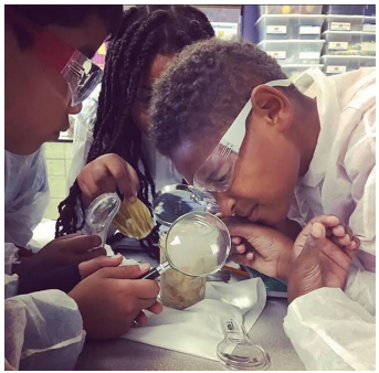
So I reached out to community centres in Lexington and asked them if I could host a science night to do activities with kids. I just wanted to garner some interest in my community, to show them at least why I like science, because I think science is like magic, but a magic where you get to know how it works. All my activities cost less than $20 and used materials that you could readily get in your own home. That way, kids could go home and recreate the activities at home, showing their families and their parents. When I first started, I might have had 10 kids, but after doing science night for about a month, I had 45 kids just at one community centre, and they were bringing their siblings and parents: all wanted to come and see what I was going to do.
I would ask the children, why don’t you like science? ‘School is boring, it’s nothing like what you do Miss Ceecee!’ But surely there’s no way science can be boring? ‘No, it really is boring!’ So I reached out to the elementary school in my neighbourhood, and asked if I could come shadow the science class. When I left I was like, oh, science is boring, they were right! So then the question is, why? Initially, we started partnering with the science teachers – they would teach the curriculum and we would design activities that fit into their curriculum to help get the point across. We started with fourth grade (in our city, students only get science in fourth and sixth grade, and then biology in ninth grade). I realised the reason there’s no fun in science class is because there’s no science foundation for these students to begin with – everything seems overwhelming and hard if you don’t have a good foundation to stand on.
The next year, the school voted to make STEM Lab an elective: all the grades got it once a week, the same way they got gym or art or computing. We started to design our own project-based learning activities and come all through the school year, and at the end of the year hosted a community night where the students would put up booths and teach the science to their families and their community. That gave them a way to be in control of the excitement of science. And from then, it just started to generate so much momentum.
When people ask me what we do in NERD SQUAD Inc, I say we teach kids how to think. It’s not a skill that you’re born with, it’s something you learn, and yet we treat it like it’s just innate – we don’t give our students the tools to actually learn how to use their brains.
Why is it so important to make the science activities relevant to the daily lives of these children?
I’ll give an example. One of the big parts of the science curriculum is the weather, and when I went to a science class, the teacher was teaching them about avalanches. How could you perceive an avalanche when you live in Kentucky? Can you even get an avalanche with two inches of snow? It’s just so hard to grasp. But in Kentucky it rains a lot, so why don’t you start with a subject or an activity that they can relate to? I taught the teacher how to make clouds in a jar so that she could do something the kids would actually be able to relate to.
If we want people to understand why science is important, not only to us, but to the world, we have to teach it so the students can draw the connections between it and their own lives. The bigger picture is important, but we break it down into the smaller things – things they can be like oh, I understand that, I didn’t realise that was science. The kids totally freak when they find out brushing your teeth is a chemical reaction. I tell them every morning, you’re doing science when you brush your teeth – it blows their minds!
As the NERD SQUAD Inc grows and develops, some of your earliest members have now started college. It must be quite rewarding to follow their lives?
It is exciting, but sometimes it’s scary, because I actually work in this field. I’m getting all these little black girls so interested, so excited and so ambitious to be in this field, but then they have to come here and actually survive. If I’m honest, it’s a double-edged sword.
Why is it important that academia recognises the importance of outreach?
Take coronavirus, for instance: people don’t believe us, and they don’t believe us because they can’t relate to us. We aren’t coming down into the community and getting them to understand why what we do is important, and why they should believe us. I think that outreach gives us a platform: it’s a scientist’s direct action. These are the issues, this is the problem, this is how I’m trying to help, this is why what I do is important; we just don’t do enough of that, and so we become more and more distant from the communities and the people that we’re trying to help.
“Outreach gives us a platform: it’s a scientist’s direct action“
What can organisations do to help the position of underrepresented minorities in science?
Academia, for instance, has to take some steps back and actually try to create an environment supportive of students of colour. It’s not that we can’t do the work – most of the struggle that I had during my PhD was actually trying to figure out whether it is a place that I want to go and work. Do I want to spend my life trying to prove that I’m capable, trying to prove that I belong here, all the while feeling like I don’t, feeling like nobody will take me seriously and welcome me in this space? I just want to be able to show up as a scientist and be a scientist. But that’s not what I get – I have to always prove that I’m more than just a good scientist. It’s the kind of environment that puts me in positions where I literally have to say to people ‘well, that’s not appropriate, that’s not cool, why would you say that?’ Who wants to go to work every day and always have to tell people that the things they say and do make you feel insignificant and invisible? It’s hard. And on top of that it’s really hard in this field to express yourself because one wrong statement, one wrong move could ruin your whole career. You’re worried about speaking up for yourself because of how that could influence your career.
When it comes to journals and magazines, they need to do a better job of highlighting scientists of colour. I came to graduate school without ever seeing another scientist of colour. How do I know that this is a possible career if I’ve never seen anybody that looks like me? Who says I want to be the first? You can’t oblige me to think that that’s the journey I should have to take, especially when I know I’m not the first. So you should give me examples so I know what this looks like, so I know what’s possible.
So role models are just as important in universities as in elementary schools?
They are. I got my PhD in the University of Kentucky, a tier one research university. They have about 200 research scientists, and this year there’s no black faculty. I was literally the only person of colour in the whole building. It didn’t feel like there was much pressure (either internal or, for example, from funding agencies) to make a change or require institutions to do anything that would help diversity or inclusion in science. I feel like comfortable people don’t change – if they don’t need to make changes in order to get the funding, why would they care?
Do you see yourself continuing in academia long term?
Yes, I do. I think that it is hard, and it is a lot of work, but at the end of the day, I already took on this role as a role model and an example. No one asked me to do it, so now I’m here, if I have the opportunity to create space for others, I’ll create space for them.
Our final question: when you’re not doing science, what do you do?
We’re outdoors people – we rock climb, visit waterfalls, hike, and just like to be outside. New Hampshire is different to Kentucky in a lot of ways, but it’s pretty rural too, and we fit into that side of it. There’s so much water here, every turn it’s a river, a lake, a pond, a waterfall. It’s been beautiful so far.
By Rachel Bonnington, Carla Lloret Fernández and Laura Molina García
Classically, developmental programmes were believed to be a one-way linear process in which cells progressively acquire their differentiated identities, each with a distinct and highly specialised morphology and function. Since differentiated post-mitotic identities are usually stable, differentiation was first thought to be irreversible. However, there are now many examples that appear to subvert this idea, both in nature and during forced reprogramming experiments, where differentiated cells are able to switch their identity into distinct differentiated cell types (Lambert et al. in press). This process is called transdifferentiation (Eguchi & Kodama, 1993; Selman & Kafatos,1974). In our recent paper (Molina-García et al., 2020), we describe how a glial cell transdifferentiates into a neuron in a sexually dimorphic manner to optimise male mating performance in the nematode worm C. elegans. Our findings demonstrate how genetically-programmed transdifferentiation acts as a developmental mechanism to allow flexibility in innate behaviour.
C. elegans is an excellent model for single cell studies
Two main challenges of studying transdifferentiation are that two distinct, stable identities need to be unambiguously defined, both before and after the proposed cell type conversion, and a direct lineal relationship must be established between them. This remains extremely difficult to determine in most model organisms, in which cellular lineages are highly variable and poorly defined. However, C. elegans conveniently overcomes these barriers, allowing us to study natural cellular reprogramming events in live animals. In fact, the first transdifferentiation in C. elegans was described nearly fifty years ago by Sir John Sulston, one of our scientific heroes, who observed that the rectal-epithelial cell Y converts into the PDA motor neuron (Sulston & Horvitz, 1977). This was possible because development in C. elegans is highly stereotyped and the number of cells in the adult is fixed. This allowed Sulston and colleagues to fully resolve the somatic lineage of the worm, which describes all cellular identities and their positions, for the two sexes: males and hermaphrodites (Sulston & Horvitz, 1977; Sulston et al. 1980; Sulston et al. 1983). C. elegans is also transparent, and transgenics are readily made, so cells can be imaged at a single-cell level, making it possible to unambiguously follow cell-type conversion events. No wonder then, that C.elegans is our model organism of choice in the Barrios and Poole labs at UCL!
PHso1 is a glial cell that changes identity in the worm
Previous work from the Barrios and Poole labs described how a pair of glial cells, called amphid socket (AMso) cells, undergo an identity change into neurons in males, providing the second example of transdifferentiation in the worm. The AMso cells form the cuticular pore of the amphid chemosensory organ in the head of the worm that allows sensory neurons to contact and respond to the external world. In this case, transdifferentiation occurs alongside asymmetric cell division that leads to a self-renewal of the AMso glial cells and the production of a previously unidentified male-specific pair of interneurons, the MCMs (Sammut et al. 2015). The AMso cells retain their structural role in forming the socket of the amphid, while the MCM neurons are involved in male-specific learning.
Following on from our discovery of this glia-to-neuron transdifferentiation, we were interested to examine the sensory pore of the equivalent sensory organ in the tail of worms, the phasmid sensillum. Unlike the amphid, the phasmid is made up of two pairs of socket cells (the sister cells PHso1 and PHso2), which together form a bilayer hollow pore in the cuticle. Again, during his description of the C. elegans lineage, John Sulston noted a difference between the phasmid sockets in males and hermaphrodites, observing that in adult males, PHso1 cells appear to retract from the hypodermis, and that they contain basal bodies (a structural component of cilia – in C. elegans, the only ciliated cells are sensory neurons) (Sulston, 1980). However, no other neuronal characteristics were noted and PHso1 cells continued to be classified as glia.
We were therefore hugely excited when we analysed the lin-48/OVO1 transgenic reporter, known to be expressed in PHso1, and noticed a long, axon-like projection in males, extending towards the pre-anal ganglion, a key neuronal centre in the nematode tail. This strongly suggested a neuronal identity for PHso1!
Could this be the smoking gun we were looking for, proving that in males the anterior-most phasmid socket glial cells (PHso1) were in fact being transformed into neurons? To determine this, we examined these lin-48-expressing cells during different developmental stages in males by time-lapse and compared them to hermaphrodites. We found that during the early stages of larval development, PHso1 cells appear indistinguishable between males and hermaphrodites. During the course of male sexual maturation, PHso1s in males undergo radical remodelling from a socket-glial morphology to a neuron-like morphology. By contrast, PHso1 cells remain as sockets in hermaphrodites (see Figure), thus corroborating John Sulston’s earlier observations of sexual dimorphisms in PHso1 morphology.
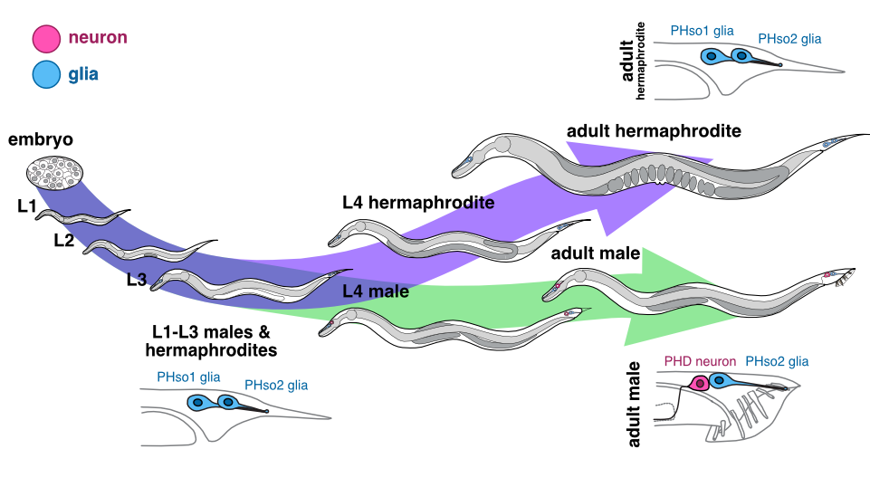
A simplified life cycle of the nematode worm, C. elegans, showing a lateral view of the Phasmid socket glia pairs (PHso1 and PHso2) in both males and hermaphrodites. Both sexes go through four larval stages (L1-4), and during L4 animals become visibly sexually dimorphic. During male sexual maturation, the PHso1 glia undergo a direct glia-to-neuron transdifferentiation, resulting in the PHD neurons, while in hermaphrodites they remain sockets for the whole life of the animal.
Male PHso1 glia transdifferentiate into PHD neurons
At the gene expression and ultrastructural levels, male PHso1 cells also acquire a number of uniquely neuronal characteristics, which we never observed in either the neighbouring PHso2 cells, nor the PHso1&2 cells of hermaphrodites. These include the expression of pan-neuronal genes and genes involved in neuronal transport and communication (assessed by fluorescent reporter expression) as well as the presence of synaptic vesicles, dense-core vesicles and ciliated structures (assessed by electron microscopy). Importantly, at the same stage of sexual maturation, when these neuronal markers are firing up, we see dimming of glial genes, such as the panglial microRNA mir-228 and the AMso and PHso glial subtype marker grl-2, in male PHso1s, before they fully switch off. Together, these results demonstrate that PHso1 glial cells transdifferentiate into a previously undescribed class of neurons that we call PHDs (Phasmid neuron D). This updates the anatomy of the C. elegans male, increasing the number of neurons from 385 to 387 (of which 93 are male-specific) and decreasing the total number of glia from 92 to 90.
The PHso1-to-PHD transdifferentiation seems to occur without a division. It does not require wholesale DNA replication or cell division followed by programmed cell death of one of the daughters, a common strategy used throughout development (reviewed in Conradt et al. 2016). To our surprise, however, we sometimes observed that PHso1 divides in a background-dependent manner to give rise to two apparently equivalent neurons (PHD1 and PHD2). Moreover, when we genetically manipulated the biological sex of the cell (i.e. we masculinised PHso1 in an otherwise hermaphroditic background) we observed a similar transdifferentiation of PHso1 to a PHD-like cell. This suggests that, as we previously published for the AMso, PHso1 is poised to transdifferentiate, awaiting cell-autonomous activation by the sex determination pathway.
We next sought to investigate if the molecular mechanisms known to regulate the Y-to-PDA transdifferentiation mentioned earlier could also control the transdifferentiation of the PHso1 and AMso cells. The Greenwald and Jarriault labs were the first to fully characterise the Y-to-PDA transdifferentiation (Jarriault et al., 2008). They later showed that a complex of conserved NODE-like factors (CEH-6/Oct, SEM-4/Sall and EGL-27/Mta) together with the transcription factor SOX-2, known to regulate mammalian embryonic stem cell pluripotency, are required for the initiation of the process (Kagias et al. 2012). Interestingly, these factors seem to be largely dispensable for the PHso1 and AMso cell identity switches. This could point towards independent mechanisms of transdifferentiation rather than a shared program. It will be interesting to determine if, as for Y-to-PDA, chromatin remodelling (Zuryn et al. 2014) also plays a role in AMso and PHso1 transdifferentiation, or whether transdifferentiation of these cells rely on completely different strategies. The new AMso and PHso1 cellular paradigms provide the perfect scenario for performing forward genetic screens and single-cell sequencing in order to identify, compare and contrast the molecules regulating transdifferentiation in the worm.
What is the function of PHD?
Bearing in mind that the PHD neurons arise during sexual maturation and exclusively in males, we asked ourselves what function these cells might have. First, we tried to identify the sensory stimulus that activates PHDs, by measuring neuronal activity in immobilised animals using a genetically-encoded calcium indicator. Surprisingly, we noticed spontaneous activity of the PHD neurons in the absence of any stimuli. We then realised that, despite immobilisation, some muscles near the PHD neurons were still twitching due to the defecation cycle and spasms of the spicules (the equivalent of the penis in nematodes). Perhaps the PHDs are mechanosensors and they are being activated by the internal deformations caused by those muscle contractions?
To test this idea, we measured PHD activity in worms in which muscle contractions were abolished using an inducible chemogenetic tool (an histamine-gated chloride channel transgene). Indeed, muscle silencing eliminated PHD activity, supporting our hypothesis. Furthermore, it appears that the PHDs sense muscle contractions directly and not through other neurons within the circuit because disrupting their chemical synaptic input (through mutations in genes required for synaptic transmission) did not eliminate activity. PHD neurons are thus likely to have a proprioceptive function – but which process could they be involved in?
Through reconstruction of serial electron micrographs we identified all the synaptic partners of PHD and we realised that they are not only unique to males but also highly connected to other male-specific neuronal circuits. This was highly suggestive of a role in mating. C. elegans mating behaviour is stereotyped and consists of a sequence of behavioural steps: response to a potential mate, scanning the mate’s body, turning, location of vulva, spicule insertion and ejaculation (reviewed in Barr et al., 2018). We compared the performance of each mating step in control animals and animals in which we removed PHD by laser ablation, and noticed a defect in the scanning behaviour of PHD-ablated males. In a normal mating sequence, the male scans the body of its mate moving backwards (tail-first) in a continuous manner. However, males lacking PHDs could not perform this movement smoothly, and they tended to switch directionality and pause during scanning.
As stated above, C. elegans mating behaviour is sequential and if completion of a step fails, the animal will repeat the previous step and try again. Interestingly, we noticed that wild-type males that were repeatedly unsuccessful at spicule insertion did not always return to scanning backwards in order to relocate the vulva. Instead, they performed a readjustment movement by going forwards (head-first), away from the vulva, and then returning to the vulva backwards to try to insert the spicules again (see Video 1). This was remarkable as all previously described male mating movements involve only backwards locomotion. We called this novel readjustment the ‘Molina manoeuvre’ (MM) after Laura Molina, who first observed it and to acknowledge her good eye for males!
Wildtype (Video 1) and PHD-ablated (Video 2) male worms performing Molina manoeuvres during mating with paralysed hermaphrodites (mutant for unc-51), as shown in our recent eLife paper (Molina-García et al., 2020).
When we looked specifically at MM performance, we found that animals without PHD neurons displayed discontinuous manoeuvres, often stopping at the transition from forward to backward locomotion to return to the vulva (see Video 2). Together, our data show that without intact PHD neurons, backward movement along the mating partner becomes somewhat erratic.
Are PHDs activated during backward locomotion? Consistent with the behavioural defects, we observed higher activity (i.e. rise in Ca2+ levels) in PHD during backward locomotion than during forward locomotion while scanning. The same happened during the MM, during which PHD activity also peaked just after the switch to backward locomotion. However, the highest level of PHD activation occurred during intromission. This step involves full insertion of the spicules into the mate’s vulva while sustaining backward locomotion and precedes sperm transfer. Interestingly, PHD-ablated males produced fewer cross-progeny than intact males after a single mating encounter. This suggests that intact PHDs may increase the efficiency of sperm transfer by controlling the male’s posture during intromission, which would be consistent with a putative proprioceptive role for these neurons.
Scientific significance
In summary, the previously undescribed male-specific PHD neurons are born through transdifferentiation during sexual maturation to control backward locomotion during mating. This is of high ethological relevance as the failure to complete mating implies missing a chance to reproduce and therefore failing to pass on one’s genes. Neurogenesis through transdifferentiation could facilitate strict temporal and spatial control of such finely tuned behaviours, repurposing a pre-existing cell for a newly required function, or allowing the generation of a cell only when specific structures are already in place (i.e. to ensure correct neuronal wiring).
Importantly, this is the second example of neurons arising from differentiated glial cells in C. elegans, following our previous work on the AMso. This process resembles neurogenesis in the vertebrate postnatal brain, where radial glial cells produce post-mitotic neurons (reviewed in Kriegstein and Alvarez-Buylla, 2009), raising the intriguing possibility that shared mechanisms may govern glia-to-neuron transdifferentiation in the worm and vertebrate adult neurogenesis. Identifying the mechanisms that regulate these naturally occurring switches in cell identities will improve our understanding of cellular plasticity and will help develop more efficient protocols for reprogramming cells in vitro, which is widely used for cell replacement therapies. Furthermore, a deeper understanding of how locomotion is guided by self-sensory feedback could be applied to improve the execution of behavioural sequences in artificial intelligence and robotics.
References
Barr, MM., García, LR., Portman, DS. (2018) Sexual Dimorphism and Sex Differences in Caenorhabditis elegans Neuronal Development and Behavior. Genetics 208(3): 909-935; https://doi.org/10.1534/genetics.117.300294
Conradt, B., Wu, YC., Xue, D. (2016). Programmed Cell Death During Caenorhabditis elegans Development. Genetics 203(4): 1533-1562; https://doi.org/10.1534/genetics.115.186247
Jarriault, S., Schwab, Y., Greenwald, I. (2008). A Caenorhabditis elegans model for epithelial-neuronal transdifferentiation. Proceedings of the National Academy of Sciences of the United States of America, 105(10), 3790–3795. https://doi.org/10.1073/pnas.0712159105
Kagias, K., Ahier, A., Fischer, N., Jarriault, S. (2012). Members of the NODE (Nanog and Oct4-associated deacetylase) complex and SOX-2 promote the initiation of a natural cellular reprogramming event in vivo. Proceedings of the National Academy of Sciences of the United States of America, 109(17), 6596–6601. https://doi.org/10.1073/pnas.1117031109
Lambert, J., Lloret-Fernández, C., Laplane, L., Poole, R. J. & Jarriault, S. (in press). On the origins and conceptual frameworks of natural plasticity – lessons from single cell models in C. elegans. Current Trends in Developmental Biology.
Molina-García, L., Lloret-Fernández, C., Cook, SJ., Kim, B., Bonnington, RC., Sammut, M., O’Shea, JM., Gilbert, SP., Elliott, DJ., Hall, DH., Emmons, SW., Barrios, A. & Poole, RJ. (2020). Direct glia-to-neuron transdifferentiation gives rise to a pair of male-specific neurons that ensure nimble male mating. Elife 9:e48361. https://doi:10.7554/eLife.48361
Sammut, M., Cook, S. J., Nguyen, K., Felton, T., Hall, D. H., Emmons, S. W., Poole, R. J., & Barrios, A. (2015). Glia-derived neurons are required for sex-specific learning in C. elegans. Nature, 526(7573), 385–390. https://doi.org/10.1038/nature15700
Selman, K., Kafatos, F.C., 1974. Transdifferentiation in the labial gland of silk moths: is DNA required for cellular metamorphosis? Cell Differ. 3, 81–94. https://doi.org/10.1016/0045-6039(74)90030-X
Sulston, J. E. & Horvitz, H. R. (1977). Post-embryonic cell lineages of the nematode, Caenorhabditis elegans. Developmental Biology, 56(1):110-156. https://doi.org/10.1016/0012-1606(77)90158-0
Sulston, J. E., Albertson, D. G., & Thomson, J. N. (1980). The Caenorhabditis elegans male: postembryonic development of nongonadal structures. Developmental biology, 78(2), 542–576. https://doi.org/10.1016/0012-1606(80)90352-8
Sulston, J. E., Schierenberg, E., White, J. G., & Thomson, J. N. (1983). The embryonic cell lineage of the nematode Caenorhabditis elegans. Developmental Biology, 100(1), 64–119. https://doi.org/10.1016/0012-1606(83)90201-4
Zuryn, S., Ahier, A., Portoso, M., White, E. R., Morin, M. C., Margueron, R., & Jarriault, S. (2014). Transdifferentiation. Sequential histone-modifying activities determine the robustness of transdifferentiation. Science (New York, N.Y.), 345(6198), 826–829. https://doi.org/10.1126/science.1255885
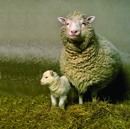
In the latest Genetics Unzipped podcast we chat with author Helen Pilcher about how humans have shaped the evolutionary trajectory of species on earth, find out how genetics is used in conservation Alex Ball from the RZSS WildGenes project , and meet Bill Ritchie, the embryologist who cloned Dolly The Sheep at the Roslin Institute in the 1990s.
If you enjoy the show, please do rate and review on Apple podcasts and help to spread the word on social media. And you can always send feedback and suggestions for future episodes and guests to podcast@geneticsunzipped.com Follow us on Twitter – @geneticsunzip
I joined the Node and Development out of a postdoc in 2016 and now after five enjoyable years I’m moving on. I’ll write something a bit more reflective closer to my leaving date in June but here I thought I’d let you know what the job entails, since we’re searching for my replacement.
If you’re interested in science communication and helping the developmental biology community in various ways, this job might be ideal for you. And if you know anyone who is thinking of hanging up their pipettes but still wants to stay in touch with science and scientists, please send the job ad their way.
Here’s a collection of the kind of things you’d get up to:
Run the Node
Commission content – research stories, interviews, meeting reports etc.
Edit / give feedback on drafts for authors
Write your own content
Run competitions and series of posts
Maintain the site (including the now 900+ strong Node Network)
Develop the site’s future – we’re now 10 years old and well-established, but there are always opportunities for innovation
Social media
For the Node: Twitter (where all the fun stuff happens) and Facebook
For Development: Twitter, Facebook, more recently Instagram, and on our YouTube channel you’ll have opportunities for video editing
With these accounts you reach thousands of people, specialists and non-specialists, to spread the word about developmental biology
For Development
Write Research Highlights (usually one a week)
Conduct ‘The people behind the papers’ interview series (usually one per issue), and longer standalone interviews (could be a Nobel laureate or an up-and-coming star)
Write and manage press releases for topical papers
Development presents… – help out with our webinar series
Attend Editor meetings and strategy sessions – insight into the publishing world
Conferences
In a normal year, go to perhaps six conferences, some abroad, usually focusing on society events. Meet people, hear new science, promote our work, write meeting reports, interview prize winners
Help out at Development’s biennial human development meetings and Company of Biologists Workshops
Working for The Company of Biologists
Provide cover for and collaborate with the two other community sites, preLights and FocalPlane
Promote our work as a not-for-profit publisher
Work with 50 other great colleagues near Cambridge (/home office)
During their practical classes the students produced some beautiful images and so, as we did in 2018, we’re going to use them in a competition to find a Development cover. Participating is easy – just vote for your favourite image from the following selection (click to get full size images, voting below the pictures). The winner will then be immortalised in print and on screen in a future issue of the journal – testament to the bright future of developmental biology in Latin America.
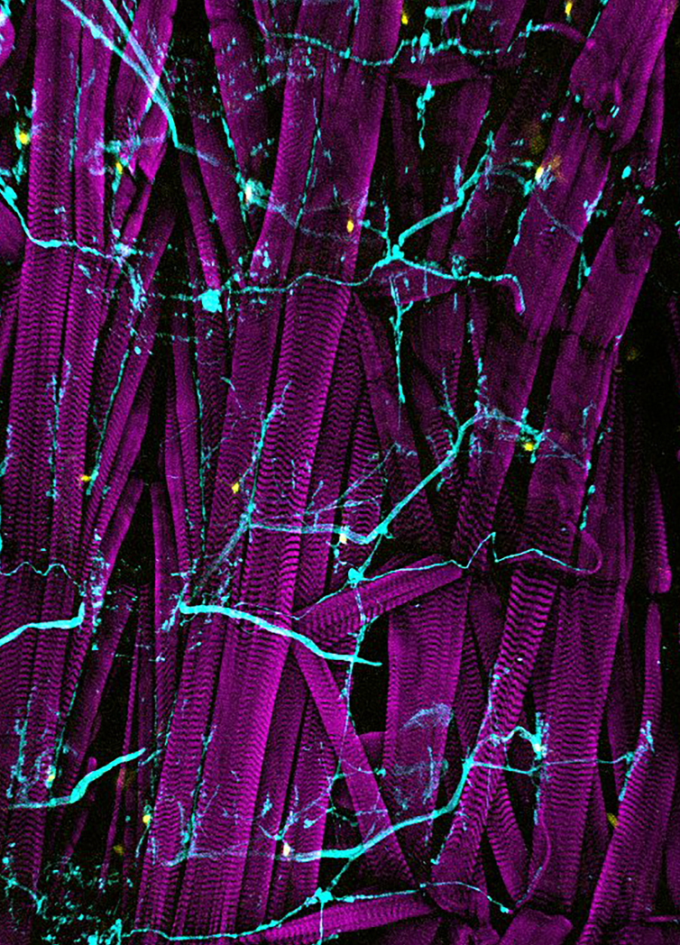
2. Drosophila larval body wall. Third instar larva body wall muscle with nervous system innervation. Phalloidin (magenta), HRP (cyan), ppk > GPF (yellow). By Pablo Guzman Palma
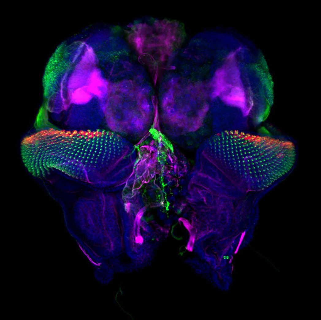
3. Drosophila eye discs and brain lobes. In blue: nuclei (DAPI); in red: ganglion mother cells and neurons (PROSPERO); in magenta: F-actin (Phalloidin) and in green: all neurons (HRP)). By Tonatiuh Molina Villa.
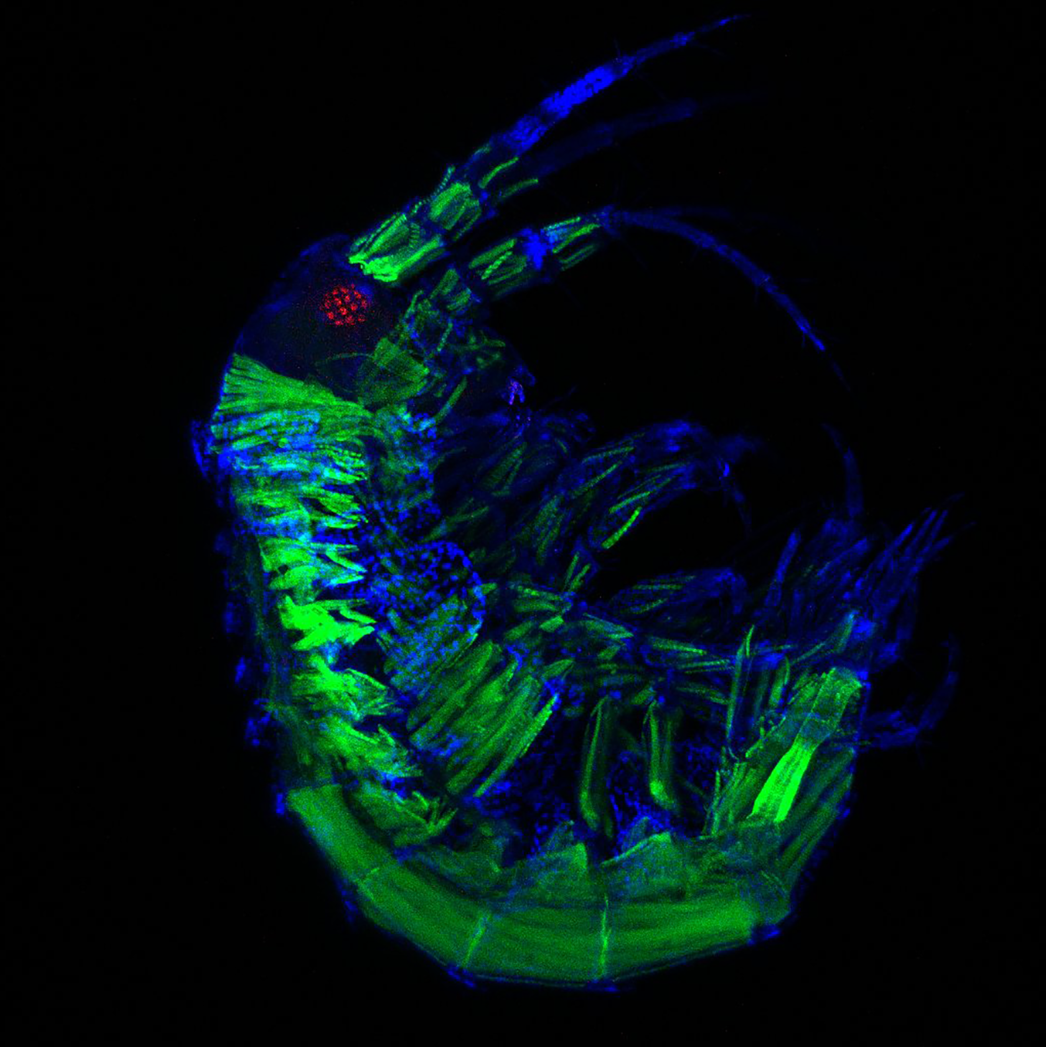
4. Parhyale(amphipod crustacean). F-actin (green-Phalloidin), nuclei (blue, DAPI) and eye (red-Elav). By Diana Carolina Castañeda-Cortés, Nicolas Eduardo Cumplido Salas, Felipe Andres Gajardo Escobar
5. Sea urchin late blastula. Green: Pax3/7; red Hoecsht. By Shurti Purushothaman
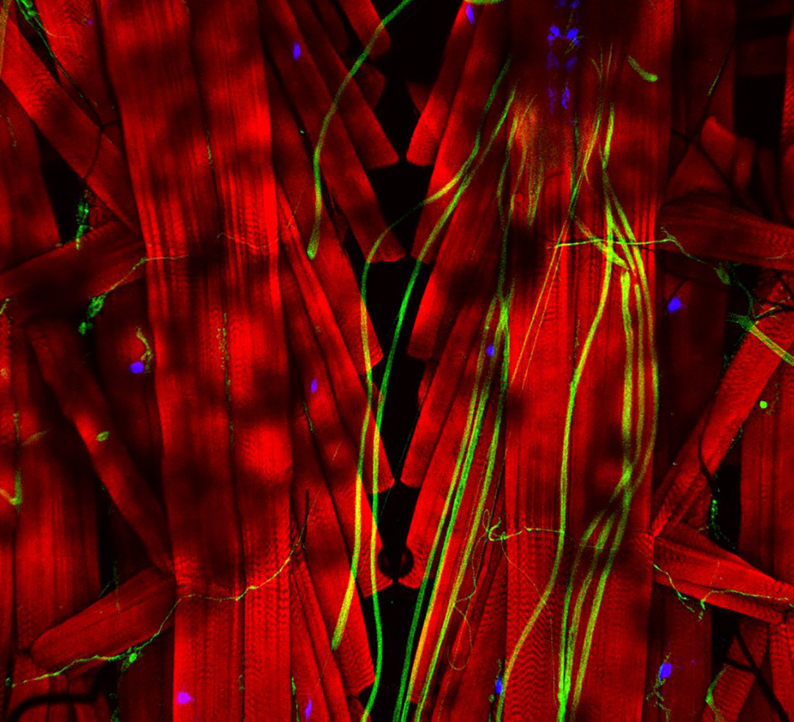
6. Drosophila larval body wall. Phalloidin (red), HRP (green), ppk > GPF (blue). By Marycruz Flores Flores, Felipe Berti Valer, Emiliano Molina.
7. Parhyale embryo. Labelled with DAPI and a membrane marker. By Marycruz Flores Flores, Felipe Berti Valer, Emiliano Molina
One vote per person – voting closes Monday 29 March!
Last week, we held the sixth webinar in our series, which was chaired by Development editor Thomas Lecuit (IBDM).
Below you’ll find recordings of the talks and live Q&A sessions.
Hongzhe Peng (from Bo Dong’s lab at Ocean University of China) ‘Ciona embryonic tail bending is driven by asymmetrical notochord contractility and coordinated by epithelial proliferation
Camille Curantz (from Marie Manceau’s lab at Collège de France) ‘Cell shape anisotropy and motility constrain self-organised feather pattern fidelity in birds’
The annual Young Embryologist Network Conference is happening this year on May 4th! Put the date in your calendars now!
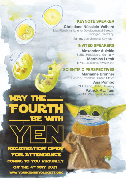
YEN is thrilled to announce that Nobel Laureate Christiane Nüsslein-Volhard from the Max Planck Institute of Developmental Biology in Tübingen, Germany will present this year’s Sammy Lee Memorial Lecture. We are also honoured to host two special speakers: Matthias Lutolf from EPFL in Lausanne, Switzerland and Alexander Aulehla from EMBL in Heidelberg, Germany. Finally, we are delighted that Marianne Bronner from Caltech, USA, Ana Pombo from MDC Berlin, Germany and Patrick P.L. Tam from CMRI, Australia will share invaluable insights from their life as a scientist in our “Scientific Perspectives” session.
YEN 2021 will be held entirely online, which will allow for unprecedented international participation. Spread the word, and let us take the Young Embryologist Network worldwide!
Talks will be held throughout the day, beginning at 9.15am (BST). We will be using the online conference platform Remo, enabling us to recapitulate that in-person networking feeling of conferences that we are all missing. Interactive poster sessions will also be held via Remo, with further discussions taking place in dedicated Slack channels both during and after the event.
Register here to submit an abstract or to attend as a delegate (and hear what your fellow young embryologists are up to). Registration is free and the deadline for abstract submission is April 4th, so be sure to sign up as soon as possible!
In 2020 the Node turned 10 and, along with a virtual networking birthday party and a Development editorial, we ran a community survey for advice on what to improve and where to go next. We gathered some fantastic ideas for content that we’re going to develop soon but also heard many suggestions for things we’d done before. This made us think about how we could better promote historical Node content (going all the way back to 2010), pieces that are currently quite hard to find in the archive. We also felt that the homepage needed a refresh – it hadn’t been updated since 2015 – and identified a few more tweaks we’d like to implement, both from reader and author perspectives. These discussions happened to coincide with a necessary upgrade in our WordPress system to their Gutenberg editor, which gives a lot more freedom in terms of page design, and also changes the user experience for writing posts. And so, towards the end of 2020 we started working on giving the Node a new look: not a full on revolution, more an upgrade, which we’re happy to launch today. Here are the main features we’ve changed:
Homepage tweaks
One of our first decisions was to refresh our header images. This being the Node, we tapped our greatest resource: the developmental biology community. A competition in February led to over fifty entries which we winnowed down to the final five (you can skip through by refreshing your page). Congratulations to competition winners Markus Schliffka, Rory Cooper, Evan Bardot, Gonzalo Aparicio and Daniel Castranova – you can find out more about their images in our ‘About us’ page.
We’ve also removed the static ‘Featured posts’ bar and replaced it with a moving carousel above the blog posts – we hope this better showcases the diverse range of our recent content. The new, more flexible, homepage will also allow us to better highlight other content and information – you can expect to see the homepage evolving further over the coming months.
Something we discovered in the survey was that many of you still don’t know just how easy it is to contribute to the Node – all you need to do is register for an account, and you’re then free to post without the need for our ‘official’ approval (though we are of course always happy to provide feedback to people interested in writing for us). Hopefully the new ‘welcome’ message at the top of the page reemphasising the fact that the Node is your site will encourage even more community engagement.
Topic pages
To help readers navigate our extensive archive of content, we are now collating blog posts on particular themes into one place – its own topic page. Here are some examples:
A day in the life… Our series of posts detailing what it’s like to work with a particular model organism
Behind the paper stories. We regularly commission scientists to tell us the stories behind their new publications.
Forgotten classics. A series on unjustly neglected papers in the literature.
SciArt Profiles. Profiles of scientists who do art, or artists who dabble with science.
You’ll find links to the topics pages in the ‘Archive’ tab at the top of the page, and we’ll continue adding more pages as they become relevant – if you have an idea for a new collection, just get in touch.
Jobs page
The jobs page now only shows active job adverts – once a job advert expires, it goes into the archive (all job adverts posted before today can be found in the archive – if you want to see your own advert back on the jobs homepage, simply post it again). We’ll soon make job adverts filterable by categories like location and position – watch this space.
The author experience
If you’re a returning author, you’ll notice a few changes in how posts are created, as we’ve upgraded to a newer version of WordPress that uses their Gutenberg Editor. This uses a ‘block’ system – blocks can be headers, paragraphs, images, YouTube links, and more, and can be used in any order. We hope that creating a post will still be relatively self-explanatory, but we have a walk through video and a written ‘how to’ over on our FAQ page. If you have any issues, just email us.
Posting a job is now different to posting a blog post – for example, you need to include an expiry date. Just check out our FAQs for more information.
We hope you enjoy our new look and, as ever, would love to hear your ideas for where we can take your community site.
 (No Ratings Yet)