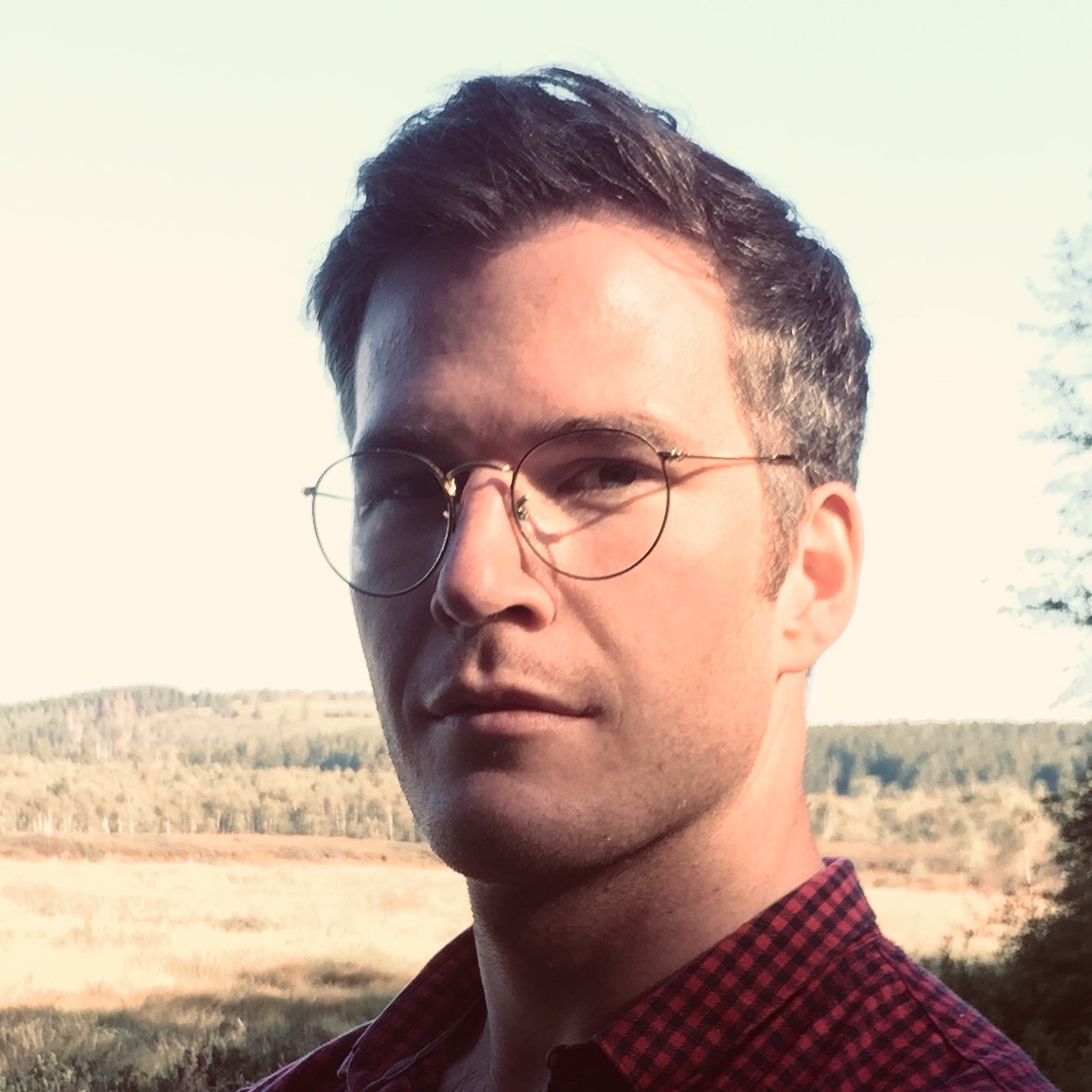
An interview with BSDB Beddington medal winner Rory Maizels
Posted by the Node, on 2 September 2025
The Beddington medal is awarded by the British Society for Developmental Biology (BSDB) for the best PhD thesis in developmental biology, defended in the year previous to the award. The 2025 winner was Rory Maizels, who completed his PhD with James Briscoe at the Francis Crick Institute in London, UK. In this interview, we hear about Rory’s career path, his PhD work and what he is excited about in developmental biology.
Where were you born and where did you grow up?
I was born and raised in Edinburgh, Scotland.
When did you first get interested in science?
My parents tell me, perhaps jokingly, that one of my first complete sentences, spoken as I dropped a rubber duck into the bath, was “why is gravity?” I guess this suggests a degree of scientific curiosity from a young age. A career in research was always in my sights: the main reason I chose to study biology over physics is that I believed, aged 16, that the biological research landscape had more opportunities for progress.
How did you come to do a PhD with James Briscoe at the Francis Crick Institute?
As an undergraduate at Oxford, I was interested in studying the ways that cells can perform communication and computation. The most fascinating example for me was the Trp operon, a gene regulation system in bacteria that controls the expression of tryptophan synthesis genes. This system is a crazily elegant example of how molecular systems can perform precise computation, which I found to be a pretty eye-opening idea.
Unfortunately, in animal systems things tend to be a bit more complicated than in E. coli. Instead of neat little operons, we have vast networks of interacting parts, where one signal seems to interact with almost all cellular processes, depending on context.I wanted to understand human biology in the way we understand the Trp operon; that eye-opening kind of understanding, that ‘oh wow, of course’intuition; but it seemed in many cases, we hadn’t got there yet. This realisation seemed, to me, pretty good motivation to go and do some research myself.
So, these tryptophanic interests led me to James’ lab in two ways: first, James’ work on patterning in the spinal cord captured exactly my interest in understanding the logic and computation of molecular systems. Second, on a more pragmatic level, after my undergraduate I went to study computational science & engineering at Harvard, funded by a Frank Knox Fellowship. I focused on mathematical modelling and data science methods and at the same time, the Briscoe lab were publishing a number of theoretical papers modelling the function of gene regulatory networks, along with more data-driven papers performing single-cell analysis. So, it seemed a perfect fit.
Can you talk more about your PhD project?
The aim for my project was to build methods for mechanistic analysis of cell fate decisions from single-cell data. To go beyond descriptive time-courses and population-level descriptions, to construct models that can simulate the gene expression dynamics of cellular transitions and, in doing so, connect early variations in expression to downstream differences in cell fate.
Key to this, in my mind, was the concept of dynamics: if we want to understand mechanism — the temporal ordering of events and the causal interactions between components — we need a clear picture of the dynamics that these mechanisms create. The value of single-cell resolution is that you can capture a picture of the spectrum of states that are possible as cells transition between types. This allows a range of different ‘pseudo-temporal’ approaches that can create expression time-series between your system’s beginning and end. But to study the mechanism driving these transitions, this sort of population-level time series analysis is insufficient: to actually model and simulate cell fate decisions, we needed to capture dynamics at single-cell level as well.
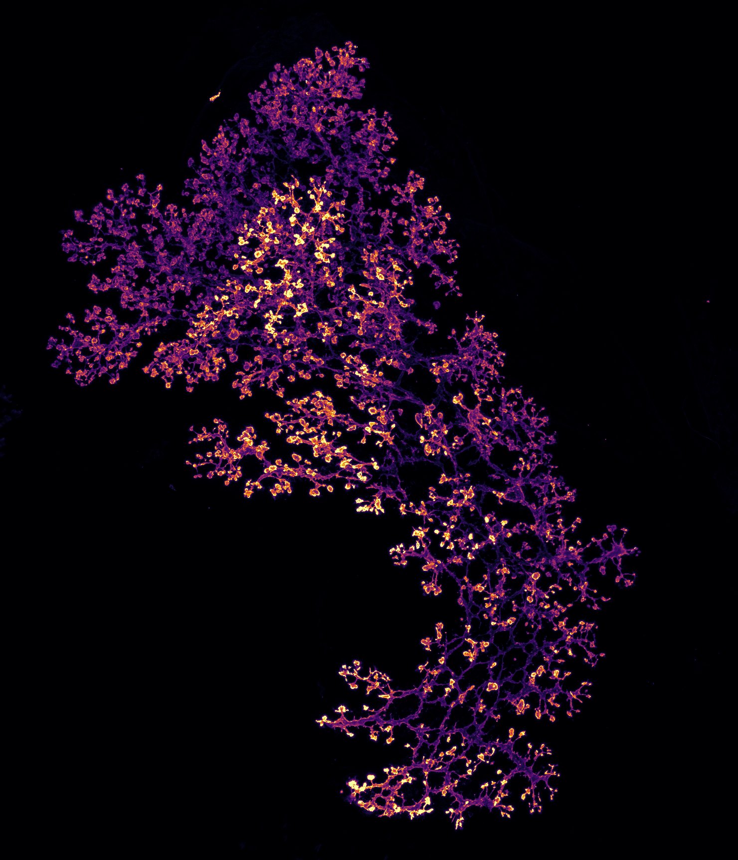
To tackle this, I established and optimised a time-resolved transcriptomics method that integrated two methods: metabolic labelling, which uses a uridine analogue 4sU to label nascent transcripts such that one can distinguish new from old reads in the sequencing data; and single-cell combinatorial indexing, a method for single-cell RNA sequencing that is compatible with fixed cells (necessary for the temporal labelling) and requires no bespoke microfluidic devices.
I applied this approach to in vitro differentiation of mouse stem cells into neural and mesodermal cells. The noisy, high-dimensional nature of the resultant sequencing data would usually be prohibitive for dynamical systems modelling (trying to learn a vector field in thousands of dimensions is no simple task…). So, to handle this I built a machine learning framework that models the dynamics of cell fate decisions with an abstract, low-dimensional vector field embedded in a ‘latent’ representation of the data. A biophysical model of transcription and labelling is embedded in the model, connecting this abstract vector field to the observed labelling data. The result was a model that could simulate the differentiation trajectory of each progenitor cell in the dataset, producing a distribution of trajectories that linked early variations to later fate decisions. Through these simulations, I identified that modulators of Shh signalling show early differences between fates, suggesting a previously unappreciated level of feedback between signal interpretation and cell fate decisions.
How did the project get started?
In the early weeks, I was deliberating over whether I should focus on a more theoretical project that applied dynamical systems theory to the study of gene regulatory networks, or a more data-driven approach that worked with single-cell data. I presented this conundrum to James, who just looked at me and asked, “why not both?”
Were there any frustrating moments?
The original aim of the project was to take existing protocols and computational methods and apply them to our system and our questions. The final product of the project was a study of why existing protocols and computational methods did not work, and the development of improved methods that did. This should be indication enough that there were frustrating moments aplenty!
If you took one abiding memory with you from your PhD, what would it be?
One moment that stands out is analysing the data from our first successful pilot of the homemade single-cell protocol. It was a simple pilot with unremarkable samples, but seeing that the experiment worked, seeing the expected cell types appear and genes being expressed in the right place came with a huge sense of excitement, almost a feeling of disbelief that this crazy, painful protocol was actually working.
Did you work on other projects during your PhD?
I sporadically got distracted and detoured, but the real exploration came at the end of the thesis, with exciting off-shoot projects that are still on-going!
What have you been working on since you completed your PhD? What’s next for you?
Towards the end of the PhD and afterwards, I started some very fun projects where we increased the throughput and affordability of the sequencing pipeline by an order of magnitude or two. In this way, we were able to massively increase the sophistication of our experimental designs, allowing us to map the function of developmental gene regulatory networks from input signals to output cell fates. Now, I’ve joined EMBL EBI and the Sanger Institute as an ESPOD postdoctoral fellow, where my postdoctoral project will be a fun mix of synthetic and systems biology, engineering cells to understand their decision making!
What techniques or areas in developmental biology excite you the most?
We’re really very good at measuring things in developmental biology. We’re creating datasets with millions of observations, tens of thousands of variables across multiple ‘modalities.’ We’re getting pretty good at perturbing things too: approaches to knockout genes, introduce mutations or alter enhancers at the scale of thousands of knockouts/mutations/alterations at a time hold a lot of promise. But we’re not so good at distilling these thousand-dimensional perturbational datasets into clear understanding.
Our brains are not thousand-dimensional. Our vision is 3D; our working attention can hold onto four things at once. To understand, rather than just observe developmental biology, we need to create intuitive, mechanistic representations of these complex systems that can actually fit into our minds.
Many people are excited about the application of AI in biology. For me, the exciting prospect is that the neural networks in AI models are really useful for learning abstract functions. This means if we want to take a thousand-dimensional dataset, abstract away all the details and just learn four key parameters of our choosing, AI is a pretty good tool for that. Neural network models can learn abstract representations of datasets that can be flexibly constrained by biological information/inductive biases, depending on the specific biological question.
So, the most exciting area of developmental biology for me is the study of emergent properties; dynamic properties of a system that are only apparent when considering the system as a whole, rather than examining the system’s individual components. If we can identify the key emergent properties of a gene regulatory system, can we use AI-driven approaches to model these key properties, such that we can build a simple, intuitive understanding of developmental systems and their many thousands of observable dimensions?
Outside of the lab, what do you like to do?
My ideal day off would involve a swim in a pond in the morning, a few hours reading an overlarge book in the afternoon, and a jaunt down to the pub in the evening.
 (No Ratings Yet)


 (7 votes)