The ELISIR program provides a stellar fresh PhD graduate the possibility to head a small team of scientists without going through a traditional post-doc, much in the spirit of analogous programs at MIT, Harvard or UCSF, to name but a few. This is a non-tenured position that can be held up to five years.
Within the context of the DFG research unit “Morphodynamics of Plants” (FOR2581) a Ph.D. position is available in the lab of Prof. Kay Schneitz, Dept. of Plant Developmental Biology, Technical University of Munich in Freising/Germany.
The research unit “Morphodynamics of Plants” is a multi-disciplinary consortium of nine groups (biologists, physicists, computer scientists) located in Munich/Freising, Heidelberg, and Cologne that want to understand how plants shape their organs. In the past the Schneitz lab contributed to the machine learning-based digital image analysis pipeline “PlantSeg” [1]. Using PlantSeg the lab succeeded in creating a digital 3D atlas of ovule development in Arabidopsis thaliana with cellular resolution [2]. The ovule is the predecessor of the seed and the major female reproductive organ in higher plants. Its extreme curvature represents a particularly fascinating morphogenetic process.
The successful candidate will take advantage of the digital 3D atlas of ovule development and combine genetic, cell biological and computational approaches to understand the genetic, cellular and mechanic basis of integument morphogenesis and ovule curvature. Starting date is June 2021 but is negotiable. Funding is at the usual TV-L E13/2 level.
We are looking for a highly motivated scientist trained in molecular and cell biology and/or biophysics with a strong interest in interdisciplinary work at the interface of bioinformatics, advanced confocal microscopy, image processing, 3D computer visualization, modelling, and cell and developmental genetics. The person should have good problem-solving skills and be able to work independently. Fluency in English is a must. Computer affinity is a must. Programming skills (Python, R) are a plus. Applicants must have a University degree equivalent to a German MSc degree.
References:
[1] Wolny et al. (2020) Accurate and versatile 3D segmentation of plant tissues at cellular resolution. eLife 9: e57613.
[2] Vijayan, Tofanelli et al. (2021) A digital 3D reference atlas reveals cellular growth patterns shaping the Arabidopsis ovule. eLife 10: e63262.
The regenerative biologist Richard Goss wrote a half century ago: ‘If there were no regeneration, there would be no life. If everything regenerated, there would be no death. All organisms exist between these two themes.’ (Goss, 1969). Tissue regeneration is on display in natural phenomena known to most professional and lay biologists, such as the renewal of tails dropped by lizards to distract predators, or the reproductive fission of flatworms that creates a new head and tail and turns one animal into two. Humans are often casually referred to in the literature or in discussion as unable to regenerate. However, tissues like liver, blood, skeletal muscle and intestinal epithelia possess tremendous regenerative potential. Other tissues such as pancreas, heart, brain and kidney lie lower on this spectrum. With regenerative capacity described as shades of gray rather than black or white, virtually all tissues have some low or latent regenerative capacity that might be stimulated by experimental or therapeutic manipulation.
The field of regeneration has been intertwined with developmental biology from its onset, so to consider tissue regeneration as nothing other than central to developmental biology undervalues both disciplines. Viewed through a regenerative biologist’s lens, fertilization triggers the most spectacular regenerative event of all – the growth and morphogenesis of an entire organism from a single cell – and the terms ‘regeneration’ and ‘reproduction’ were once used interchangeably by biologists. A rich history of luminary scientists who shaped biology in multiple ways over their careers included regeneration as a topic of their investigations. Thus, any student who readies themself with scissors or scalpel today against worm, fish, frog or salamander will be repeating an exercise from centuries ago. René-Antoine Ferchault de Reaumur described experiments examining appendage regeneration in crayfish in 1712 (Reaumur, 1712), building on observations of Jean-Baptiste Du Tertre decades before and contributing with others to debates on predestination, the existence of miniaturized tissue reserves or germs, and the nature of the soul. A founding father of laboratory animal model systems, Abraham Trembley brought hydra from the field into his university in the early and mid-18th century to bisect and disorganize (Trembley, 1744). Vertebrate regeneration fell under the magnifying glass of Lazzaro Spallanzani who, in the 1760s, described regeneration not only in snails and worms, but also in amphibians, most notably salamanders – cementing limb regeneration in newts and axolotls in the pantheon of regenerative events (Spallanzani, 1769). At the turn of the 20th century, Thomas Hunt Morgan explored regeneration in a host of creatures from flatworms to killifish. Descriptions of his experiments and those of others, showcased in his classic Regeneration, were profoundly thoughtful and rigorous, challenging controversial views on the extent to which regeneration is a product of adaptation (Morgan, 1901).
Over the past two decades, regenerative biology has grown exponentially as a field, making this an exciting time indeed. Transgenesis and knockout technologies for mice applied initially to fields of embryology, immunology, cancer and neuroscience found a willing partner in regeneration. Key regulators of events such as skeletal muscle regeneration, liver regeneration and axon regeneration began to emerge, and source cells for regeneration in many tissues were implicated by genetic lineage tracing. The past decade has seen the use of these techniques, most recently accompanied by genome editing, perfuse additional model systems, many of which have been reinvigorated from the classic era.
This is a crude and filtered representation of a great history, but we are supremely fortunate today to have available dozens of animal model systems and injury contexts, and methodologies some of us could not have dreamed of two decades ago. Lineage-tracing experiments, complemented by molecular trajectories inferred from single-cell RNA-sequencing, have rooted out cellular subpopulations and states, and enable conclusions on developmental plasticity during regeneration. New imaging platforms and cell tracking software facilitate direct visualization and quantification of cell behaviors in live regenerating tissues, either in accessible surface tissues or by intravital imaging to probe internal tissues. CRISPR-Cas9-based tools have brought genetics to the masses, facilitating mutant creation and analysis in familiar species and those new on the scene. Induced pluripotent stem cells can be used to generate ex vivo regeneration models for human tissues, or can be a source of resident stem cells for engineering applications such as skin or corneal therapy.
The current and next generations of regeneration scientists have many key questions to attack and goals to consider. Among these, to what extent do gene regulatory networks of regeneration overlap with those involved in initial development, growth or homeostasis of tissue? Why does wound healing trigger regeneration in some contexts but scarring in others, and what roles do inflammatory cells and fibroblasts play? How do we genome- and epigenome-edit for safe, effective regenerative therapies, and can regeneration programs be harnessed for particularly ambitious goals such as a functional blastema for mammalian limb stumps? What is altered during senescence to impact physiological regeneration and injury responses, and how are these targets best modulated to slow aging? And, as Morgan wondered, can we understand how and why regeneration has been retained or lost during evolution? Addressing questions like these will not only require the innovation of new tools and technologies, but continued inclusion of new animal species in which we study regenerative events.
Which takes us (finally) to the point of this editorial. When we started our laboratories, given the miniscule size of the community, talks on regeneration represented a small fraction of coverage in conferences centered on developmental biology, tissue disease or animal model systems. Poster presentations were sprinkled among various session themes, and we liked to joke that our regeneration talks were relegated to those necessary, end-of-conference sessions. Now, thanks to recognition and embrace of its re-emerging importance by major societies such as the Society for Developmental Biology, the International Society for Stem Cell Research and others, regeneration is a cornerstone and plenary topic of many meetings worldwide. Yet, no formal community for regenerative biologists exists – nor has one ever to our knowledge – and regular meetings are infrequent, every 2 or 4 years. We are thus pleased to launch the International Society for Regenerative Biology (ISRB) this year, to promote research and education in the field of regenerative biology (isrbio.org). This effort will formalize an inclusive and integrated community of scientists that studies all aspects of regeneration in invertebrate and vertebrate model organisms. The ISRB will support and enhance key existing meetings, while organizing its own main conference and virtual events such as webinars. Another function of the ISRB will be to convey the importance and impact of regeneration research to the greater scientific and lay communities, by highlighting regenerative biologists and their discoveries and through outreach activities and educational resources. It will promote regenerative biology by giving awards for discoveries and service, and by advocating for research and community funding. One of us (K.D.P.) will serve as Founding President, and the other (E.M.T.) will serve as President-Elect. Regular elections will fill positions for other Officers, and a diverse Board of Directors will advise and oversee operations.
We recognize that regenerative biologists have existing commitments with societies, and we emphasize that ISRB will complement and collaborate with these societies rather than compete with them. A primary role for the ISRB will be enabling cross-species comparisons to understand the commonalities and divergences in regenerative capacity and the mechanisms of regeneration – toward an integrative framework. We envisage the ISRB as the central society to promote and help achieve this next frontier for our field. Regular opportunities will be available for investigators using different models and injury contexts, but with the common goal of understanding how and why regeneration works, to share their discoveries and form new collaborations. Junior scientists will be a focus of the ISRB and will be provided with opportunities for interactions, visibility and career support.
Our first meeting will be virtual and concise, consisting of exciting scientific talks and an open community discussion on April 8-9, 2021 (isrbio.org). Some of these talks will be chosen from abstracts of unpublished work submitted by graduate students and postdoctoral researchers. We await our first in-person conference at a stimulating location in 2023, most likely to be hybrid with virtual features. We warmly and eagerly encourage you to join as a member, and, if you are a group leader, to encourage your group members to join. Participate in the launch meeting this April to see great science, and get involved! This has been a long time coming.
Acknowledgements
We thank A. Dickson, H. Roehl, K. Echeverri and S. Eming for comments on the manuscript.
References
Goss, R. J. (1969). Principles of Regeneration. New York: Academic Press.
Morgan, T. (1901). Regeneration. New York: Macmillan.
Reaumur, R. A. F. (1712). Sur les diverses reproductions qui se font dans les Écrevisses, les Omards, les Crabes, etc., et entr’autres sur celles de leurs jambes et de leurs écailles. Mem. Acad. Roy. Sci., 223–245.
Spallanzani, L. (1769). An Essay on Animal Reproductions. London: T. Becket and P. A. de Hondt.
Trembley, A. (1744). Memoires pour servir a l’histoire d’un genre de polypes d’eau douce a bras en forme de cornes. Leiden, The Netherlands: J. & H. Verbeek.
Hox genes instruct positional identity along the anterior-posterior axis of the animal body. A new paper in Development addresses the question of how similar Hox genes can define diverse cell fates, using mouse motor neurons as a model. To hear more about the work, we caught up with the paper’s two first authors, PhD students Milica Bulajić and Divyanshi Srivastava, and their respective supervisors Esteban Mazzoni (Associate Professor of Biology at New York University, USA) and Shaun Mahony (Assistant Professor of Biochemistry & Molecular Biology at Penn State University, USA).
Milica, Divyanshi, Esteban and Shaun (clockwise from top L).
Esteban and Shaun, what questions are your labs trying to answer, and how did you come to collaborate on this project?
EM: To understand cell differentiation, we focus on investigating how transcription factors control transcription and establish long-lasting epigenetic memories. With this knowledge, we then aim to control cell fate at will for clinical applications.
SM: We develop machine learning applications to understand gene regulatory systems. We particularly focus on understanding how transcription factors find their binding sites and drive regulatory responses in dynamic contexts such as development.
EM & SM: We began collaborating as postdocs more than a decade ago when ChIP was emerging (back when it was ChIP-chip!), and there were few computational tools. Even back then, we collaborated at a distance, with EM in New York and SM in Boston. EM was developing cellular models to understand cell differentiation at scales and purity compatible with the technology, and SM was developing tools to analyse the data, extract meaningful information and generate hypotheses. This cycle has been going strong ever since: the analyses carried out in SM’s lab have proposed hypotheses about transcription factor selectivity that EM’s lab has tested, and the systems and technologies developed in EM’s lab have inspired many of the computational tools developed in SM’s lab.
Milica and Divyanshi – how did you come to work in the Mazzoni and Mahony labs, and what is the main drive behind your research?
MB: I finished my undergraduate studies in Molecular Biology at the University of Belgrade, Serbia, where I am from. I joined the PhD program at the Department of Biology at New York University in 2014. After spending my first year rotating in different labs, I joined the Mazzoni lab because I really liked the research and enjoyed my rotation project, which was Hox related. I knew that I wanted to continue working on Hox genes and felt supported by Esteban in choosing questions to work on.
DS: When I started my PhD at Penn State, I was keen to work on computational regulatory genomics. I am very excited by the potential of novel computational methods to elucidate complex biology. Therefore, the Mahony lab was a great fit, with Shaun’s expertise in computational biology and the Mazzoni lab’s exciting work on the regulatory biology of cellular differentiation!
How has your research been affected by the COVID-19 pandemic?
EM: Like most institutions, we closed down with two days’ notice. The situation really dawned on me when we turned off equipment for the first time since I opened the lab. However, the hiatus made us focus and plan, and execute the most informative experiments now that we are at 50% output. Thus, it has had a positive side effect.
MB: We were out of the lab for about 3 months so there were some experimental delays, but I’m very lucky that I didn’t lose any work, or need a long time to start things up again. I also had plenty of data to analyse and manuscript edits to incorporate so that has been keeping me busy.
SM: As a computational lab, we were fortunate that we could continue making progress when others lost access to their facilities. But it has still been challenging to adapt to remote research; we miss the conversations and spontaneous debugging sessions that drive computational research forward. As with many others, I’ve personally found it difficult to devote enough time to research while also dealing with remote elementary school and adapting my own courses to a remote format.
DS: COVID-19 has been challenging due to the remote nature of all computational work, but I was fortunate that we had continued access to computational resources, as well as a supportive lab environment, which made it easier to work through the more difficult days.
What was known about the relationship between Hox binding and chromatin accessibility prior to your work?
MB, DS, SM, EM: When we planned these experiments, not much was known about their differential ability to bind inaccessible chromatin. Soon after that, in 2016, Robert White’s group described how some Drosophila Hox factors bind to chromatin. And then, around the time we were writing our paper last summer, a few relevant papers came out. The White group published a more extensive evaluation of all Drosophila Hox proteins showing that accessibility has a role in Hox selectivity, and out of all of the central and posterior Hox proteins, Abd-B stood out in having a higher ability to bind inaccessible sites. This was really interesting for two reasons: first, Hox proteins do have different abilities to bind to inaccessible chromatin; second, it primed our work – how do vertebrate posterior Hox genes (Hox9-13), all of which are fly Abd-B orthologues, behave? Coincidentally, Marie Kmita’s group published a preprint showing that Hox13 paralogs are required to open specific sites during limb development. Finally, Denis Duboule’s group showed similar results in genital development. Thus, the field was coming together.
Can you give us the key results of the paper in a paragraph?
MB, DS, SM, EM: We investigated the binding, transcriptional targets, sequence and chromatin preferences of seven different mammalian Hox proteins in a relevant cell type patterned by Hox genes. We discovered that the ability to engage with inaccessible sites is an important factor that drives Hox binding specificity. This ability seems to be driven by the DNA-binding domain and C-terminus. These results show that Hox specificity models should incorporate sequence preference, co-factor interactions and intrinsic abilities to bind inaccessible chromatin. We believe this can be extended to other homeobox genes (and perhaps other paralogous transcription factor groups) as a binding diversification strategy.
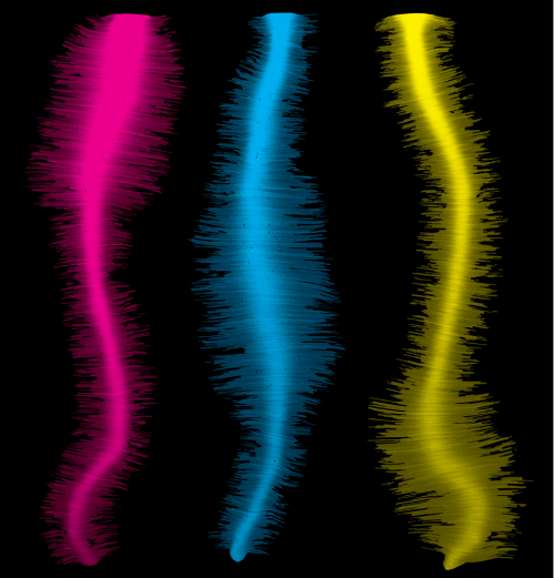
This piece of art was made by Dylan Iannitelli, a PhD student in the Mazzoni lab, from ChIP-seq data for Hox binding.
Where Hox proteins show high affinity for inaccessible chromatin, do you think they are acting as so-called ‘pioneer’ factors?
MB, DS, SM, EM: Our results and other studies show clearly that some Hox proteins play a role in ‘opening’ some regions of relatively inaccessible chromatin during differentiation. However, in the strict sense, the term ‘pioneer factor’ is reserved for those transcription factors that have been demonstrated to bind to DNA wrapped around nucleosomes, which subsequently evict nucleosomes. Our data is compatible with some posterior Hox proteins acting as pioneers, but it is now a good hypothesis to test.
What explains the different chromatin affinities – even among paralogs – of the various posterior Hox proteins?
MB, DS, SM, EM: We used multiple different approaches to characterize sequence preferences and found no evidence that sequence explains the different chromatin affinities. For example, we found no sequence preference differences between HOXC9 and HOXC10, or HOXC9 and the other HOX9 paralogs. Our results with the chimeras, made by swapping HOXC10 and HOXC13 DNA-binding domains, show that chromatin affinities seem to be controlled by the homeodomain and C-terminus. As shown with the bHLH family, the different homeodomains could engage the DNA-nucleosome complex in slightly different ways.
When doing the research, did you have any particular result or eureka moment that has stuck with you?
MB: I think for me, the most impactful thing was seeing the binding results for HOXC13, and finding that it binds to very inaccessible chromatin. Similarly, when I made the chimeric Hox proteins, seeing that this ability is controlled by the DNA-binding domain and C-terminus.
DS: For me, observing the difference in chromatin accessibility at HOXC9-only sites compared with other differentially bound Hox transcription factor sites was an exciting moment. And of course, the binding results for HOXC13 were striking.
Observing the difference in chromatin accessibility at HOXC9-only sites compared with other differentially bound Hox transcription factor sites was an exciting moment.
And what about the flipside: any moments of frustration or despair?
MB: Waiting for reviews during the publication process can be stressful. There are always ups and downs when writing a paper, but when it’s finally written and then accepted for publication, it’s a great feeling.
DS: It was challenging to design a differential binding strategy for multiple transcription factors. We took a long time to arrive at analyses that were robust and reproducible, and that could overcome biases related to technical and experimental noise.
What next for you two after this paper?
MB: I am writing another manuscript and scheduling my PhD defence for early 2021. I’m also in the process of looking and applying for jobs.
DS: I am working on developing computational approaches that can interpretably model transcription factor binding sites. I also plan to defend in early 2021, and pursue research-related positions after my PhD.
Where will this story take the Mahony and Mazzoni labs?
EM: For us, it has two logical future paths. First, gaining insights into Hox-dependent positional identity allows for the precise control of in vitro differentiated motor neuron positional fate. Second, it opened a new dimension within homeodomain transcription factor diversification. The small sequence preference variation was always hard to reconcile with their diverse functions. Now, we hypothesize that the ability to engage inaccessible sites provides an orthogonal mechanism for homeobox genes to diversify their binding and, thus, gene regulation.
SM: This project has really brought home the importance of pre-existing chromatin environments in determining transcription factor binding specificity during development. In a parallel project, Divyanshi has also developed neural networks that can interpret how sequence and pre-existing chromatin features predict the binding specificity of a transcription factor. So, the use of these types of approaches to understand how chromatin shapes transcription factor binding (and vice versa) will continue to be a big focus in our lab, especially in terms of being applied to understand the dynamic systems studied in Esteban’s lab.
Finally, let’s move outside the lab – what do you like to do in your spare time in New York and Pennsylvania?
MB: Going for long walks and hikes, and sitting in a park with a good book.
EM: I am an avid sailor, taking me beyond the lab, the city and the continent. Last October, I participated in a trans-Atlantic race.
DS: I like to go cycling, with the rolling hills of central Pennsylvania providing some lovely terrain.
SM: We’re very fortunate in central Pennsylvania to have lots of beautiful parks and trails, and that’s where my family and I like to spend our spare time.
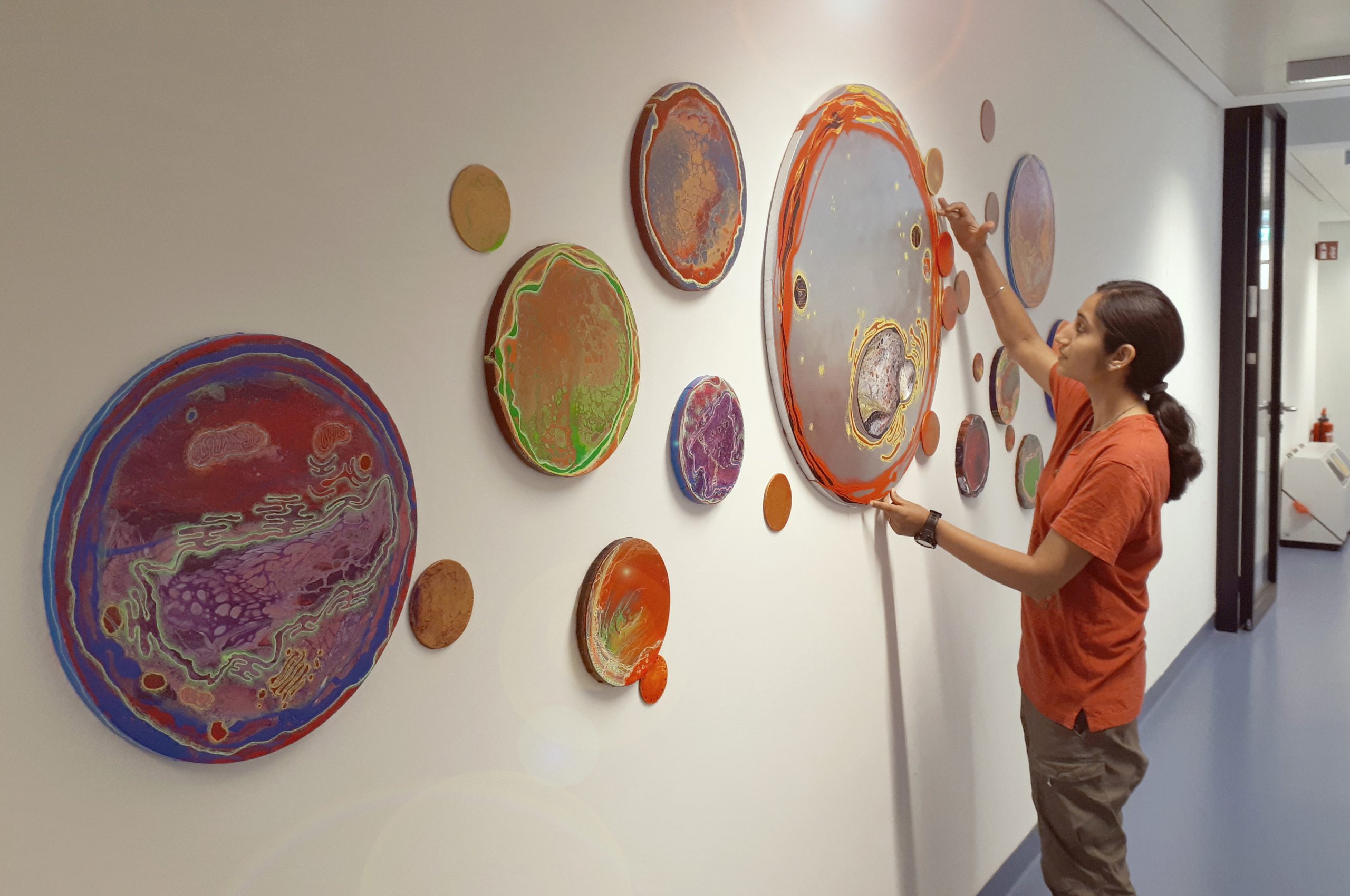
In our fourth SciArt Profile we meet Priyanka Oberoi, an illustrator, artist and photographer whose work often features scientific themes.
Priyanka with a wall of her art.
Where are you originally from and what do you work on now?
Originally I am from India, and am an illustrator and photographer by profession. After spending five years studying art in the College of Art in New Delhi and National Institute of Design in Paldi I started my career as a staff photographer for the magazine ‘Sports Illustrated’ in New Delhi, India. From there I moved on to freelancing as an illustrator and photographer full time. This gave be creative freedom to explore and time to increase my skill set. Studio pottery and wall murals are some of the few skills that I added to my portfolio. I started making scientific illustrations 4 years ago in Germany and currently continue the same in Brussels with my husband and five month old baby boy.
When did science first come in to your life?
I studied science and math in school about fifteen years ago. Deriving physics equations and finding the square root from a hypotenuse taught me that I was better suited for something else. While being an artist was never a childhood dream, it definitely became so after school, and I feel extremely fortunate to have found my forte in art.
Science came back in my life when a few scientist friends asked for some drawings. What started with scientific illustrations for journal papers went on to posters and merchandise for conferences, journal covers, gifs explaining various scientific concepts, lab wall art and more. The scope of scientific communication has really caught my imagination.
Who are your artistic influences?
The impressionists with their bold and confident brush strokes have always left me in awe, and Monet, Manet, Van Gogh and Surat adorn my home walls. The Indian artist from Goa Mario Miranda is someone I look up to for inspiration for my pen and inks. The pop colours in Andy Warhol’s screen prints gives me confidence to go crazy with my colour palettes.
What do you think of the relationship between science and art?
I am not a scientist, but the process of creating art certainly has an impact on scientific process. With art, one needs to distil an idea into a single, coherent visual representation. For instance, when I work with client to create a cover art or graphical abstract, a lot of thought goes into which elements faithfully represent the finding, not just for the author’s peers but for the broader audience. This certainly helps streamline scientific thinking. In-fact, I would suggest scientists create graphical abstracts of their work or progress, as it would help with understanding the major focus of the work and improving the planning of experiments.
“With art, one needs to distil an idea into a single, coherent visual representation”
How do you make your art?
Pen and inks are my specialty when it comes to illustrations. While I make most of my scientific illustrations digitally the comfort of the simple pen and paper are undeniable.
Be it a commissioned work of art or an idea that I want to bring to life, I always start with my little sketch book. I start by writing key words of a particular project, and if the client has any specific requirements then I note them down too, but if not I start with my favourite, a blank canvas. Then comes the mood board: this generates the basic feel of the artwork which includes the colour palette, sketch style, canvas shape and many such details. This gives the client an idea of the route I will take for the project. With these approvals I start with the artwork. Rough sketches start to adorn my sketch book. Some concepts take days to formulate while some just click instantly. These steps give me a clear passage into my final work of art.
I work on a variety of techniques. Sometimes if the requirement demands a painterly quality that I cannot achieve via digital platforms I go back to the traditional canvas or paper with high resolution scans. The bottom line is that the process is super intoxicating and so when one project comes to an end I cannot wait to begin the next!
What are you thinking of working on next?
I am currently working on a board game for the Deutsch museum in Munich – acompletely new challenge with its new set of rules.
Also filling up my sketch book pages is a self initiated project called ‘Danio & Rerio’. A weekly comic strip that I recently started on twitter that embarks on the adventures of two zebrafish and their stints with various fun science experiments.
Apart from commissioned works of art I conduct team building and exploratory workshops. One such workshop includes traditional wooden blocks that I got made in India. These wooden blocks are science themed with a twist used for block printing on bags, t-shirts and pretty much any surface. These workshops are open to any lab that want some reclamation time. This inexpensive activity is fun and refreshes all the members of a lab without planning a formal retreat. One set of workshops are going to be offered to an international school in Brussels for their extra curricular spring and possibly summer sessions.
Conference poster: Molecular origins of Life, Munich 2021
Conference poster: Molecular origins of Life, Munich 2020
Conference poster: Engineering Life from origins to organs, Dresden 2019
Scientific conference posters
Lab Wall Art: ‘Bottoms up life forms’. Commissioned by Dr James Saenz, B CUBE – Center for Molecular Bioengineering, Dresden
Danio & Rerio – My weekly comic strip based on two zebrafish and their adventures with various scientific and fun experiments (more of this can be seen on my twitter handle- PriyankaObero16)
Model Organism Illustrations- These are three of my best seller illustrations available on sale as prints on t-shirts, mugs, phone covers and more on Redbubble
Model Organism Illustrations- These are three of my best seller illustrations available on sale as prints on t-shirts, mugs, phone covers and more on Redbubble
Model Organism Illustrations- These are three of my best seller illustrations available on sale as prints on t-shirts, mugs, phone covers and more on Redbubble
Corona Mandala- I made this illustration in April 2020 during the lockdown when I was confined to the space in my home like the rest of the world, I could only see and hear Corona. Like a mandala it started as a small dot and spread to what feels never ending even today.
The joint CiM-IMPRS graduate program of the International Max Planck Research School – Molecular Biomedicine and Muenster’s Cells in Motion Interfaculty Centre offers positions to pursue PhD projects in the areas of biology, chemistry, physics, mathematics or computer science. We are looking for young scientists with a vivid interest in interdisciplinary projects to image cell dynamics from the subcellular to the patient level. PhD projects range from the analysis of basic cellular processes to clinical translation, from the application of novel biophysical approaches and the generation of mathematical models to the development of new imaging-related techniques and compounds.
Research areas:
Cell and Molecular Biology. Developmental and Stem Cell Biology
Vascular Biology. Immunology
Microbiology. Neurobiology
In vivo Imaging. High Resolution Optical Imaging
Biophysics. Chemical Biology
Label Chemistry. Mathematical Modelling
and more.
Applications for the PhD program can be submitted from 10 February to 4 April 2021. Projects start in October 2021 (earlier starts are possible if desired). Applications can only be submitted via our online system.
For online application and further information go to
www.cim-imprs.de.
We offer 16 fully financed PhD positions. More positions financed by work contracts may be offered depending on availability. Excellent scientific and transferable skills trainings, competitive work contracts or tax-free fellowships as well as support with administrative matters, accommodation, and visas are part of the program. There are no tuition fees. The program language is English. We invite applications from highly qualified and motivated students of any nationality from biological sciences, chemistry, mathematics, computer sciences and physics. Be part of CiM-IMPRS, a program run jointly by the University of Muenster and the Max Planck Institute for Molecular Biomedicine.
We, Jose Casal and Peter Lawrence, have an opportunity to hire a suitably qualified research assistant to help us for 6 months starting anytime from March Ist. Unfortunately we do not yet know if the job could be extended further than that, but if there is anyone who knows flies, is skilled with the confocal microscope and would like to join us briefly we can pay them at the going rate from the remnant of our current grant until the money runs out.
We are in the Zoology Department of the University of Cambridge and work on planar cell polarity in Drosophila
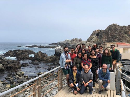
As astronomers look up to the sky to analyze the infinite universe, developmental biologists look at life unfolding, revealing itself under the light of the microscope. Stars above us, embryos below, we wonder about the possible worlds hindered from our sight. These reflections took place at “The Marine Biology Laboratory Practical Course in Developmental Biology – Quintay 2020” in Chile, where a total of 18 students from 9 different countries (Argentina, Brazil, Chile, Colombia, Ecuador, India, Mexico, Puerto Rico and Uruguay) met, fascinated with the broad diversity of animals, range of experimental techniques, and the huge quality of faculty available in this course.
Despite the fact that most of us were working with one of a small number of “model organisms”, we had the unusual chance to observe, describe and perform experiments with flatworms, sea urchins, fruit flies, Zebrafish, African frogs, and chicken embryos. It was an intense 2 week-practical and theoretical course at the facilities of the Quintay Marine Research Center (CIMARQ) of the Andres Bello University. This immersive experience right next to the Pacific seashore of Chile, together with a remarkable array of experts in different fields of developmental biology in a friendly environment, encouraged us to explore some of the frontiers and unanswered questions in developmental biology.
Students at The MBL Practical Course in Developmental Biology in Quintay Marine Research Center (CIMARQ) in 2020.
The course began with the Drosophila module, coordinated by Profs. Nipam Patel (MBL, USA) and John Ewer (University of Valparaiso, Chile). We were introduced to the basic aspects of invertebrate embryogenesis, genetics, and the powerful impact of the Drosophila model in Developmental Biology. Nipam was a very engaging lecturer, transmitting us the beauty of visualizing embryos under a confocal microscope, and giving us the most useful advice for nice immunostainings: “You just need to Wash! Wash! Wash! …and have faith”. We got stunning expression patterns of Hox genes in Drosophila embryos that will become part of a joint publication with results from previous versions of this course. Next, Nipam introduced us to Evo-Devo, the possibility of exploring evolution through the lens of development, by analyzing the gene regulatory networks that establish the anterior-posterior axis of the crustacean Parhyale hawaiensis in comparison to Drosophila.
In sea urchin module, directed by the Prof. David McClay (Duke University, USA) or Uncle Dave as he would say, we took advantage of the CIMARQ-Quintay red sea urchin facility, learning some of the classical experiments of early embryogenesis and the morphogenetic events occurring during sea urchin gastrulation. Dave provoked us with inspiring discussions about developmental biology, and enchanted us with his passion earned through his many years in this field. His lectures about how sea urchin embryo development has been understood were like hearing a nine year old kid telling you about his favorite Christmas presents.
We continued with Prof. Cecilia Moens (University of Washington, USA), who engaged us with an inspiring talk about the early development of the zebrafish brain, and then guided us through a debate about employing gene editing tools such as CRISPR/Cas9 for dissecting early developmental processes, not only in Zebrafish but in almost all research organisms present in the course. This discussion was insightful since nowadays we have a vast array of tools for manipulating gene expression at our fingertips. We were able to compare the advantages and limitations of each one, highlighting the importance of conducting the proper experimental controls.
In the planarian module, Prof. Alejandro Sanchez-Alvarado (Stowers Institute, USA) challenged us to test the regenerative capacities of planarians. He proposed we perform experiments from different kinds of amputations to even tissue transplants, experiments that we would follow for the rest of the course. But his module projected outside the lecture room and the lab. He took us to the closest beach to CIMARQ to get samples of the living creatures that inhabit the Quintay coast. We were impressed by the rich animal diversity that lived there of a variety of shapes and colors. For some of us, it was the first time that we could see, touch and study marine organisms such as sea stars, snails, anemones and a variety of worms including planarians.
The first week ended with a lecture by Prof. Alfonso Martinez Arias (University of Cambridge, UK) on gastruloids and mouse stem cells. He engaged us in a discussion about the philosophical aspects of developmental biology. “Do you think a machine can be an embryo?” he said, to our astonished faces. These sorts of questions lit a heated debate. We discussed the manipulation of human stem cells and embryos, the possibility to compute embryonic development and the limitations of modeling biological phenomena. A key idea that emerged from the conversation, was that we should start talking about “research organisms” instead of “model organisms” because ultimately a model organism only models itself. The group was surprised and motivated by the questions, and the conversation was inspiring: at the end of the day, nobody was indifferent.
Led by Profs. Sally Moody (The George Washington University, USA), Roberto Mayor (University College of London, UK), and Fernando Faunes (University Andres Bello, Chile) the Xenopus module then came into the picture. After enlightening and fulfilling lectures, the practical activities were free and diverse. We were able to choose from a variety of different experiments to learn about axis development and the dorsal organizer inductive properties in Xenopus. Starting with different practical techniques to manipulate embryos – even using an eyebrow as a tool – we were challenged to graft neural crest cells from a fluorescent donor embryo into a wild-type host embryo. Despite the high handling complexity of this experiment, many of us succeeded and were able to record the neural crest migrating in living embryos. This module ended up with the presentation of our results and a funny awards ceremony.
To conclude the organism modules, Profs. Andrea Streit (King’s College London, UK), Claudio Stern (University College London, UK), and João Botelho (Pontificia Universidad Católica de Chile) guided us through the fascinating world of chick development. In very didactical, histrionic and immersive lectures with Claudio, we studied concepts such as regulative development and cell states during early chick embryo development. Then, Andrea brought our attention to non-coding regulatory regions in DNA and regulation of gene expression in the context of sense organ development. In the laboratory, we did ex ovo culture of primitive streak stage embryos and we injected DiI or DiO in Hensen´s node and could follow cell fates and see a fluorescent notochord the next day. We also did in ovo culture experiments and tried methods such as electroporation, to introduce morpholinos, and adding beads to the embryos to study limb development. Inspired by the organizer transplant experiments in Xenopus we asked to do something similar with chicken embryos. Andrea, who is an expert in node transplantation, quickly taught us this technique and the next day we were able to discuss the inductive capacity of the node according to the region where it had been transplanted.
A week into the course we had the opportunity to attend the “Developmental Biology Symposium-Quintay 2020”. Researchers from different universities of Chile came to Quintay to share their work, integrated with talks of some professors of the course. It was an amazing event to get to know the high quality and engaging science developed in Chile.
During the course, the most important complementary activities were the student talks in the evenings. Here, we had the opportunity to introduce our own research projects to the whole group in a very comfortable and relaxed setting, enjoying drinks and snacks during the presentations. The variety of research organisms was amazing, from Drosophila, Zebrafish, Medaka, C. elegans, passing through Xenopus, Axolotls, and even Cestodes and wild Planarian species. Even the experimental approaches varied from one to another, from molecular biology to very robust bioinformatics. The discussion that came up was very helpful, adding different classical and new approaches that we could use in our projects. On many occasions, it was so interesting that we kept talking about it in our lab nights, where the fun lasted until late and we would finish our experiments and record our results with a confocal microscope, with essential help from Jaime Espina (University Andres Bello).
Although all these activities sound tough and demanding, occasions to give our minds a break and enjoy a relaxed conversation were not missing. Besides the lunch time and student talks, we were able to organize a BBQ with our professors and fellows. Here, we got to know each other better, discuss in a relaxed atmosphere, and why not?, laugh with some jokes and chat about life (our life, not the embryo’s!). Another memorable activity was a visit to a neighboring beach, where together with some faculty we were able to enjoy a nice picnic next to ocean. All of this highlights how engaging this course is, how interaction between students and professors, even outside a purely academic context, lies at its heart.
To close these awesome weeks, the closing ceremony was presided over by Prof. Ángela Nieto (CSIC-UMH, Spain), with a lecture of the most recent findings of her laboratory, including the blended study of gene expression profiles, the physical, and cellular variations controlling normal development, metastasis, and cell proliferation. Afterwards, the professors awarded Ailen Cervino and Nicolas Cumplido with a well-deserved reward, which will enable them to attend the next “Embryology: Concepts & Techniques in Modern Developmental Biology” MBL course. Finally, we had lunch together on the Quintay coast, enjoying such good company and filled with energy, looking ahead for our own goals.
A year has passed since “Quintay 2020” took place, a few months before the Covid-19 pandemic broke out. We didn’t know then how fortunate we were to carry out face-to-face discussions, share the bench with other students and professors, or even enjoy a BBQ with people from all over the world! Even though much effort has been placed in order to continue with courses and meetings online, being able to experience a practical course like this one in real life, we believe, has transcendental effects on its students, both at the personal and professional level. Hopefully, new generations of Developmental Biologists will draw on the “Quintay experience” in the near future.
Students (co-authors) & Talks:
Juan A Sanchez. Growth coordination within tissue in Drosophila. Felipe Berti Valer. The Irre cell Recognition Module and ovarian development in Drosophila: the role of the Roughest protein. Marycruz Flores Flores. Characterization of cell recruitment mechanism driven by vestigial in the imaginal wing disc of Drosophila melanogaster. Alison Julio. Structural aspects and evolutive conservation of Calpain action in early insect embryogenesis. Pablo Guzman. The Slit/Robo pathway is required in different stages of the development of the Drosophila lobula plate. Emiliano Molina. Differential requirement of the t6A modification in tRNAs, between undifferentiated and differentiated cells in Drosophila melanogaster. Emilio Oviedo. Regeneration in Ecuadorian land and freshwater planarians. Cristian Reyes. Reprimo genes in cancer and development, what do we know so far? Nicolas Cumplido. From Devo to Evo: Hox genes and the shaping of the zebrafish caudal fin. Sruthi Purushothaman. Fgf-signaling is compartmentalized within the mesenchyme and controls proliferation during salamander limb development. Aitana Castro Colabianchi. The role of Notch1 during the early development. Ailen Cervino. A conserved role of Furry in cell polarization and morphogenesis. Diana Carolina Castañeda Cortés. Crossover between stress and tyroid hormone axes in stress-induced sex reversal. Felipe Gajardo. Transposable elements in zebrafish hypoxic response: What the data has to tell us. Oscar Javier Ortega Recalde. DNA methylation memory: Understanding epigenetic reprogramming in vertebrates. Tonatiuh Molina. Betaglycan, a multifunctional accessory. Jimena Montagne. Cell differentiation and tissue reorganization during the larval metamorphosis of cestodes. Juan Rodriguez. Regulation of embryonic cell fate decision by histone methylation.
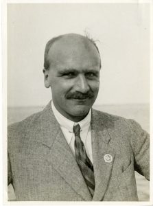
In the latest episode of Genetics Unzipped we’re taking a look at the life of JBS Haldane, whose work, writing and dominant personality made him one of the most interesting characters of 20th century genetics.
As well as being an insightful scientist, fearless self-experimenter and artful communicator, Haldane’s political leanings also affected his approach to science – even at the expense of the scientific rigour that he usually applied to his endeavours.
If you enjoy the show, please do rate and review on Apple podcasts and help to spread the word on social media. And you can always send feedback and suggestions for future episodes and guests to podcast@geneticsunzipped.com Follow us on Twitter – @geneticsunzip
Yesterday we held the fifth webinar in our series, this time chaired three members of the preLights team – Sundar Naganathan, Irepan Salvador-Martinez and Grace Lim – in celebration of the site’s third birthday.
Below you’ll find recordings of the talks and live Q&A sessions.
MichèleRomanos (from Bertrand Benazeraf’s lab at the Centre de Biologie Integrative in Toulouse)
‘Cell-to-cell heterogeneity in Sox2 and Brachyury expression ratios guides progenitor destiny by controlling their motility.’
 (4 votes)
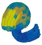
 (1 votes)
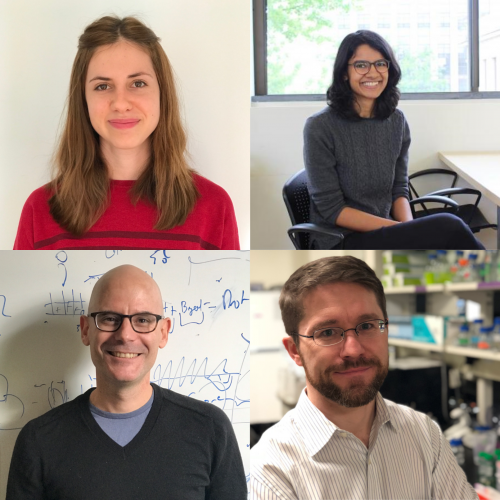


 (7 votes)