PhD student position in neurobiology at Stockholm University atthe Department of Molecular Biosciences, The Wenner-Gren Institute.
Research at the Department of Molecular Biosciences, The Wenner-Gren Institute (MBW) experimentally addresses fundamental problems in molecular cell biology, integrative biology, and infection and immunobiology. State-of-the art and advanced methodologies are applied in a professional research environment characterized by its well-established international profile. The institute has 30 research groups with a research staff of 170, of which 60 are PhD students. Read more about MBW at www.su.se/mbw.
Project description
This doctoral student position is available in the laboratory headed by Associate Professor Qi Dai. The Dai lab exploits the combination of molecular, genetic and genomic approaches to gain mechanistic insights on how cell fates are specified. The project aims to uncover novel regulatory mechanisms controlled by chromatin and transcription factors during neural cell fate decisions in the developing mouse brain. Candidates with strong background at molecular genetics, developmental biology and biochemistry are welcome to apply.
Qualification requirements Graduate studies in molecular biosciences requires a completed university degree at an advanced level or at least 240 credits of university education (240 hp in the Swedish universities), including at least 120 hp at the bachelor level in molecular biology, biology, chemistry or similar subject. Candidates should have successfully completed courses at the advanced level in molecular biosciences or equivalent subject (at least 60 hp), of which at least 30 hp represent independent research project work. Candidates who have other documented qualifications, obtained in Sweden or elsewhere, that are judged to provide equivalent knowledge are also qualified.
Selection The selection among the eligible candidates will primarily be based on the ability to obtain education at the research level. The following criteria will be used to assess this capacity: the candidates’ documented knowledge in a relevant field of research, written and oral proficiency in English, the capacity for analytical thinking, the ability to collaborate, as well as creativity, initiative, and independence. Experience in mouse genetics, biochemical and genomic approaches will be an asset. The assessment will be based on previous experience, grades and publication record, the quality of the degree project, references, relevant experience, interviews, and the candidate’s written motivation for seeking the position.
Terms of employment
The term of the initial contract may not exceed one year. The employment may be extended for a maximum of two years at a time. However, the total period of employment may not exceed the equivalent of four years of full-time study.
Doctoral students should primarily devote themselves to their own education, but may engage in teaching, research, and administration corresponding to a maximum of 20 % of a full-time position.
Please note that admission decisions cannot be appealed.
Stockholm University strives to be a workplace free from discrimination and with equal opportunities for all.
Contact
Further information about the position can be obtained from Qi Dai, telephone: +46 8 16 4149, qi.dai@su.se
Application This is a non-official advertisement. The documents for initial application should be sent directly to qi.dai@su.se.
Please include the following information with your application
Your contact details
Your highest degree
Your English language skills
Contact details for 3 references
and, in addition, please include the following documents
Cover letter in which you explain why you are interested in the project described in the advertisement and what makes you suitable for the project in question
CV – degrees and other completed courses, work experience and a list of degree projects/theses
Degree certificates and grades confirming that you meet the general and specific entry requirements (no more than 6 files)
Letters of recommendation (optional, referees’ contact details are required instead)
The Echeverri lab at the MBL seeks a highly motivated individual to join the Eugene Bell Center for Regenerative Biology and Tissue Engineering as a Postdoctoral Researcher. The successful candidate will work on in vitro models for neural regeneration. .The specific goal of the project is to examine how pathways that are essential for regeneration have evolved in different species with different regenerative capacity and to translate those findings into an in vitro model of spinal cord injury.
Applicants should have a Ph.D. in a biology related field. Must have prior experience working in the field of neuro, cell or developmental biology, as well as experience with cell culture and molecular biology. Must be independent, enthusiastic, self-motivated, productive, and enjoy working in a highly collaborative environment. The ideal candidate will have direct experience working with cells in vitro or organoids.
Required documents:
1. Cover letter explaining specifically why you are interested in joining our lab to work on this project and what positive qualities you would bring to our team.
2. Curriculum vitae.
3. List of 3 references (Please do not have letters sent at this time. Letter writers will be contacted directly by the PI)
Apply online at :
https://recruiting.ultipro.com/MAR1033MBL/JobBoard/4c3007c3-6354-41de-a13f-d95be60d91e9/OpportunityDetail?opportunityId=8353d1ab-73d0-4bcd-a257-99dbc5f4d10a
The Beddington Medal is the BSDB’s major commendation to promising young biologists, awarded for the best PhD thesis in Developmental Biology defended in the year previous to the award. It was announced in 2002 [letter #23/2] and first awarded in 2004.
Rosa Beddington was one of the greatest talents and inspirational leaders in the field of developmental biology. Rosa made an enormous contribution to the field in general and to the BSDB in particular, so it seemed entirely appropriate that the Society should establish a lasting memorial to her. The design of the medal, mice on a stylised DNA helix, is from artwork by Rosa herself.
Nominations for the Beddington Medal
The eligibility period covers PhD dissertations which were defended during the calendar year previous to the award (i.e. until end of December 2020). Furthermore, applicants need to have at least one paper accepted or close to acceptance. Nominations should be in two parts:
From the candidate, up to 2 pages A4 describing the thesis and supplemented with up to 1 extra page of figures from the thesis to illustrate key results, plus a 1 page CV, including statement of prizes/awards already received. These should all be in the form of a single pdf file of no more than 1 MB. A candidate exceeding these limits risks having to resubmit their application. In addition, candidates should supply formal documentation of the date of submission of the thesis.
From the candidate’s PhD thesis supervisor, a letter of support, sent independently, consisting of no more than 2 pages A4, describing why the student was deserving of this award. This letter should explicitly comment on the status of publications arising/expected from the thesis work, and also on any unusual circumstances, including duration of study.
Candidates can be of any nationality, must be BSDB members at the time of nomination, and at least one of their supervisors must be UK-based. Nominees must be able to attend the BSDB Spring Meeting, where the winner is to present a plenary talk.
All nominations received will be considered and voted upon by the Committee and the winner invited to present the Beddington Medal lecture on their thesis work at the following BSDB Spring Meeting.
We are seeking a highly motivated candidate strongly interested in interdisciplinary science, to join our groups (van de Pavert and Guignard labs) and work on a project at the crossroad between Computer Science and Developmental Biology as a Ph.D student.
Quantification and modelling of embryonic lymph node organogenesis at the single cell scale.
The role
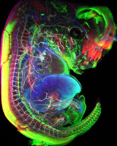
The project aims at studying lymph node (LN) formation during mouse embryonic development. LN formation requires hematopoietic lymphoid tissue inducer cells (LTi) to interact with mesenchymal cells at precise locations within the embryo, where they subsequently form aggregates. We have postulated that the peripheral nervous system outgrowth initiates the earliest events in LN formation. Indeed, preliminary data show that LTi aggregates morphology and cell density is affected in mouse embryos lacking neuronal subsets.
To understand the relationship between neuronal outgrowth and lymph node formation, the successful candidate will work between the van de Pavert lab and the Guignard lab to develop new computational methods to reconstruct and quantify LTi aggregates and peripheral nervous system morphology at the single cell scale. The reconstructions will then be used to develop a machine learning framework to systematically quantify phenotypes in perturbed mouse embryos. These quantifications will in turn allow to model the effects of neuronal outgrowth on LN formation.
The successful candidate will work towards:
building a library of whole-mount mouse embryo images
developing computational methods for the analysis of the generated library, including:
reconstruction and mapping of the neuronal network
quantification of LTi aggregate morphologies and positions
developing computational methods to automatically stage mouse embryos
modelling LN formation in relation to neuronal network morphology
Keywords
Quantitative embryogenesis, whole-mount analysis, peripheral nervous system, immune system, image analysis, machine learning, big data analysis
Whom would we like to hire?
The student should be enthusiastic, creative and ambitious, have good communication skills and be eager to learn. A master degree with major or minor in computer science is required. Affection for developmental biology is preferred. Some experience in developmental biology is also preferred but not required. Note: exception can be made for students who have not studied computer science if the student can prove coding skills.
The offer
3-year Ph.D. position in the van de Pavert lab and the Guignard lab
Call open for applications: January 15 – February 26
Interviews of shortlisted candidates by the evaluation committee: April 20 to April 22
The pre-selection process will be based on qualifications and expertise reflected on the candidates CV and motivation letter. It will be merit-based. All candidates will be informed whether they have been pre-selected or not.
We are a group of computer scientists with a strong interested in biology in general and more specifically in embryonic development. We develop novel computational methods and models that allow the analysis of very large 3D movies of animal embryonic development (up to 2TB per movie). We work in close relationship with biologists to tailor our methods so that they help to address fundamental biological questions.
The developmental biology question that mainly but not only animates us is to better understand the mechanisms driving embryogenesis to robustly form a complex organism despite genetic polymorphism and variable environmental conditions.
We are interested to study the first cues required to form lymph nodes at specific locations within the embryo. We are fascinated by the interactions between different cell types, such as hematopoietic cells, mesenchymal cells and neurons which eventually will generate a highly organized LN. To study these interactions, we use a multi-disciplinary approach and combine techniques such as 3D immunofluorescence imaging, flow cytometry and single cell sequencing on embryos from different mouse models.
The Turing Centre for Living Systems (CENTURI) is an interdisciplinary project located in Marseille (France).
CENTURI aims at developing an integrated interdisciplinary community, to decipher the complexity of biological systems through the understanding of how biological function emerges from the organization and dynamics of living systems.
The project federates 15 teaching and research institutes in biology, physics, mathematics, computer science, engineering and focuses on Research, Education and Engineering, 3 missions that hold interdisciplinary as their core principle.
The research and training programmes implemented under the auspices of CENTURI will foster new collaborations, will transform practices, will attract new talents and thereby contribute to making the Luminy campus a leading site for the interdisciplinary study of biological systems.
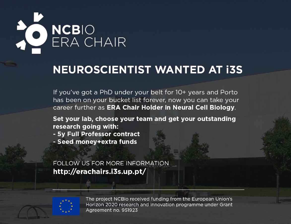
i3S – Institute for Research and Innovation in Health (Porto, Portugal) is looking to recruit a senior researcher with an established international reputation in Neural Cell Biology and strong expertise in securing, managing and leading collaborative research projects and teams/institutional units.
NCBio offers:
5-year contract as Research Coordinator, research equivalent to Full Professor, with an internationally competitive salary;
Relocation expenses, along with support concerning housing, legal/immigration issues, spouse’s employment, kindergarten/schooling, etc;
250 thousand euros in seed money in the first year of operation for the group’s research activities and to cover initial operational costs;
Total scientific freedom including the selection of a research team (four additional researchers – 2 at the Associate Professor level, 1 Postdoctoral Fellow, 1 Technician) paid directly by the NCBio project throughout its course.
Admission requirements include:
PhD degree obtained at least 10 years before application;
Established international reputation based on research excellence in the field of Neural Cell Biology;
Strong record of publication of influential papers;
Large experience in leading research groups;
Proven track-record in securing and managing significant funding;
Experience in establishing collaborative relationships with relevant industry R&D stakeholders;
Track-record in workshop and conference organising committees and delivering invited talks.
The successful applicant will be expected to commit to the position for the grant’s full duration.
Call opens on February 1st and the preliminary deadline is May 31st.
For any inquiries, please contact us at erachairs@i3s.up.pt
The recruiting process will be conducted following the Portuguese labor code, and selection criteria that prioritise merit and transparency, as well as a non-discrimination and equal access policy.
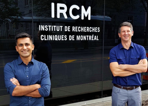
The mammalian retina contains a variety of functionally distinct cell types that are generated by progenitor cells in a specific chronological order. A new paper in Development probes the role of the POU-homeodomain factors Pou2f1 and Pou2f2 in the timely generation of cone photoreceptors in mice. We caught up with first author and PhD student Awais Javed and his supervisor Michel Cayouette (Director of the Cellular Neurobiology Research Unit at the Montreal Clinical Research Institute, Professor at the Université de Montréal and Adjunct Professor at McGill University) to hear more about their work.
Awais (L) and Michel (R).
Michel, can you give us your scientific biography and the questions your lab is trying to answer?
MC: I obtained my PhD in Neurobiology from Université Laval, in Québec city, Canada, working on viral vector-mediated gene transfer approaches in mouse models of retinal degeneration. Towards the end of my PhD I became very interested in understanding how all the beautiful cell types I was looking at under the microscope were generated, and decided I would study neural development during my postdoc. I contacted several labs and, fortunately for me, the famed developmental neurobiologist Martin Raff, whom I admired greatly, offered me the last postdoc spot available in his lab before he retired. Realizing this unique opportunity, my wife and I decided to move to London where I spent 3 years studying asymmetric cell divisions in retinal progenitors and the relative contribution of intrinsic and extrinsic signals in cell fate specification. When Martin retired, I was in the middle of a project that I wanted to finish before looking for an independent position. I then joined Ben Barres’ lab at Stanford University, USA, where I continued to work for 2 years on the project, which was finally published in 2003. In November 2004, I started my independent career in Montreal.
Ever since the beginning, my lab has been focused on studying how cell diversification is achieved during nervous system development, with the long-term goal of using this knowledge as the basis to develop regenerative therapies. We are particularly interested in understanding how neural progenitors know when it is time to make a specific combination of cell types and, once a temporal window has been defined, how progenitors choose between alternative cell fates available to them at that time. We primarily use the mouse retina as a model system to address these questions, but we have also studied myelination and the inner ear to ask questions related to cell polarity, which we also study in the context of asymmetric cell division in the retina.
And Awais – how did you come to work in Michel’s lab and what drives your research today?
AJ: In the final year of my undergraduate degree at University College London, I was revising the course material for the end of term developmental biology exam, including the temporal competence cascade in neuroblast ventral nerve cord specification in Drosophila. I found it fascinating how the same set of genes could regulate cell fate in different parts of the ventral nerve cord, and I wondered if there was a similar cascade in the vertebrate central nervous system. A quick PubMed search led me to the work of the Cayouette lab, who showed that Ikaros, the fly hunchback homolog, was a temporal competence factor in the mammalian retina. As it so happens, I was planning to move to Canada for personal reasons and the first lab I looked up was Michel’s. I started my PhD trying to find similarities between vertebrate and invertebrate neurodevelopment, but now I feel more driven by the complexity biology has to offer and how different the systems can be.
How has your research been affected by the COVID-19 pandemic?
AJ: I was very fortunate because on the last day before the lockdown; we received the next-gen sequencing results for a number of experiments I had submitted in February. The first 2 months of the shutdown, I learned how to analyse these datasets using R and Python. I had no previous knowledge of using any programming language, so it was a true test of perseverance. When the lab finally opened up, I was just happy to be back at the bench: I didn’t think I would ever say this but I definitely missed genotyping!
I didn’t think I would ever say this but I definitely missed genotyping!
MC: The lab has been completely shutdown with only essential activities allowed for almost 2 months. Like Awais, people in the lab tried to use this difficult period to make some progress. Some wrote a draft of their paper, analysed large datasets or counted cells, while others took online classes and learned how to code. I was very proud of my group – they were amazing and really used this time as best they could, despite all the challenges associated with the situation. But of course, everything was delayed in the lab. Most difficult for us was having to scale down our animal colony, as the animal facility staff was reduced. We are just now getting back to normal after several months. This also meant it took longer than expected to carry out the revisions for our paper, but the Development editors were helpful in guiding us to prioritize experiments and were flexible with the time allowed for us to do these, which was appreciated!
Why is timing critical for the generation of cellular diversity in the retina?
AJ & MC: While it is not always clear why specific neurons must be generated before others, it is generally accepted that cell birth order is critical to ensure proper neural circuit formation. Just as the foundation of a house must be built first because other parts of the house sit on it, certain types of neurons constitute the foundation of a given circuit, as they receive inputs from neurons produced later. This tightly regulated chronology is critical for the generation of highly complex tissues.
Can you give us the key results of the paper in a paragraph?
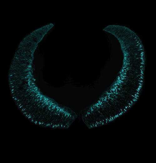
AJ & MC: In this paper, we investigated the role Pou2f1 and Pou2f2 in the developing mouse retina. We show that both genes are necessary and sufficient for cone photoreceptor cell fate specification during retinal development. We further report that Ikzf1, an early temporal identity gene that we previous identified in the retina, upregulates Pou2f1, which in turn represses the late temporal identity factor Casz1, thereby defining a temporal identity window conducive to cone photoreceptor production. Mechanistically, we show that Pou2f1 activates Pou2f2, which then represses expression of the rod-promoting factor Nrl in postmitotic photoreceptor precursors by binding to a POU-specific site in the promoter, thereby favouring the cone fate. As Pou2f1 and Pou2f2 are orthologues of fly pdm, which is well known for its role in neuroblast temporal patterning, our results, together with previously published studies, suggest that some aspects of this cascade are conserved in vertebrates. This work also establishes a link between temporal identity genes and cell fate determinants in the mammalian central nervous system.
E15 mouse retina immunostained for Rxrg, which labels newly generated cones at the apical side and ganglion cells at the basal side of the retina.
Loss of Pou2f2 reduces, but does not eliminate, cone production: how are cones determined in its absence?
AJ & MC: It is of course possible that other genes compensate for the loss of Pou2f2. Potential candidates include Onecut1 and Onecut2, as a partial loss of cones, similar to what we observed in this study, is observed in double knockout animals. Another possibility is that abolishing the temporal window for cone production induces progenitors to generate cones outside the normal window using alternative pathways. Finally, we have previously suggested that loss of temporal identity in progenitors does not lead to complete absence of any given cell type, but simply to a reduced probability of generating these cell types, which might explain why we do not observe a complete loss of cones in this study.
Do you know what restricts Pou2f1 expression to early RPCs? Is there evidence of mutual inhibition with other temporal factors?
AJ & MC: This is a very interesting question. We show in our paper that Ikzf1 upregulates Pou2f1 expression in early RPCs, but it remains unknown what actually turns off Pou2f1 expression in later progenitors. An obvious candidate is Casz1, which might repress Pou2f1 at later stages, similar to what is observed in Drosophila neuroblasts, but our preliminary experiments looking at this possibility do not appear to support this model. Another possible regulator of Pou2f1 is Foxn4, which was recently discovered as a regulator of Ikzf1 and Casz1 in the mammalian retina. More work is needed to fully elucidate this issue.
When doing the research, did you have any particular result or eureka moment that has stuck with you?
AJ: The main result that comes to mind was actually one of the first observations that led us down the path of this paper. Initially, immunostainings of Pou2f1 showed strong expression in ganglion cells, as well as other cell types, which we later found out to be cone and horizontal cells. When we first tried gain-of-function experiments with Pou2f1 using a retroviral vector, we did not initially consider that cone production would be induced. Out of sheer curiosity, I added S-opsin as a marker for cones and, to my surprise, I found S-opsin+ cells in the overexpression condition. My first thought was that I had mixed up the viruses or the immunostainings, but when it started to repeat, I was convinced it was a real result. It wasn’t a eureka moment per se, but it felt quite good to stumble onto a finding that would later turn out to be the central hypothesis of the paper.
And what about the flipside: any moments of frustration or despair?
AJ: The ChIP-qPCR experiments, without a doubt. It is running joke in the lab at this point because of how tired I was during lab meetings when I was working on those experiments. It was very labour intensive because I used embryonic retinal tissues and I had quite a bit of optimization to do before I could get any results. But it was well worth it, when I finally got the experiment to work. In the end, it was a fantastic lesson learned on perseverance.
What next for you after this paper?
AJ: I am finishing up a few projects in the lab and hoping to graduate in early 2021. I am currently looking for postdoc opportunities and I am very excited to continue investigating cell fate specification in other parts of the central nervous system.
Where will this story take the Cayouette lab?
MC: We are becoming increasingly interested to determine whether temporal factors could be used to promote tissue regeneration. The idea is simple, as early temporal factors are sufficient to reprogram late-stage progenitors into generating early-born cell types, we wonder whether these factors might also be able to reprogram differentiated cells into neural progenitors. We would also like to determine whether progenitors invariably go through all the temporal identity windows defined by the temporal factors we have studied so far in the retina (Ikzf1, Pou2f1, Pou2f2 and Casz1) or whether some can skip a given window. Answers to this question might explain the huge heterogeneity of clonal composition observed in lineage-tracing studies in the retina. This will require the development of tools to follow temporal factor expression in real time in single cells, but I think it would be very cool to address this question, as it has far-reaching implications. Finally, detailed mechanistic understanding of temporal patterning remains poorly understood, even in flies. We are becoming increasingly interested in this question. It is likely that temporal identity is defined by specific epigenetic landscapes, and whether and how temporal factors shape chromatin conformation and nuclear architecture is a problem we will likely focus on in years to come.
Finally, let’s move outside the lab – what do you like to do in your spare time in Montreal?
AJ: I am an avid painter and it has helped me deal with the ups and downs of a PhD. I also founded a drama club at the institute. We wrote and directed a couple of plays to a full audience at one of the institute’s auditoriums, which was an amazing experience. I did not realize that many scientists are fantastic artists!
MC: Since I was a child, I have been playing ice hockey and I reached fairly high competitive level. To this day, I continue to play once or twice a week, often with students who seem to be getting younger and faster each year… I am also a fervent cyclist and love to ride the roads of the countryside and the trails of the provincial park around our house. Finally, I love great food and wine, and since Montreal has a large choice of fantastic restaurants, it is a good place to be!
Yesterday we held the fourth webinar in our series, this time chaired by Development Editor Swathi Arur. Here you’ll find recordings of the talks and their live Q&A sessions moderated by Swathi.
The Genetics Unzipped podcast is back for 2021 with a new series of stories from the world of genes, genomes and DNA, from the history of genetics to the latest cutting-edge research.
In our first episode we take a look back at the discovery of messenger RNA, or mRNA, in the 1960s. There are some big names involved – Francis Crick, Sydney Brenner, Francois Jacob and more – but who actually discovered this vital molecular messenger? And why did nobody win a Nobel Prize for it?
Then we come right up to the present day to look at mRNA vaccines for COVID-19, which have been developed at breakneck speed to tackle the pandemic. We find out how mRNA vaccines work, how they were developed so fast for COVID-19, and how this new technology might change the face of immunization and public health in the future.
If you enjoy the show, please do rate and review on Apple podcasts and help to spread the word on social media. And you can always send feedback and suggestions for future episodes and guests to podcast@geneticsunzipped.com Follow us on Twitter – @geneticsunzip
The laboratory of Pamela Geyer invites mature, highly motivated candidates to apply for a position as postdoctoral fellow in the Department of Biochemistry at the University of Iowa. These fellows will join a research team interested in understanding the impact of nuclear architecture in stem cell survival and aging. Prominent changes in nuclear morphology occur during physiological aging and in disease. Our recent data demonstrate that the asymmetrically dividing germline stem cells (GSCs) in the Drosophila ovary employ a non-canonical mode of mitosis that sensitizes these stem cells to defects in proteins comprising the nuclear lamina, a protein network that build contacts with the genome to regulate transcription, replication and DNA repair. Current studies are focused on exploring the mechanisms involved in execution of this non-canonical mitosis and implications for stem cell health and longevity. In addition to driving science at the bench, the fellow will be introduced to the network of collaborative scientists with shared interests in defining biological processes critical for development and disease. The Geyer laboratory is located in a state-of-the-art facility that features a collaborative atmosphere to foster innovation, and is part of the Carver College of Medicine, a highly ranked biomedical research institute. The University of holds a long tradition of diversity, equity, and inclusion, a transition that began upon its founding with being the first public university to admit men and women on an equal basis (1855). Iowa City is a medium-sized, culturally rich city with vibrant communities in the arts, sciences, and literature. Interested applicants should contact Pamela Geyer (pamela-geyer[at]uiowa.edu), including a letter of interest, a CV and contact information of three references.
Cellular differentiation describes the process by which embryonic cells become different from one another, acquiring distinct identities and specialised functions. These cells are responding to signals that modify the dynamics of the interacting key genes controlling the cell’s state. The arrival of more and better single cell data opens up new opportunities for better understanding of this cellular decision-making. However, to realise these opportunities, new mathematical and statistical tools are needed to characterise the cellular dynamics and to organise, analyse and visualise such data. This workshop aims at reviewing the current work in this area.
 (No Ratings Yet)
 (1 votes)

 (3 votes)