The next webinar in our Development presents… series will be a little different: rather than being chaired by a Development editor, the preLights team will be in control. preLights is the preprint highlights service run by the biological community and supported by The Company of Biologists, and in February they celebrate their third birthday.
We brought together three preLighters with interests in developmental biology – Sundar Naganathan, Irepan Salvador-Martinez and Grace Lim – who have each invited authors of recent exciting preprints to give talks. The preLighters will chair the talks and the Q&As, and we’ll also hear from outgoing preLights Community Manager Mate Palfy about three years in the life of a preprint-focused community. We hope to see you there!
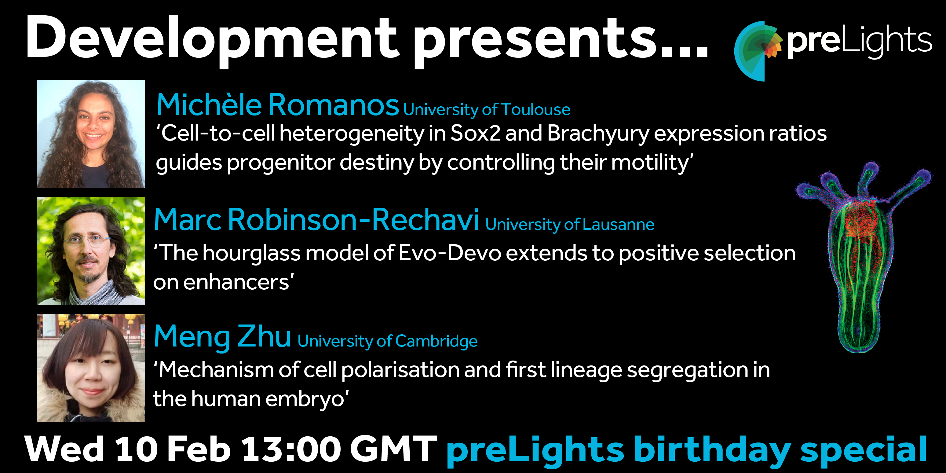
Wednesday 10 February 2021 – 13:00 GMT
MichèleRomanos (from Bertrand Benazeraf’s lab at the Centre de Biologie Integrative in Toulouse)
‘Cell-to-cell heterogeneity in Sox2 and Brachyury expression ratios guides progenitor destiny by controlling their motility.’
The webinar will be held in Remo, our browser-based conferencing platform – after the talks you’ll have the chance to meet the speakers and other participants at virtual conference tables. If you can’t make it on the day, talks will be available to watch for a couple of weeks after the event (look out for details on the Node).
For more information about what to expect in Remo, go to
The employment is for 4 years, as research assistant is for 1 year and as PhD fellow for the following 3 years, and is scheduled to start in July or upon agreement with the chosen candidate.
The Novo Nordisk Foundation Center for Stem Cell Biology (DanStem) at Faculty of Health & Medical Sciences at the University of Copenhagen is looking for a Research assistant subsequent appointed as PhD fellow to join the Żylicz group starting July 2021 or upon agreement with the chosen candidate.
The position as Research assistant is for 1 year. The position as PhD fellow is for 3 years.
This position is available in the group of Jan Żylicz at DanStem. The team studies fundamental mechanisms of early mouse development and stem cell biology. The Żylicz group wants to understand how metabolic and epigenetic mechanisms cooperate to regulate transcription during early development. In particular the team is interested in how metabolism regulates histone modifiers, and how these in turn affect lineage choice and embryo growth at around the time of implantation. To achieve this, the group utilizes both in vivo mouse models as well as in vitro stem cell culture systems and state-of the-art ultrasensitive transcriptomic, epigenomic and metabolomic techniques. This research project will employ a multi-disciplinary approach to understand how first lineage choices are influenced by metabolic and chromatin states. Starting date
for this position is the 1 July 2021, or upon agreement with the chosen candidate.
Job description:
We are seeking a highly motivated and ambitious pre-doctoral candidate with a strong educational background in either stem cell and developmental biology and/or metabolomic research and/or bioinformatics. The candidate will investigate the molecular mechanisms controlling first lineage choice of early development using both in vivo and in vitro stem cell models. By employing genomic engineering methods the candidate will go beyond description of epigenetic and metabolic states and uncover novel functional regulatory mechanisms at the interplay of epigenetics and metabolism.
About DanStem:
The Novo Nordisk Foundation Center for Stem Cell Biology – DanStem – addresses basic research questions in stem cell and developmental biology and translates results from basic research into new strategies and targets for the development of new therapies for diabetes and cancer. DanStem was established as a result of a series of international recruitments coupled with internationally recognized research groups focused on insulin producing beta cells and cancer research already located at the University of Copenhagen.
Qualifications:
Candidates must hold a master’s degree in biotechnology, bio-informatics, biology or similar, and possess a strong background in developmental and stem cell biology and/or metabolomics and/or bioinformatics.
Previous practical experience in bioinformatics, analysis of epigenomic data and/or software programming is considered of great advantage.
Previous experience using rodents as a research model, stem cell culture, embryogenesis and/or next-generation sequencing are considered an advantage.
Practical project experience in basic lab techniques is considered beneficial.
Good English communication skills, both oral and written, are prerequisite for the successful candidate.
A solution-oriented, organizational and positive mindset is required. The ability to work in a highly diverse and collaborative environment both independently and as part of the team is essential.
Terms of salary, work, and employment:
The employment is for 4 years, as research assistant is for 1 year and as PhD fellow for the following 3 years, and is scheduled to start in July or upon agreement with the chosen candidate. The employment as a PhD student is conditioned upon a positive assessment of the candidate ́s research performance and enrolment in the Graduate School at the Faculty of Health and Medical Sciences.
Salary, pension and terms of employment are in accordance with the provisions of the collective agreement between the Danish Government and AC (the Danish Confederation of Professional Associations). In addition to the basic salary a monthly contribution to a pension fund is added (17.1% of the salary).
The PhD study must be completed in accordance with the ministerial orders from the Ministry of Education on the PhD degree and the University ́s rules on achieving the degree.
The place of work is at DanStem, University of Copenhagen, Blegdamsvej 3B, Copenhagen.
Questions:
For further information please contact Associate Professor Jan Żylicz, jan.zylicz@sund.ku.dk.
The application must include:
Motivation letter
Curriculum vitae incl. education, experience, previous employments, language skills and other relevant skills
List of two references (full address, incl. email and phone number)
Copy of diplomas/degree certificate(s)
How to apply
The application, in English, must be submitted electronically by clicking APPLY below.
Application deadline: 21 February 2021
Only applications received in time and consisting of the above listed documents will be considered.
Applications and/or any material received after deadline will not be taken into consideration.
The University of Copenhagen wishes to reflect the diversity of society and welcomes applications from all qualified candidates regardless of personal background.
The application will be assessed according to the Ministerial Order no. 284 of 25 April 2008 on the Appointment of Academic Staff at Universities.
Assessment procedure
After the expiry of the deadline for applications, the authorized recruitment manager selects applicants for assessment on the advice of the Appointments Committee. All applicants are then immediately notified whether their application has been passed for assessment by an expert assessment committee. Selected applicants are notified of the composition of the committee and each applicant has the opportunity to comment on the part of the assessment that relates to the applicant him/herself. You can read about the recruitment process at http://employment.ku.dk
Københavns Universitet giver sine knap 10.000 medarbejdere muligheder for at udnytte deres talent fuldt ud i et ambitiøst, uformelt miljø. Vi sikrer traditionsrige og moderne rammer om uddannelser og fri forskning på højt internationalt niveau. Vi søger svar og løsninger på fælles problemer og gør ny viden tilgængelig og nyttig for andre.
Info
Application deadline: 21-02-2021
Date of employment: 01-07-2021
Working hours: Full time
Department / Place: The Novo Nordisk Foundation Center for Stem Cell Biology
From the almost identical faces of monozygotic twins, we can appreciate that facial appearance is encoded for the most-part in the DNA of our genomes. Therefore, changes to this DNA sequence can contribute to the variation in facial appearance seen between humans, and at the more extreme end of the spectrum can cause human disease. Consequently, improving our understanding of how genetic sequence encodes for our physical traits remains an exciting and important question in developmental biology.
Interestingly, many of the genetic changes that contribute to facial variation or dysmorphology in disease occur in the non-gene encoding parts of the genome. And this raises the question how non-coding DNA mutations can impact development and drive morphological change. To answer this question, in the Wysocka lab we study facial progenitor cells called the cranial neural crest which give rise to the majority of the facial structures, and investigate how genetic changes can drive alterations in physical traits.
At the beginning of our recent study, published last year in Cell Stem Cell, we set out to determine the molecular mechanisms that cause a human craniofacial disorder, called Pierre Robin sequence (PRS) [1]. In this disorder, under-development of the lower jaw (micrognathia) leads in sequence to posterior displacement of the tongue, and in some cases cleft palate [2, 3] (Figure 1). Several labs had previously utilised genomic methods to identify a number of large deletions and chromosomal translocations in PRS patients that cluster in a gene desert region far upstream of the SOX9 gene .
SOX9 is an important DNA-binding transcription factor that plays crucial roles in the differentiation of numerous cells types, and is expressed broadly during development including in chondrocytes, testis, pancreas, heart valve, lung, kidney, liver, hair follicle stem cells, and progenitors of the face (cranial neural crest) [8]. In keeping with its broad roles during development, heterozygous loss-of-function mutations in the SOX9 gene cause a devastating multisystemic syndrome called campomelic dysplasia. Patients typically exhibit skeletal abnormalities including bowing of the long bones, disorders of sex determination and facial dysmorphism, and occasionally have additional malformations of the heart, lung, kidney and pancreas suggesting differential sensitivity to SOX9 gene dosage (Figure 1). Importantly, selective knockout of Sox9 in mouse facial progenitors results in developmental defects of the facial skeleton [1, 9] emphasizing the importance of Sox9 in the development of the face. Given the non-coding nature of the mutations in PRS patients, it had been suggested that gene regulatory elements called enhancers may be perturbed [4–7]. However, it remained to be functionally demonstrated how these mutations impacted genome function, and whether SOX9 was the target gene to cause this disorder.
Figure 1. Non-coding mutations far upstream of the SOX9 gene are associated with an isolated congenital craniofacial abnormality called Pierre Robin sequence (PRS).
Enhancers are non-coding DNA sequences elements that can act across large genomic distances to regulate gene expression in a cell-type specific manner. And key developmental genes are often associated with complex regulatory landscapes with many such context-dependent enhancers [10]. Therefore, unlike mutations in coding sequences which will alter gene function in all cell-types in which that gene is expressed, mutations in enhancers will have a more developmental stage and cell-type specific impact leading to tissue-restricted phenotypic consequences.
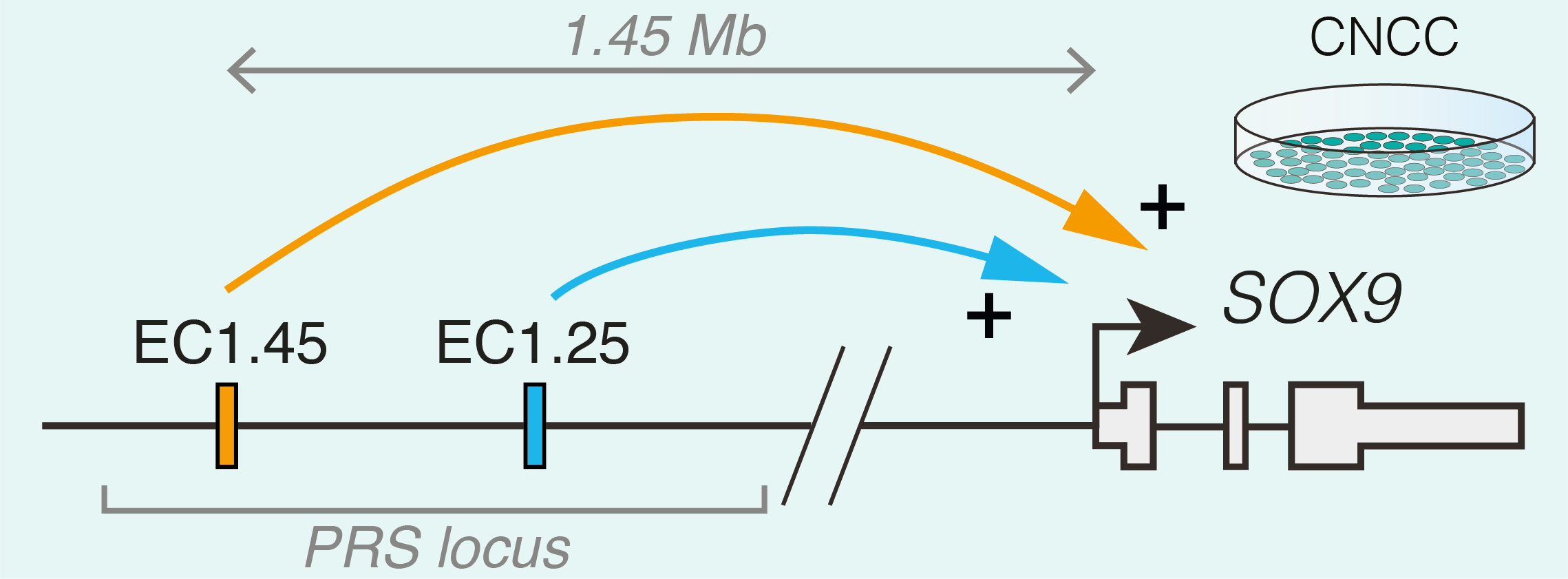
Studying human facial development presents a challenge, as formation of the face occurs early during gestation. However, leveraging a robust in vitro differentiation model of cranial neural crest cells (CNCCs) developed in the lab we have been able to model human craniofacial disorders in culture [11–13]. Utilising this model, and genomic enhancer profiling, we identified three clusters of enhancer elements overlapping the human PRS mutation region. We rationalised that these elements may regulate SOX9 expression during development, and that their loss could cause mis-regulation of SOX9 in the neural crest with detrimental consequences for facial development. Indeed, using CRISPR/Cas9 genome editing we demonstrated that SOX9 expression is specifically perturbated by PRS mutations in the neural crest (and not during cartilage formation when SOX9 is also expressed), defining a developmental window for disease causation in this disorder (Figure 2).
Figure 2. Two clusters of enhancers overlapping the PRS locus regulate SOX9 expression across extreme long-distance, up to 1.45 Mb, specifically in cranial neural crest cells (CNCCs).
To further understand the morphological impacts of enhancer ablation, we set out to generate mouse models to investigate Pierre Robin sequence disease mechanisms. However, we soon appreciated the challenges associated with modelling human genetic disorders of the non-coding genome in mouse. Unlike genic regions, which make up 1-2% of the genome and are deeply conserved, the remainder of the genome is highly divergent between human and mouse. Indeed across mammalian species, enhancers can vary greatly in their location, sequence composition and activity [14]. Therefore, while one of the PRS-associated enhancer clusters (named EC1.45) was at least partially conserved in activity during mouse facial development, the second enhancer cluster (EC1.25) was not, limiting our further morphological investigation of PRS mutations to EC1.45 alone. Given that directly addressing the functional impact of specific human non-coding mutations in model organisms poses a huge challenge due to a limited sequence conservation of the non-coding genome, future studies exploring the genotype to phenotype connection in human development and disease will greatly benefit from improved models of human development, including advances in organoid models.
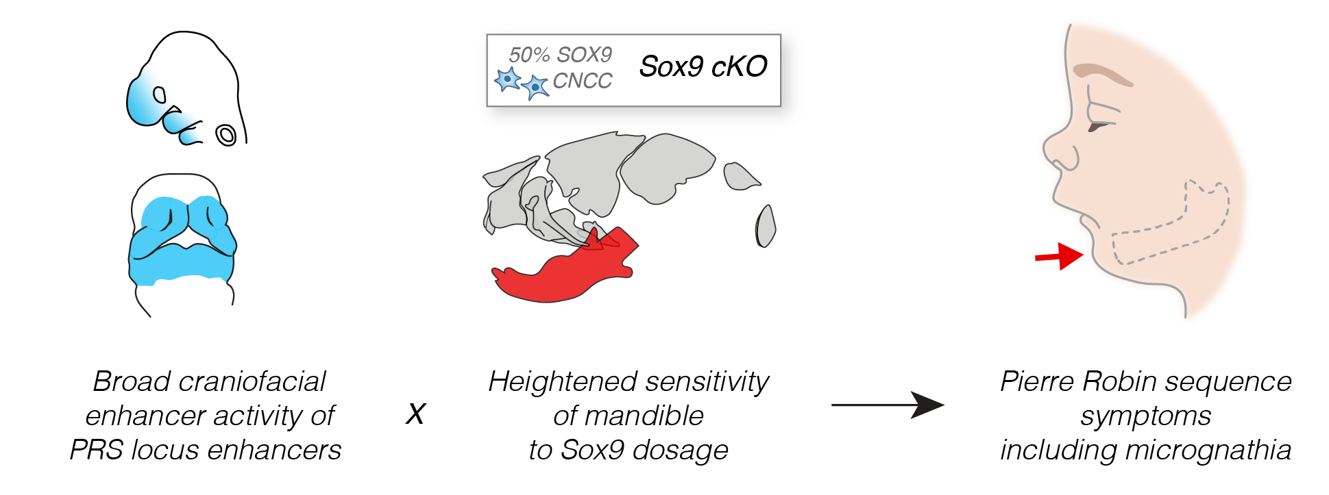
Despite the limitations, our models of Sox9 perturbation in mouse led to a number of intriguing observations. Firstly, we determined that lower jaw development is highly sensitized to reduction in Sox9 gene expression compared to the rest of the skull. Therefore, despite the PRS enhancers being widely active across the developing face, this regional sensitivity in the lower jaw to Sox9 gene dosage may drive the phenotypic specificity seen in patients (Figure 3). Secondly, we observed that even small changes in Sox9 expression during development lead to alterations in lower jaw morphology, demonstrating how subtle changes in enhancer activity may drive morphological change and fitness between members of the same species and across evolutionary timescales.
Figure 3. The specificity of the lower jaw phenotype in Pierre Robin sequence (PRS) patients derives from a combination of ablation of broad craniofacial enhancers, and a heightened sensitivity of the lower jaw to Sox9 reduction of gene dosage (PRS locus enhancer activity in facial prominences during development is highlighted in blue).
Together, our study uncovers the workings of a complex regulatory domain that controls SOX9 expression during a narrow window of facial development. We further explore the sequence motifs and trans-regulatory factors that regulate enhancer function at the PRS locus, and the conservation of enhancer activity across vertebrate and recent hominin evolution – and we invite you to see our paper for more details. Importantly, the enhancer clusters we characterised at the PRS locus represent the longest-range human enhancers involved in congenital malformations described to date and their deletion in mouse illustrates how small changes in gene expression can lead to morphological variation.
Combined, our work provides a clear illustration and mechanistic details how large deletions and translocations in the non-coding genome can cause human congenital disease. This joins a number of recent studies that have explored how genetic aberrations such as inversions, duplications and topological domain boundary perturbations can either displace enhancers away from their target gene, or can lead to de novo enhancer co-option and ectopic gene expression [15–17].
Looking forward, it is exciting to speculate how such long-range gene regulatory elements (at up to 1.45 megabases away) such as those at the PRS locus find their target gene. From our study, we speculate that perhaps a third element at the PRS locus may play a structural role to bridge these long genomic distances. The developmental importance of this genetic feature, and whether this is a generalisable feature of long-range regulation will be an exciting avenue for future research.
Where are you originally from, where do you work now, and what do you work on?
I am originally from Alsace in France. I got a Master’s in developmental biology which I ended with a 6 months internship at EMBL in Heidelberg. This successful internship and the interesting Masters courses led me to pursue a Ph.D. at the Institute of Science and Technology in Austria at IST Austria.
Unfortunately this Ph.D. had to end prematurely due to emergency brain surgery I had to undergo in the third year. but at the same time as doing my Ph.D., IST Austria had given me the opportunity to discover the exciting field of science communication. The experiences I took part in made me realize that this was the job I wanted to do, and therefore I decided to enrol in another program to get the set of skills needed in science communication.
I am currently in the process of getting a Master’s degree from the University of Bordeaux Montaigne in France. Because I want to help to bridge the gap between research and innovation, I’ll do a 6 months internship at the scientific research and innovation board of Loreal.
Were you always going to be a scientist?
Science was my first love. From very early on I was fascinated by the biological processes which govern life. I couldn’t imagine myself feeling as happy and useful in any other discipline. So I hesitated for a long time before commuting from academia to science communication but this change has been really positive so far and allows me to keep updated with scientific discoveries in various fields. It is very rewarding.
And what about art?
I started drawing at a very young age, mainly drawing my favourite animals, especially horses and dogs. In recent years I started making illustrations for the new papers of my fellow colleagues. I really enjoy conveying scientific discoveries through art. Asking questions and listening to the scientists somehow triggers my artistic imagination in a way that I always end up having tons of illustration ideas.
“I really enjoy conveying scientific discoveries through art”
What or who are your artistic influences?
I love surrealism, and artists such as Salvador Dali, Yves Tanguy, René Magritte and Frida Kahlo.
How do you make your art? How do you approach making a new piece of art?
I used to start with a simple pencil and paper but after a few years of experience with numerical drawing, I directly started drawing on my tablet or my laptop with the touchpad. I start with a broad idea, a drafty sketch, which I turn very dim in the background. I draw a more realistic vision on top of it with larger and darker brushes, making use of many layers on top of each other to go more in detail.
Does your art influence your science at all, or are they separate worlds?
Science certainly influences my art. I love to make illustrations of beautiful data, something catchy which attracts the reader’s attention. Nature is so perfect and beautiful, it’s a never-ending source of inspiration.
We’re looking for new people to feature in this series throughout the year – whatever kind of art you do, from sculpture to embroidery to music to drawing, if you want to share it with the community just email thenode@biologists.com (nominations are also welcome!).
Our group is seeking a highly motivated candidate strongly interested in interdisciplinary science, to join the Guignard lab and work on a project at the crossroad between Computer Science and Developmental Biology as a Postdoc fellow.
Automatic reconstruction of Drosophila average embryonic development at the single-cell scale
The role
We are seeking a highly motivated candidate, strongly interested in interdisciplinary science, to join our group and work on a project at the crossroads between Computer Science and Developmental Biology.
The project aims at developing computational methods and models to better understand how robustness to biological noise is achieved during the development of Drosophila embryos. This will be done by first quantifying and characterising Drosophila embryogenesis variability at the single cell scale.
The successful candidate will be tasked to build statistical representations of the morphogenesis of Drosophila embryos at the single cell scale by combining image analysis, big-data science and data visualisation. To this end, the candidate will first develop novel image and big-data analysis algorithms. These algorithms will be first applied to 3D movies of Drosophila embryos acquired with state-of-the-art light-sheet fluorescence microscopes. The successful candidate will then develop novel algorithms to combine the set of recorded embryos together to build a single-cell scale, in-toto, atlas of Drosophila embryogenesis.
Depending on the advancement of the project and the candidate preferences, the second part of the project will then focus around either integrating complementary single-cell omic data to the atlas or developing machine-learning based methods to analyse, detect and classify cell patterns in the developing embryo.
The candidate must have at least one of the following expertises:
Computer Science: Image Analysis, Graph Theory, Data Science
Developmental Biology (sole experience in Developmental Biology requires to be able to show good coding skills)
Education and training
You hold a master degree in Bioinformatics, Computer Science, Developmental Biology (with good computational skills) or equivalent
Competences
You are eager to learn
You are creative
You have good communication skills
You want to work as part of a collaborative team
Languages
You have French or English fluency (at least B2 on the CEFR)
The Offer
Contract duration: 2-year position, extension possible
Target start date: from July 2021
The salary will be based on Aix-Marseille University’s salary scale, depending on the candidate’s profile and experience
Application Procedure
All applications must include:
A motivation letter addressed to Léo Guignard.
A complete CV including contact details.
Contact details of at least two referees.
All applications must be sent to Léo Guignard by email with the mention [Job-2021] in the subject at the address leo.guignard+lab[at]gmail.com.
Selection Process
Pre-selection: The pre-selection process will be based on qualifications and expertise reflected in the candidates CV and motivation letter. It will be merit-based. All candidates will be informed whether they have been pre-selected or not.
Interview: Pre-selected candidates will be contacted to coordinate a set of interviews with a set of selected members of CENTURI (including Léo) and a seminar. The interview will include a computational skill test (no specific coding language is required).
We are a group of computer scientists with a strong interest in biology in general and more specifically in embryonic development. We develop novel computational methods and models that allow the analysis of very large 3D movies of animal embryonic development (up to 2TB per movie). We work closely with biologists to tailor our methods so that they help to address fundamental biological questions.
The developmental biology question that mainly animates us is to better understand the mechanisms driving the reproducibility of embryogenesis.
The Turing Centre for Living Systems (CENTURI) is an interdisciplinary project located in Marseille (France).
CENTURI aims at developing an integrated interdisciplinary community, to decipher the complexity of biological systems through the understanding of how biological function emerges from the organization and dynamics of living systems.
The project federates 15 teaching and research institutes in biology, physics, mathematics, computer science, engineering and focuses on Research, Education and Engineering, 3 missions that hold interdisciplinary as their core principle.
The research and training programmes implemented under the auspices of CENTURI will foster new collaborations, transform practices, attract new talents and thereby contribute to making the Luminy campus a leading site for the interdisciplinary study of biological systems.
Our group is seeking a highly motivated candidate strongly interested in interdisciplinary science, to join the Guignard lab and work on a project at the crossroad between Computer Science and Developmental Biology as a Ph.D student.
Automatic reconstruction of Drosophila average embryonic development at the single-cell scale
The role
We are seeking a highly motivated candidate, strongly interested in interdisciplinary science, to join our group and work on a project at the crossroads between Computer Science and Developmental Biology.
The project aims at developing computational methods and models to better understand how robustness to biological noise is achieved during the development of Drosophila embryos. This will be done by first quantifying and characterising Drosophila embryogenesis variability at the single cell scale.
The successful candidate will be tasked to build statistical representations of the morphogenesis of Drosophila embryos at the single cell scale by combining image analysis, big-data science and data visualisation. To this end, the candidate will first develop novel image and big-data analysis algorithms. These algorithms will be first applied to 3D movies of Drosophila embryos acquired with state-of-the-art light-sheet fluorescence microscopes. The successful candidate will then develop novel algorithms to combine the set of recorded embryos together to build a single-cell scale, in-toto, atlas of Drosophila embryogenesis.
Depending on the advancement of the project and the candidate preferences, the second part of the project will then focus around either integrating complementary single-cell omic data to the atlas or developing machine-learning based methods to analyse, detect and classify cell patterns in the developing embryo.
The candidate must have at least one of the following expertises:
Computer Science: Image Analysis, Graph Theory, Data Science
Developmental Biology (sole experience in Developmental Biology requires to be able to show good coding skills)
Education and training
You hold a master degree in Bioinformatics, Computer Science, Developmental Biology (with good computational skills) or equivalent
Competences
You are eager to learn
You are creative
You have good communication skills
You want to work as part of a collaborative team
Languages
You have French or English fluency (at least B2 on the CEFR)
The Offer
Contract duration: 3-year position, extension possible
Target start date: from July 2021
The salary will be based on Aix-Marseille University’s salary scale, depending on the candidate’s profile and experience
Application Procedure
All applications must include:
A motivation letter addressed to Léo Guignard.
A complete CV including contact details.
Contact details of at least one (for Ph.D. candidates) or two (for postdoc candidates) referee(s).
All applications must be sent to Léo Guignard by email with the mention [Job-2021] in the subject at the address leo.guignard+lab[at]gmail.com.
Selection Process
Pre-selection: The pre-selection process will be based on qualifications and expertise reflected in the candidates CV and motivation letter. It will be merit-based. All candidates will be informed whether they have been pre-selected or not.
Interview: Pre-selected candidates will be contacted to coordinate a set of interviews with a set of selected members of CENTURI (including Léo) and a seminar. The interview will include a computational skill test (no specific coding language is required).
We are a group of computer scientists with a strong interest in biology in general and more specifically in embryonic development. We develop novel computational methods and models that allow the analysis of very large 3D movies of animal embryonic development (up to 2TB per movie). We work closely with biologists to tailor our methods so that they help to address fundamental biological questions.
The developmental biology question that mainly animates us is to better understand the mechanisms driving the reproducibility of embryogenesis.
The Turing Centre for Living Systems (CENTURI) is an interdisciplinary project located in Marseille (France).
CENTURI aims at developing an integrated interdisciplinary community, to decipher the complexity of biological systems through the understanding of how biological function emerges from the organization and dynamics of living systems.
The project federates 15 teaching and research institutes in biology, physics, mathematics, computer science, engineering and focuses on Research, Education and Engineering, 3 missions that hold interdisciplinary as their core principle.
The research and training programmes implemented under the auspices of CENTURI will foster new collaborations, transform practices, attract new talents and thereby contribute to making the Luminy campus a leading site for the interdisciplinary study of biological systems.
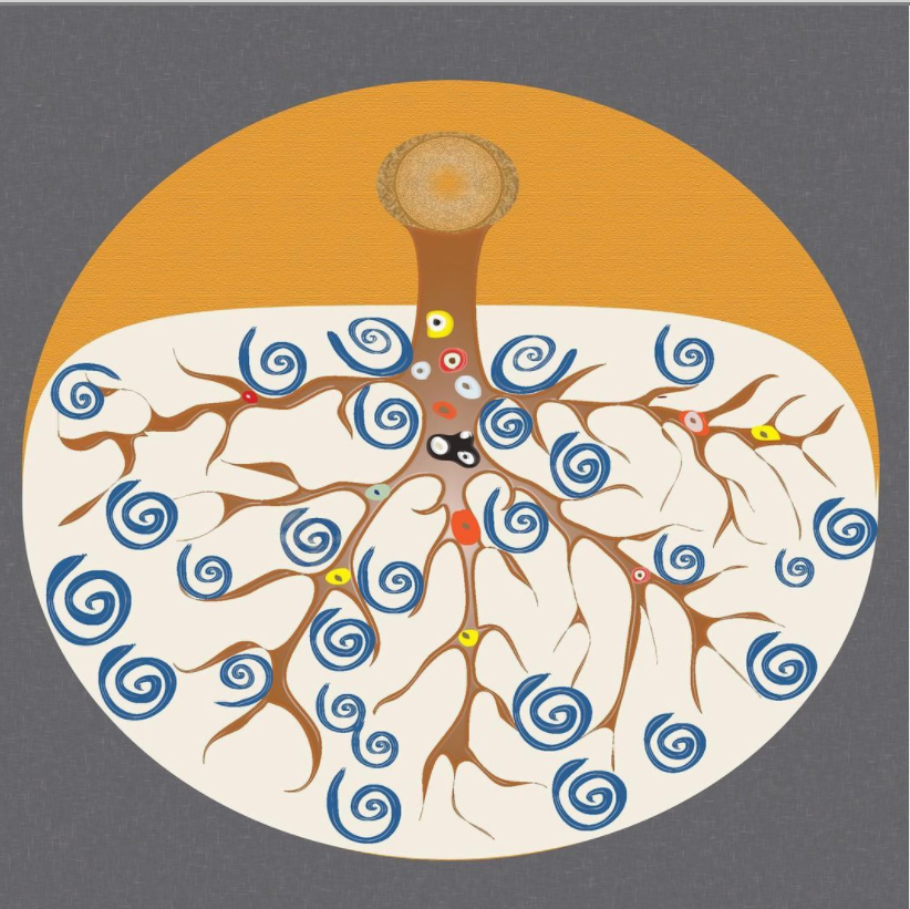
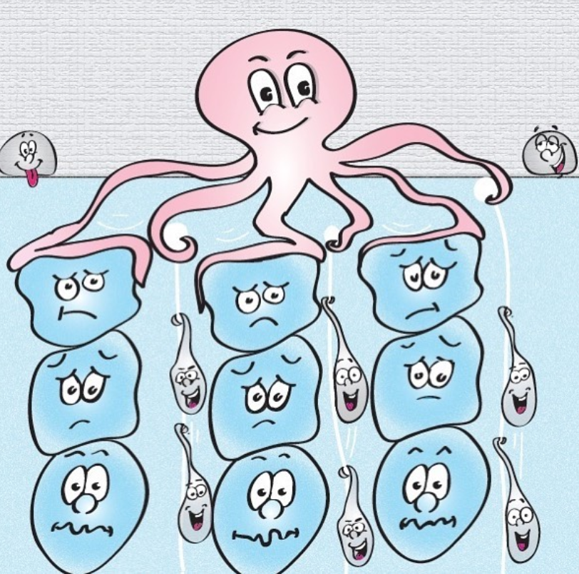
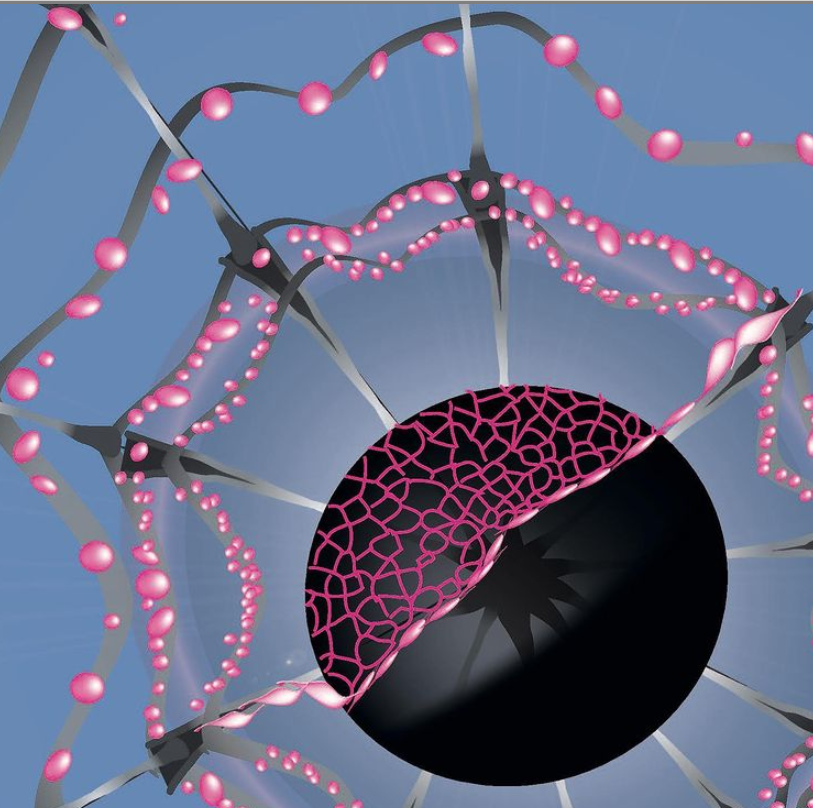
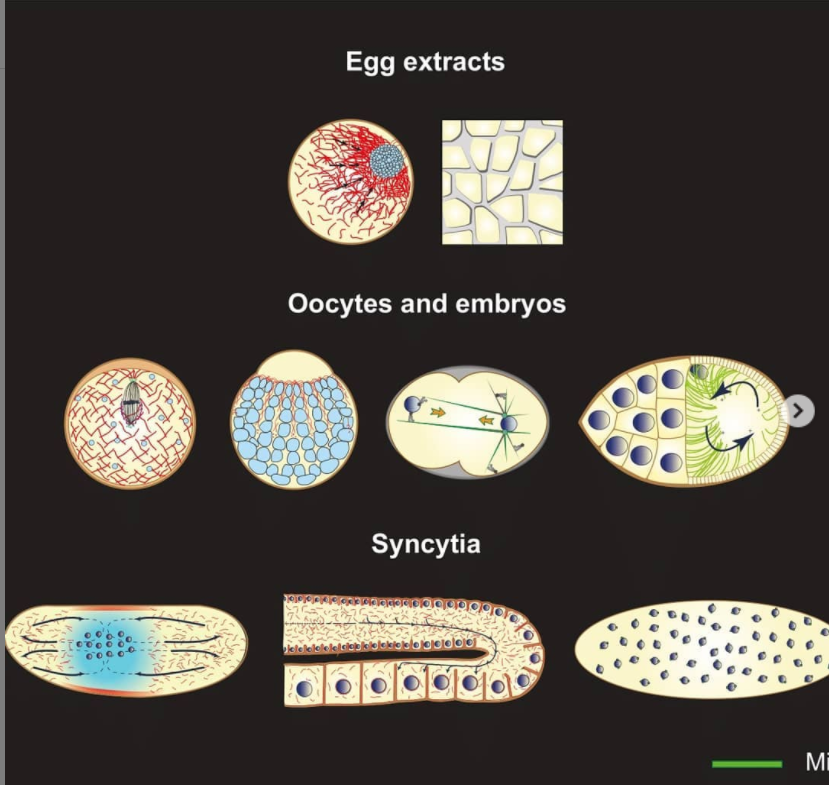
We’ll soon be launching a newly designed Node homepage to help make our historical content easier to find and improve navigation through the various parts of the site. To accompany this change we’re going to refresh our header image – that’s the letterbox-shaped image you see above the menu bar. Currently we circulate between these four images:
…and now we need four more. And where better to source beautiful developmental biology than the community?
So, do you have a developmental biology image that would be a good replacement? Winning images will be seen by thousands of readers a month from all over the world (better, surely, than languishing unseen in a folder within a folder on your desktop!).
Competition details
The image must be croppable to 1140×190 pixels (that’s 6:1), or be submitted at those dimensions.
The higher quality the better – if cropping leads to pixellation, we won’t pick it
Subject can be anything related to developmental biology – any organism, any system, any imaging platform.
As you see above, the image could be a striking image, a close up of an embryo, or a repeated pattern
If there is a background to your image, please make it black
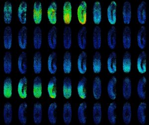
During their journey from zygote to adult, embryos experience several symmetry breaking processes. Structures which are not isotropic (equal in all directions) are formed, creating the inside-out axis, forward-backwards axis, etc. Each of these patterns increases the information required to describe a multicellular system. As Maynard Smith put it: sometimes the “extra” information is stamped by maternal cues (gradients) and other times it emerges from self-organized processes. Decades of research have accrued a lot evidence for self-organized processes in development, from the theoretical to the experimental. More recently, synthetic biology has provided an avenue for enquiry by allowing us to create invivo and de novo patterning mechanisms.
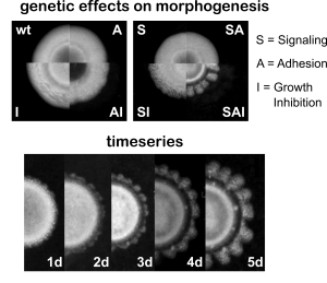
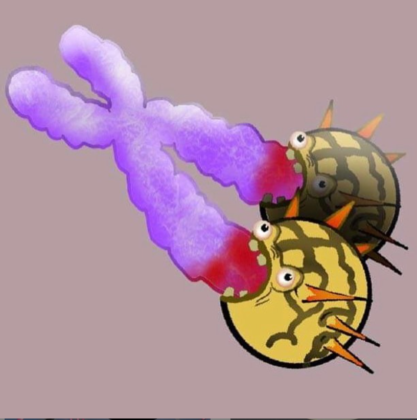
(above) Effects of gene expression on pattern formation. (below) Temporal dynamics of synthetic gene morphogenesis.
In our recent publication we take an engineering approach by inducing a symmetry breaking process in growing bacterial colonies. Without external factors like lack of nutrients or chemoattractants, E. coli colonies develop with remarkable symmetry into circular shapes. Their propagation front is isotropic and homogenizing diffusion is the leading morphogenetic drive, erasing any patterns in cellular density.
We induced pattern formation using four synthetic genes relating to adhesion, signaling and growth inhibition. These would make cells stop dividing and attach to one another, but only once a certain local cellular density threshold had been surpassed. With this we hoped that differences in cell density would be amplified with short range activation and long range inhibition, and that the patterns in space would be preserved from diffusion through adhesion.
When we put these elements together in growing colonies, we observe a remarkable symmetry breaking process. Colonies now develop into flower-like shapes with “petals” forming at a characteristic scale (constant wavelength). Regions with increased cell density inhibit the growth of neighboring cells and delay front propagation. This emergent pattern is not found in any other possible combination of our synthetic genes.
We believe these is just a first step into developing an engineering research program of artificial developmental processes. Using mechanical processes inherent to cellular embodiment allows us to make robust patterns that emerge from many interacting agents. Hopefully, armed with bioengineering tools we will gain a better grasp on the sometimes counter intuitive emergent interactions that create order in real developing embryos.
We are looking for two postdoctoral researchers (3 or 5 years) to work in Roberto Mayor laboratory at University College London, UK.
The scope of the positions is quite open and the project will be agreed with the candidate according to background and research interests. The general aims of the project is to study the interplay of mechanicaland chemical cues on cell migration and differentiation in vivo, using zebrafish and Xenopus embryos and cells cultured in vitro.
Project will involve tools to:
measure and modify mechanic properties of tissues in the embryo and ex vivo, such as AFM nanoindentation, optogenetics, traction force microscope (Nature. 2018, 554, 523; Science. 2018, 362, 339; Nat Commun. 2020, 11, 472; Dev Cell. 2018, 45, 565)
Analyse cell migration and differentiation in vivo such as 4D live imagining, transgenesis (J Cell Bol.204, 206, 113)
Study cell shape, migration and differentiation in vitro using micropatterning (Dev Cell. 2015, 34, 421)
The selected candidate will join an interdisciplinary group with possibility of multiple collaborations in a stimulating and high quality international scientific environment. Salary will be determined upon experience and scientific background. There is flexibility in the start date of the position. Candidates interested could contact Roberto Mayor (r.mayor@ucl.ac.uk) for more details.
In December, The Company of Biologists hosted its first virtual workshop on Cell State Transitions: Approaches, Experimental Systems and Models. Ten early-career researchers were given the opportunity to participate along with several invited senior scientists. Instead of meeting in-person at the Wiston House, we used the Remo platform to convene online, utilizing a virtual stage for presentations and individual tables to facilitate smaller discussions. The variety of model systems represented, from cell culture to organoids to embryos, was only rivaled by the areas of expertise of the speakers, which included mathematicians, physicists, and cell biologists. Some key themes of discussion included defining cell identity and heterogeneity, autonomous and non-autonomous regulation of cell behavior, as well as advancements and limitations of current technologies. As some of the early-career researchers invited to participate, we each provide our perspectives on this incredible workshop below and collectively express our gratitude to the workshop organizers, Kevin Chalut and Austin Smith, as well as the Company of Biologists, for this experience.
Taylor Medwig-Kinney
PhD student, Stony Brook University, USA
This workshop could not have come at a better time for me, as my research has recently become more focused on cell fate acquisition and maintenance. I feel very fortunate to have been selected to participate alongside this impressive cohort of junior and senior scientists. From the very start of the workshop, we began collectively thinking about difficult, perhaps philosophical, questions related to our field, including “how does one define a cell state?”. Each presentation spawned inspiration, including different tools and analyses to utilize and new experimental questions and approaches to explore. At the conclusion of each day I was excited to return to the laboratory to apply what I had learned. Finally, the workshop ended with lively themed discussions surrounding open questions in our field. The result of these, as Sally Lowell put, is that “we still don’t really understand cell state transitions, but we now understand more about why we don’t understand them.”
In addition to the content of the workshop, I really enjoyed the format. Unlike most conferences, where junior scientists can feel like small fish in a large pond, early-career researchers like myself made up about a third of the group and were given priority to encourage our active participation. In fact, the topics of the themed discussions at the conclusion of the workshop were decided based on our suggestions. Additionally, there were plenty of opportunities for intimate discussions during breaks and “meet the speakers” sessions. I had the opportunity to have one-on-one chats with several brilliant scientists from across the world from the comfort of my own home. While I certainly can appreciate the benefits of in-person meetings, the virtual format was convenient for me personally as the mother of a young child. Thus, I hope that, even post-COVID, organizers of conferences and workshops will consider the benefit of a virtual option for parents and caretakers.
Merrit Romeike
PhD student, Max Perutz Labs, Austria
When the application deadline arrived in spring of 2020, I never imagined how this year would play out. I was still naively optimistic, that by fall we would be “back to normal”. However, it quickly became clear that a pandemic lasts longer than just a couple of weeks and that the workshop could not take place in the original in-person format. There was the perspective to postpone it for two years, but especially as an early career researcher this was only little comfort. Therefore, I was happy that the workshop was moved to a virtual format at the end of a turbulent year. I do not think that a virtual setting can fully replace face-to-face interactions and especially informal conversations, but the way this workshop was organized was the best proxy I have experienced so far.
Even though the workshop was called “Cell State Transitions”, one fundamental question came up repeatedly and was even one focus during the themed discussions at the end of the workshop: How do we define a cell state? And, maybe even more important: How can such a definition not be just semantics, but aid our understanding of biology in development and disease?
It is clear that the last years have seen fantastic advances in single cell methods, which have changed the traditional concept of cell state based on characteristics like function or morphology to the consideration of multiple modalities. However, my biggest take away message from this workshop is the need to place this single cell knowledge back into the broader perspective: To understand dynamic cell fate transitions, we have to consider the spatial and temporal context of each cell. Can there be the same cell state in different contexts? Can we distinguish cell autonomous from non-autonomous effects? How can we integrate our generated knowledge across experimental scales?
These questions will surely drive the field for several years to come, and I am grateful for the opportunity to be part of the discussion.
Ani Amar
Postdoctoral research fellow, Bar-Ilan University, Israel
As an early-career researcher, I was excited to be offered a place at this great workshop. For me, it was the first opportunity to present my recent unpublished research, which was one of the bright sides of 2020. Looking at the smiling face of my 3-month-old baby, I can say that last year I took “cell state transitions” research one step ahead ?.
I have always dreamed of visiting the south coast of England. Picturesque villages, greenery and sweeping valleys, and iconic landscapes – I didn’t get to see them this time. What I got was a chance to meet a group of exceptional senior and early-career scientists in a fruitful and mutually advantageous virtual environment, which encouraged them to share their distinct experiences and provide honest feedback to each other. The format of the pre-recorded presentations allowed me to reconsider each slide in order to make it more relevant, concise, and tailored for the audience.
While I looked forward to hearing the talks on stem cell decision-making, which is one of my research interests, it was exciting to see how other computational scientists like me present their research. I really enjoyed the research topics that, to some extent, were new to me, for instance inspiring synthetic biology approaches for reconstituting developmental mechanisms, as presented byMiki Ebisuya. On the other hand, I learned a lot of new aspects of mechanical signaling in controlling cell fate choice from a highly interesting talk byKevin Chalut, who described a computational approach to explain the spatial segregation of embryonic cell lineages. Lastly, I was more than glad to hear about the new experimental model of signal transduction pathways responsible for cell fate decisions during gastrulation in human ES cells, presented byAryeh Warmflash. These pathways are very well conserved and operative in mouse, and can hence be used for the construction of genetic regulatory networks and further in silico analyses in my current project.
I truly believe that the combination of experimental knowledge and computational models can provide important clues about the dynamics of cell state transitions or can reveal missing regulatory interactions that control them. Using gaps between sessions, I raised this discussion with experimentalists, encouraging them to consider multidisciplinary partnerships. This workshop was a real opportunity to broaden my network of scientific collaborators. I would like to express my sincere thanks to the organisers, Austin Smith and Kevin Chalut.
 (No Ratings Yet)
 (1 votes)
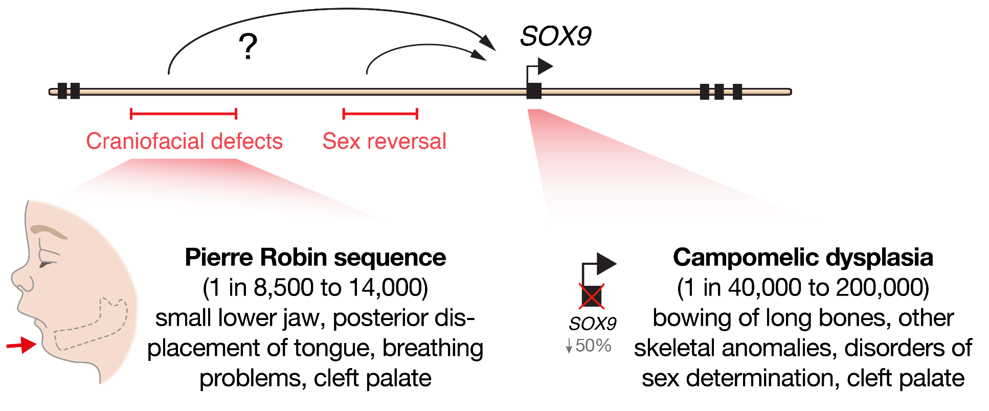
 (8 votes)