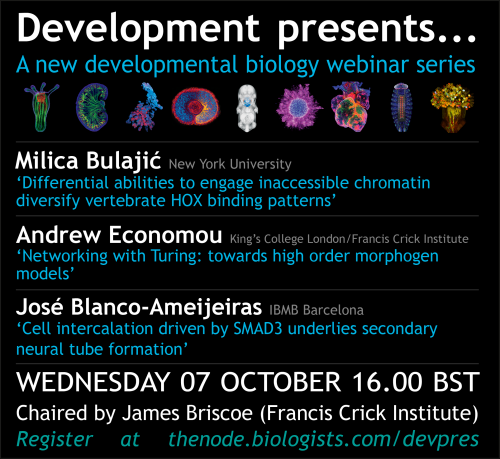
The community site for and by
developmental and stem cell biologists

The people behind the papers – Charles Sheehan, John McMahon and Debby Silver

Posted by , on 27 May 2020

This interview, the 76th in our series, was published in Development earlier this year


Interneurons are crucial to cortical function and their dysregulation has been implicated in various neurological pathologies, yet how they are generated during development is still poorly understood. A new paper in Development investigates interneuron neurogenesis in the mouse embryo and its control by Magoh, a component of the exon junction complex. We heard more about the work from the paper’s two first authors, Charles Sheehan and John McMahon, and their supervisor Debby Silver, Associate Professor at Duke University Medical Center in Durham, North Carolina.


Debby, Charles and John (L to R)


Debby, can you give us your scientific biography and the questions your lab is trying to answer?

DS I am broadly trained as a developmental neurobiologist. I have had the opportunity to pursue undergraduate research on the biochemistry of the cytoskeleton, PhD studies on cell migration in Drosophila, and postdoctoral research on neural development of the cerebral cortex and neural crest in mice. I started my independent lab at Duke Medical Center in 2010, where I am now an Associate Professor. The overall goal of my lab is to elucidate fundamental principles governing brain development and contributing to neurodevelopmental pathologies. We specifically investigate development of the embryonic cerebral cortex. A main focus is to understand post-transcriptional control, a fascinating but poorly understood regulatory layer of brain development. We have several questions of interest, including how RNA-binding proteins control cortical development at a cellular and molecular level, and the role of subcellular RNA localization and local translation in neural progenitors. We are also fascinated to understand the genetic underpinnings of human cortical evolution. We are guided by the premise that the same mechanisms at play during normal development were co-opted during evolution and, when dysregulated, can cause neurodevelopmental disease.


John and Charles, how did you come to work with Debby and what drives your research today?

JM I have always been driven to apply science to gain a better understanding of disorders, with the goal of ultimately developing novel therapies to treat a clinical need. After finishing my graduate studies, I came across Debby’s lab, which was doing fascinating work and working on very translatable science.

CS I first learned about Dr Silver’s work when I was applying for Research Technician jobs in 2017. When I interviewed with her, there was a genuine shared excitement for science that convinced me to join her lab.


What first got you interested in the role of post-transcriptional regulation in interneuron development?

JM Interneurons have been of interest to me since my graduate work on epilepsy. In my opinion they have been a largely understudied and underappreciated cell type, especially given their prominent role in numerous disorders. Likewise, there are significant gaps in our understanding of post-transcriptional regulation in both ‘normal’ and disease states. Therefore, I found exploring the role of the exon junction complex in interneuron development a particularly exciting project to work on.

DS Development of cortical inhibitory neurons is essential for establishing brain circuitry and is relevant for the etiology of many neurological disorders. Yet in contrast to the field of excitatory neurogenesis, and despite a lot of excellent work being done, I have been struck by the fundamental gaps in our understanding of the genesis of interneurons. For example, we lack a basic understanding of the role of the cell cycle in directing interneuron fates, as well as how RNA regulatory modules shape development. Given our lab’s prior work on Magoh’s role in the cortex, I felt we were in a unique position to make contributions to this field and address these knowledge gaps. I was also motivated to pursue this line of research given the increasing human genetic data linking the exon junction complex to neurodevelopmental pathologies.


Can you give us the key results of the paper in a paragraph?

JM The findings of the paper center around the integral role of Magoh in interneuron progenitors. We find absence of Magoh is detrimental to the balanced production of progeny in neural stem cells. We identify that a contributing factor to the pathogenic brain development is the activation of the p53 pathway. These findings raise important questions about the role of post-transcriptional regulation in neurogenesis.


Immunofluorescence depicting the embryonic brain and progenitors (green) of the ganglionic eminence giving rise to newborn interneurons (red) that migrate into the developing cerebral cortex


What do you think explains the difference in the effect of p53 loss in Magoh heterozygotes compared with homozygotes?

JM I think it really comes down to a dose-dependent function of Magoh, and likely the exon junction complex. We have seen dose dependence in a number of our conditional mutants, which points toward a criticality in the balance of EJC components. If a cell loses some Magoh expression, it seems to drive a less severe phenotype. A more extensive depletion results in a striking amount of cell death. My suspicion is that there is a fundamental role of the EJC components in mitosis, which we have yet to fully uncover.

CS We believe that p53 loss differentially affects Magoh heterozygotes and homozygotes because of the severity of their mitotic defects. Simply put, p53 deletion cannot rescue daughter cell death if the progenitors can’t divide. How Magoh dosage causes these differential phenotypes via transcriptome alteration and the effect that p53 loss on has on these changes would be of interest to understand.

DS What we discovered in this study, as well as in prior research, is that cells undergoing mitosis are exquisitely sensitive to Magoh levels. Losing a single copy of this gene causes progenitors to be delayed in mitosis, resulting in inappropriate activation of p53 signaling in newborn progeny, likely due to accumulating DNA damage. We postulate that in Magoh homozygotes, interneuron loss is caused by two defects: p53-dependent apoptosis of newborn cells and p53-independent depletion/death of precursors.


What does your study suggest about the role of EJC components in neurodevelopmental pathologies?

JM This study, along with others from the Silver lab, highlight that EJC components are absolutely necessary for proper function of neural stem cells, likely serving a critical role in mitosis.

CS This work suggests that neurodevelopmental pathologies caused by EJC mutations may not be caused solely by dysfunction of excitatory progenitors, but also interneuron progenitors. This finding gives a more complete understanding on how mutations in Magoh, and other EJC components, alter neural stem cell function and cause neurodevelopmental disorders.

DS Copy number variations and point mutations in EJC components are linked to intellectual disability, autism and microcephaly. Our study, together with prior work from our lab, suggests that mutations in EJC components disrupt proper generation and survival of both excitatory and inhibitory neurons. It may be that in patients carrying EJC mutations, there is imbalanced production and/or survival of these two populations or there may be qualitative aberrant defects in viable interneurons. Historically, microcephaly is linked to abnormal mitosis, with a heavy emphasis on cortical neurogenesis. Our study indicates that mutations affecting mitosis of interneuron progenitors may also be relevant for the etiology of microcephaly.


When doing the research, did you have any particular result or eureka moment that has stuck with you?

JM After a session of live imaging, reviewing the videos always provided me with tremendous insight into the dynamics of neural progenitors and cell divisions. Not necessarily a eureka moment, but very insightful and absolutely beautiful to watch.

CS I think the most memorable eureka moment was when I was imaging the p53 rescue brains. Immediately when looking at the cKO, you could tell that there was still a drastic loss of cortical interneurons, which was unexpected based on what we knew at the time.


Many who have been at the bench for a while will tell you science is not for the weak hearted


And what about the flipside: any moments of frustration or despair?

JM I think many who have been at the bench for a while will tell you science is not for the weak hearted. From what I have seen, even the best scientists suffer more failures than successes. There were plenty of late-night experiments that were revealed to be pure failures once the results were in, and plenty of time spent trouble-shooting technical issues before seeing the light at the end of the tunnel.

CS Similar to the eureka moment, the p53 KO rescue experiments were quite challenging. The mouse genetics required to obtain the samples was a difficult and long process. Even once we had the data, interpreting how p53 was functioning took several discussions and looking back to other data until we reached on our current model.


So what next for you two after this paper?

JM Since my time in Debby’s lab I have been working in regulatory affairs, and furthering my understanding of how a concept grows from an idea, to the bench, to a clinical trial, to a marketed therapy.

CS I have recently started my PhD at Duke in Cell and Molecular Biology and am currently trying to decide on a lab and mentor for this next step.

Where will this work take the Silver lab?

DS We would like to better understand the molecular mechanisms by which Magoh dosage impairs interneuron development, including its role in splicing and translation and its direct targets, as well as discriminating why Magoh loss causes such a striking mitotic defect. It is also exciting to consider whether all EJC components act similarly in development, and how the EJC fits into a larger network as a master regulator of corticogenesis. Beyond the EJC, these studies have sparked our interest in understanding fundamental aspects of interneuron development and how genetic modifications over the course of evolution have shaped this process.


Finally, let’s move outside the lab – what do you like to do in your spare time in Durham?

JM Hiking in Eno park or exploring a new brewery.

CS Durham has a lot of great local breweries, restaurants and concerts, so I spend most of my free time exploring those with friends.

DS I spend my free time mainly with my husband, two kids and our new doggie. We enjoy the outdoors, including mountain biking in our backyard, as well as the great food and music scenes in Durham and Carrboro.


Thumbs up (No Ratings Yet)
Loading...

Tags: , , ,
Categories: Interview

Navigate the archive

Use our Advanced Search tool to search and filter posts by date, category, tags and authors.

PhD Studentship in Cell and Developmental Biology: Cytoskeletal Regulation of Epidermal Stem Cells

Posted by , on 25 May 2020

Closing Date: 15 March 2021

The group of Chen Luxenburg at the Faculty of Medicine, Tel Aviv University, invites applications for a PhD student position.


Our laboratory is looking for excellent and highly motivated PhD students to study the role of the actin cytoskeleton in skin development. Our goal is to understand how cytoskeleton derived signals regulate stem cell ability to create the skin epidermis during development, maintain it in the adult, and repair it upon wounding. On top of molecular biology, tissue culture, advanced microscopy, and mouse work, we utilize state of the art technology that allows us to rapidly manipulate the function of any gene of interest in epidermal stem cells in utero. Several exciting projects are available for successful candidates.


We offer state of the art laboratory with a dynamic and international atmosphere and full financial support (tuition and stipend)


Candidates should hold a Master’s degree in Biology/Life-sciences or related fields.


For more details about our lab: www.luxenburglab.com


Interested candidates should email their CV and a brief paragraph describing their research experience and career plans to Chen Luxenburg  (lux@tauex.tau.ac.il)


Thumbs up (No Ratings Yet)
Loading...

Tags: , ,
Categories: Careers, Education, Jobs, Research

Research Highlight #3: IL-1 regulates haematopoietic stem cell pool size via PU.1

Posted by , on 25 May 2020

Today, we move to mouse adult haematopoiesis, with an elegant work on inflammation and stem cell biology. As always, feedback is more than welcome!

Chronic inflammation is one of the classic events underlying aging and infection response. When associated with loss of quiescence in haematopoietic stem cells (HSCs), it is commonly the initiating event in leukemia progression. Chavez and colleagues present the impact of chronic interleukin-1 (IL-1) exposure on the most immature HSCs. First of all, they exposed adult mice to IL-1β for 20 days, observing major changes in the HSCs transcriptome. In particular, IL-1β treatment reduced the expression of genetic programs associated with cell cycle progression and protein synthesis. In vitro short-term culture of the most immature HSCs (called LT-HSC), in the presence of IL-1β, confirmed a slower cell cycle entry. Using a genetic reporter for the myeloid master regulator Pu.1, the authors proved that IL-1 is directly linked to the transcription factor increased activity. Specifically, PU.1 is able to bind more than two-thirds of the genes downregulated by IL-1β exposure. Finally, the authors used a genetic mouse model for reduced Pu.1 activity, proving elegantly that an inefficient PU.1 opens the way to the aberrant increase in LT-HSCs cell cycle activity and protein synthesis, leading to an increased cellular pool of stem cells.

Taken altogether, those results describe an IL-1 – PU.1 system that physiologically contributes to stabilizing the number of LT-HSCs. Those results are relevant, because IL-1β prematurely initiate myeloid differentiation, and in general promote the expansion of white blood cells. How to conciliate therefore those apparently contradicting results? Here, the authors focused on a smaller subset of HSCs, and affirm that PU.1 in these cells acts as an activation barrier against unwanted proliferation, while still able to prime HSCs for myeloid differentiation. It will be interesting to assess if other signals could activate similar pool size regulation mechanisms for HSCs.


Chavez J et al. “PU.1 enforces quiescence and limits hematopoietic stem cell expansion during inflammatory stress”

Doi: https://doi.org/10.1101/2020.05.18.102830

Thumbs up (3 votes)
Loading...

Categories: Highlights, News, Research, Uncategorized

Postdoctoral position in nervous system development and function – USF Tampa

Posted by , on 21 May 2020

Closing Date: 15 March 2021

The group of Dr. Krishna Bhat at University of South Florida, Tampa, is searching for postdoc candidates interested in our areas of research:


  1. Determining the genetic basis for the self-renewing stem cell and terminal asymmetric divisions of neural precursor cells
  2. Guidance of longitudinal and commissural axon tracts
  3. Adult brain development, function, and neurodegenerative diseases.


Please refer to Manavalan et al, Science Signaling 2017 and KM Bhat, Plos Genetics 2017 for recent research.


Contact us if you have a PhD in cellular/molecular biology and have expertise working with Drosophila genetics.


To apply, email a letter of intent and a CV to Jorge Dominguez ( jdominguezba [at] mail.usf.edu ).

Thumbs up (No Ratings Yet)
Loading...

Categories: Jobs

Staying connected during the COVID19 pandemic

Posted by , on 21 May 2020

Among many other (many more important) things, pandemic times have meant an end to travel, which for the Development team means no conferences and no lab visits. For me personally, it’s meant cancelled trips to Shanghai, Kyoto, Washington and Chicago, and while I’m delighted to be keeping my carbon footprint down, I’m definitely missing the opportunity to get out and meet the developmental biology community – and hear about your latest work.

I do think, and hope, that this travel hiatus will lead to long-term changes in the way we do conferences – and a more sustainable and eco-friendly conference culture (as discussed in this recent preprint, preLight and Node post). But I also believe that nothing beats face-to-face meetings, and I’m looking forward to getting back on the road once it’s safe and practical to do so. In the meantime, we at Development have been thinking about ways to connect with and support the community while we’re all stuck at home. As James Briscoe wrote in his recent update, we’re always happy to hear from you. So if you’ve got a paper you might be interested in submitting to Development, would like to discuss an idea for a possible review, or just want to talk about the journal or publishing more generally, please get in touch. You can drop us an email and, if it’d be helpful, we can try and set up a phone or video call.

We’ve also been spending some time giving (hopefully!) useful tips to the community here on the Node: if you’ve not read them already, check out Seema’s recent post on writing reviews, and Alex’s piece on how to get involved in peer review. If there’s something you’d like us to write about, do let us know and we’ll see what we can do. But we’d like to be more interactive – we’d normally be out and about giving talks at institutes or panel discussions at conferences, so are thinking about ways of replicating some of this online.

I’m sure many of you already have far too many Zoom calls and webinars to attend, but would you be interested in joining us for webinars on various aspects of publishing? We’ve set up a brief survey for you to indicate your interest and to let us know what you’d be most keen to hear about. Please do fill it in if you’d like to get involved and we’ll try and set something up. We’re also considering the possibility of hosting a series of scientific webinars, perhaps featuring authors of Development papers – again, please let us know in the survey if you would be interested in a devbio webinar series. Finally, we’re beginning to think about whether we could replace in-person lab visits and institute seminars with virtual equivalents – we’ve got limited capacity to do this, but if you’re interested in having one of the Development team ‘visit’ your institute, let me know!

One of the joys of working in this field is the wonderful community of developmental biologists. So while we can’t meet in person, I hope we can continue to connect in other ways – and if you’ve got thoughts about other things we should be doing to help with this, I’m all ears…!

Stay well and stay safe, and see you on the other side…

Thumbs up (3 votes)
Loading...

Categories: Events, News

Genetics Unzipped podcast: From one generation to the next – the life and work of Dame Anne McLaren

Posted by , on 21 May 2020

Anne McLaren
Anne McLaren – photo courtesy of The Gurdon Institute

In the latest episode of Genetics Unzipped, Kat Arney reflects on the life and work of Dame Anne McLaren, one of the leading embryologists of the 20th century, whose work underpinned the development of the in vitro fertilisation (IVF) techniques responsible for bringing millions of ‘test tube babies’ into the world, and more besides.

Not bad for someone who ended up studying biology at Oxford University almost by accident. Apparently she picked the course because cramming for the zoology entrance exam seemed like an easier option than doing the required reading for English literature.

Anne’s curiosity about early mammalian embryos was the fire that fuelled a lifetime of research. Her groundbreaking work in the 1950s laid the foundations for in-vitro fertilisation, cloning and genetically engineered mice — technologies that have revolutionised human reproduction and biomedical research.

Later in life, she turned her attention to what she used to call “the most fascinating and deeply mysterious cells of all” — the germ cells that will become eggs and sperm in an adult, which are specified just a few days after fertilisation. Anne devoted the rest of her research career to pinning down the characteristics of these unique cells, trying to understand where they came from, where they were going, and what made them so special.

As well as being a highly respected scientist, Anne was also deeply involved in the scientific community. She was the first woman to serve as an officer of the prestigious (not to mention male dominated) Royal Society, and also sat on the Warnock Committee – the fore-runner of the Human Fertilisation and embryology Authority.

She was also a role model and mentor to countless scientists, many of them women working hard to establish a career in a male-dominated world. Anne occasionally complained about the “old boy’s network”, which she felt sometimes led to men only putting forward male friends for jobs. But she did note wryly that there seemed to be an “old women’s network” developing, at least in her field of developmental biology, which was helping to even up the balance.

As she wrote in an eloquent review paper on genetic inheritance, “history may be circular, but the history of science is helical: it repeats itself, but each time at a deeper level”. Rather than the Newtonian idea of standing on the shoulders of giants, Anne saw scientists as forming a twisted helix through time, intertwining as we pass on skills, knowledge and friendship to those who come up behind us.

Anne McLaren’s influence on the world of reproductive science and medicine corkscrews deeply back in time, and legacy will stretch for years to come in the lives of those she knew, those who knew her work, and the many, many more who benefit from it.

This story was first published in the book A Passion for Science: Stories of discovery and invention, which is packed with 20 stories about amazing women in science and is available to download as an ebook for just £1.99.

Genetics Unzipped is the podcast from The Genetics Society. Full transcript, links and references available online at GeneticsUnzipped.com

Subscribe from Apple podcasts/iTunes, Spotify and all good podcast apps to make sure you get the latest episodes and catch up on our back catalogue.

If you enjoy the show, please do rate and review on Apple podcasts and help to spread the word on social media. And you can always send feedback and suggestions for future episodes and guests to podcast@geneticsunzipped.com Follow us on Twitter – @geneticsunzip


Thumbs up (No Ratings Yet)
Loading...

Tags: , , , ,
Categories: Podcast, Resources, Societies

Getting involved in peer review

Posted by , on 19 May 2020

In pre-COVID19 times, back when conferences happened largely in person, early-career researchers (ECRs) often asked me how they could get more direct invitations to be a reviewer. Peer review is a crucial part of the publishing ecosystem and therefore it’s not uncommon for group leaders to invite members of their lab to review articles with them as part of their academic training. However, direct invitations to review mark a point at which trainees begin feel established in the field (along with moves to start their own group) and can make a useful contribution to a CV. During the current disruption, some reviewers might be feeling overburdened, and editors might be looking to expand their pool of reviewers. Here, I’ve provided a short guide with tips on how ECRs can make themselves known to editors with the hope of receiving direct invitations to be a reviewer. This post does not prescribe what makes a “good” reviewer, nor does it provide training on how to write a peer review report, but you can read advice from Elsevier’s Reviewer Hub or participate in training programmes from Publons Academy, Society for Neuroscience and Genetics Society of America.

So, to stand a chance at being invited to review a manuscript, editors need to know just two things: (1) who you are and (2) your scientific expertise. Editors can’t invite reviewers they don’t know exist or can’t find! Each editor will have a different strategy for finding potential reviewers, and these approaches might vary between professional vs. academic editors. Ideally, an editor aims for three reviewers with complementary expertise to cover the major topics in the manuscript. At Development (and presumably other journals), we also consider aspects of diversity, including career stage, geographical location and gender – see this Editorial by James Briscoe and Katherine Brown. But where do editors look for inspiration?

Author suggested reviewers
Most manuscript submission systems allow authors to suggest (or exclude) particular researchers for reviewing their manuscript. Editors will check that these suggestions are sensible, and that the suggested reviewers have the right expertise. It’s also important that there is no conflict of interest between parties, which could be negative (e.g. a direct competitor) or positive (e.g. a previous member of the lab or a recent collaborator). Note that editors are unlikely to only use reviewers suggested by the author, and it is up to the editor who they decide to invite to review a manuscript, although we do respect exclusions.

Reference list
Editors might also look through the references cited in the manuscript to see whether it heavily features the work of another group or – if they know the field well – whether certain relevant publications are missing. Authors of these publications might be invited to review the manuscript.

Journal databases
Most journals have databases containing researchers, expertise terms and records of previous reviews. These records might detail the number of reviewers a researcher has accepted (or declined), the length of time they took to complete their report, previous reports and, in some cases, a ranking system where the editor can comment on the quality of the report.

Talks, seminars, conferences and meetings
For editors, talks and conferences are a great way to meet new researchers. They also provide a broader view of the field as a whole, highlighting areas of debate, uncertainty or controversy, which is useful to know when selecting balanced reviewers.

Publications
Editors might be inspired by other articles they’ve handled recently and look to the authors of those articles to review new work. In addition, they might get ideas from publications in other journals within the field or through keyword searches of publication databases such as PubMed.

Google search
A Google search using key terms can be a useful way to widen a reviewer pool. Such key terms might be the subject area, a particular technology, specific gene name etc. Editors will explore group and departmental websites to have a clear understanding of the group’s research focus, background and publications.

Social media
Social media might be another way for editors to meet reviewers, although this may lead to bias towards particularly vocal scientists. I (and others) have also used the Node Network to find reviewers – read more about the Node Network in this announcement.

Expert advice
Usually, if an invited reviewer is unable to review an article, there is the option for them to suggest alternative researchers and it’s really helpful for them to do so! Editors might also seek the advice of their Advisory Editorial Board, either directly or for reviewer suggestions.

Editor expertise
Particularly experienced editors will have a thorough understanding of the community in their field and will be able to choose reviewers based on their own knowledge of everything above.

So, with these things in mind, here are three tips for how you can expand your own profile:

1. Get involved: Participate, publish, present and be pro-active

Participate in co-reviewing manuscripts with mentor or group leader and ask them to provide your name to the journal to acknowledge your contribution (see “get credit”’ below). If you’ve left the lab and your former mentor or group leader is unable to review a manuscript, you can ask that they suggest that the editor contact you in their place. Some publishers also have the option to “volunteer” to review, such as Elsevier’s “VolunPeer” initiative and some individual journals – such as eLife, STAR protocols, Stem Cell Research and PeerJ journals – have opportunities to sign up.

Your publication record is the primary way you can demonstrate your research interests and expertise. Although this obviously isn’t the main incentive for publishing, each publication has the added benefit of exposing your name to editors and it might get you inside a journal database.

Try to present your research at conferences, either through a talk or a poster, so that editors become familiar with you and your work. You can also use social media to disseminate your research to a wider audience or talk about subjects that interest you.

You can also be pro-active by approaching editors at meetings and conferences to talk about your work and your interests – when doing so make sure that the editors are left with a clear idea of the topics you cover in your research. Along similar lines, you can email journals and editors to ask to be added to their reviewer database – make sure to also include a list of key terms for your expertise and keep this profile up-to-date (see “get up-to-date” below). The Node Network has been set up specifically for developmental and stem cell biologists with the aim of finding speakers and reviewers that would not normally come to mind – add yourself! Other initiates also exist, such as the GoogleDoc mentioned below.

2. Get up-to-date

Ask that your institution or departmental page is kept up-to-date, easy to find and easy to navigate, or include a link to your professional/group website that you can keep on top of it yourself. Make sure that within your page you include a list of your recent publications and keep this updated. Also have a page or paragraph that outlines your research interests as specifically as possible; details of any interdisciplinary research, as well as the model organisms or technologies that your group utilises, are also useful for editors to know.

Keep your profiles on various databases and directories (such as the Node Network, Google Scholar, ORCID and ResearchGate) up-to-date with your current institution, email address and expertise. If old information is in a database, then you might miss out on an invitation; although an editor should check for that your most recent contact information is correct before they get in touch! Again, an accurate departmental website can be crucial for editors to know old email addresses from new ones.

3. Get credit

As mentioned above, if you have participated in peer review with your mentor or group leader, consider asking them to provide you with some credit. At Development, we encourage lab members to contribute as co-reviewers and have a specific part of the report where these lab members are named. The names provided here are still withheld from the authors, but the journal will contact to the co-reviewer and invite them to join the journal’s database.

You can also receive credit through initiatives that track peer review activity, such as Publons and ORCID. Some publishers, such as Elsevier, also have reviewer recognition platforms, which allow editors to acknowledge reviewers that provide particularly useful reports with a “certificate of excellence”.

Finally – and perhaps more controversially – you could consider signing the report if the journal allows you to do so. There are various arguments for and against waiving anonymity, but if the authors felt that your report was particularly constructive they might suggest you to review future manuscripts. In journals where peer review reports are published alongside the article, the recognition might go even further. Ultimately, you should do what make you comfortable and therefore allows you to produce the most constructive report possible.

I’d like to finish by saying thank you to all the researchers that participate in peer review, especially those who have taken the time to review manuscripts for Development. Although the publishing landscape is changing, and there are important discussions to be had around this, peer reviewers continue to be an essential part of moving science forward.

Thumbs up (10 votes)
Loading...

Tags: , , ,
Categories: Careers, Discussion, Resources

Last chance to take our user survey

Posted by , on 19 May 2020

We’ve had a great response so far to our tenth birthday user survey, which we hope will guide us going forwards and give us ideas for how we can better serve the developmental biology and stem cell community. If you haven’t taken it yet, it closes Thursday midday BST. Even if you’re a less-than-occasional reader, we’d love to have your input:


https://www.surveymonkey.co.uk/r/theNode2020


Thumbs up (No Ratings Yet)
Loading...

Categories: News

Research Highlight #2: Blood stem cells heterogeneity arises from accessible chromatin

Posted by , on 18 May 2020

Hi there! Today’s highlight is focusing on a major question for the haematology community: what is causing blood stem cell heterogeneity? Please do not hesitate to let me know your thoughts in the comments here or on Twitter (@BioRugby)!


Highlight #2: Chromatin accessibility is the main source of heterogeneity in foetal haematopoiesis

We have a limited understanding of the haematopoiesis in the human foetus. Particularly, it is unclear whether stem cell differentiation depends on transcriptional priming (lineage-specific transcription factors) or a broader epigenetic one. This preprint delves into this question, presenting a single-cell resolution study of foetal human blood stem and progenitor cells. The authors analysed the transcriptome and the chromatin accessibility of over 8000 cells from different organs, confirming the high heterogeneity of the stem cell compartment. They inferred some differentiation trajectories, although the most-well know transcription factors were not consistently identified. In addition, the authors combined the datasets, and with the exception of the progenitors linked to red blood cells and platelets, they have been unable to unequivocally associate chromatin accessibility to defined transcriptional signatures. Therefore, they concluded that stem cells in the developing foetus are minimally primed towards differentiation, and this priming is not due to specific transcriptional programs associated to mature cell types. Finally, they compared different organs, confirming the higher cycling tendency of stem cells from foetal liver, compared to bone marrow. In the future, it will be interesting to garner a better understanding of the signals that trigger differences between haematopoietic sites.


Ranzoni AM, Tangherloni A et al. “Integrative Single-cell RNA-Seq and ATAC-Seq Analysis of Human Foetal Liver and Bone Marrow Haematopoiesis”
doi: https://doi.org/10.1101/2020.05.06.080259

Thumbs up (No Ratings Yet)
Loading...

Tags: , , , ,
Categories: Highlights, Research, Uncategorized

Cilia, Reissner Fiber and Crooked Spines

Posted by , on 18 May 2020

Perhaps the most exciting aspect of attending any scientific meeting is the privilege of becoming aware of novel research findings in our fields of interest, prior to their appearance in published literature – and this begins as soon as we have the abstract book in hand! Sitting in my hotel room in Suzhou, and browsing through the abstracts of the first Cold Spring Harbor meeting on cilia and centrosomes which was held in the spring of 2017, I chanced upon work from Chengtian Zhao’s group (Ocean University, Qindao, China) that described the analysis of gene expression changes in zebrafish embryos deficient in zymnd10, a gene encoding a ciliary dynein assembly factor (1). Since my lab had also performed similar analyses with two other cilia mutants, ccdc103 and lrrc50 (also encoding dynein assembly factors (2,3)), I decided that I must meet up with Chengtian and discuss whether we should collaborate or at the least, co-ordinate our studies. That evening, as I settled down for a sumptuous dinner in the esteemed company of two prominent cilia researchers, Hiroshi Hamada and Cecilia Lo, I was pleasantly surprised when a Chinese gentleman came forward and introduced himself to me as Chengtian Zhao, and asked if he could join us for the meal. Conversation flowed effortlessly and the rest, as they say, is history.

Over the years, a number of studies had observed that zebrafish embryos with mutations in many different cilia genes exhibit ventrally curved body axis. More recently, work from the Burdine and Ciruna labs had implicated defects in cilia motility, causing abnormal cerebrospinal fluid (CSF) flow, in curvatures of the vertebral column in adult zebrafish (4), making this system a good model for uncovering the etiological basis of the common human spine disorder idiopathic scoliosis (IS) that affects up to 3% of children and adolescents world-wide (5). Abnormal 3D curvatures of the spine in IS can cause a considerable degree of morbidity in patients, manifest in symptoms like difficulties in breathing, postural issues and gait problems. Despite these important findings, the molecular mechanism operating downstream of cilia-driven CSF flow that ensures proper spine development had remained unclear. With the collaboration that sparked off at the dinner table of the Suzhou meeting, Chengtian’s and my lab published a collaborative paper just before Christmas of 2018, describing a possible molecular pathway by which ciliary motility, within the brain ventricles and spinal canal of the zebrafish, ensures that they develop with a straight body axis (6). We showed that cilia beating transports catecholamines (like epinephrine) in CSF, and they stimulate the expression of Urotensin-related peptides (Urp) in CSF-contacting neurons (CSF-cNs) that differentiate along the spinal canal. The Urp family consists of small cyclic neuropeptides, homologous to Urotensin II, which is known to function as a potent vasoconstrictor (7). Indeed, it was the significant decline in expression levels of the urp genes in our respective gene expression analyses of cilia mutants that prompted us to follow up on Urp signalling to uncover the mechanism acting downstream of cilia-driven CSF flow. Our current model posits that Urp peptides, secreted from CSF-cNS, brings about a certain degree of tension or contraction of the somitic slow-twitch muscles of the dorsal somites, and this is the biomechanical force for axial straightening. In keeping with this view, we also found that a Urp receptor, Uts2r3, a G-protein coupled receptor, is expressed in the dorsal slow muscles, and when it was rendered non-functional using a targeted lesion at the locus, the adult uts2r3 mutants developed with severely curved spines, resembling humans with IS (6).

More or less concurrent with our publication, Claire Wyart, Pascal Bardet and colleagues reported a very intriguing story that ascribed, for the first time, a definitive biological role for the enigmatic Reissner fiber (RF) (8). Discovered in the 19th century by the German anatomist Ernst Reissner, RF is a filamentous structure that remains suspended in CSF along the brain ventricles and the spinal canal in all vertebrates examined, including humans. RF is polymerized from a large glycoprotein, SCO-spondin (Sspo), secreted from the subcommissural organ (hence SCO) and the floor plate. RF is thought to participate in many functions of the nervous system, but the lack of genetic mutations in Sspo has precluded a firm association of RF with any of these proposed roles. Wyart et al. showed that in zebrafish embryos, RF is required for proper axial development since sspo mutants developed ventrally curved body axes (and perished at the early larval stage), closely mimicking the curved bodies of cilia mutants (8). They also demonstrated that cilia motility is required for RF biogenesis, by somehow facilitating the polymerization of the protein into the fiber. Since biochemical studies with mammalian RF had already shown that it can efficiently bind and facilitate the transport of catecholamines present in CSF (9), it dawned on me that cilia, RF, and CSF catecholamines could all be functioning via Urp signaling from CSF-cNs.

The idea that I wanted to explore is whether sspo mutants develop curved bodies because in the absence of RF, they are unable to bind CSF catecholamines and present to CSF-cNs to activate urp gene expression. It is with this view in mind that we approached Claire and Pascal, who most generously shared with us the sspo mutants. In a paper that we have just published in Biology Open (10), we now show that consistent with our expectations, sspo mutants exhibit a loss of urp gene expression from CSF-cNs: urp1 is significantly reduced, while urp2 is almost completely absent. As we had demonstrated previously for cilia mutants, culturing sspo mutants in the presence of exogenously added epinephrine in embryo medium, restored urp expression in CSF-cNs and also rescued their ventrally curved axial defects. However, one issue that continued to confound us was whether abnormalities of the embryonic axis in sspo mutants have any connection with scoliosis of the spine in adult zebrafish. If there is, our work will have relevance for furthering our understanding of the etiology of IS. The Burdine and Ciruna et al. paper, that had initially linked cilia motility and CSF flow with spine curvature, used a clever strategy to bypass the severe embryonic body curvature and associated lethality of the cilia mutants that they studied (4). They injected the corresponding (in vitro synthesized) wild-type sense mRNA into mutant eggs: this rescued the axial defects as well as lethality, and the mutants developed into adults but they exhibited severely curved spines. Unfortunately, we could not utilize this strategy to rescue sspo mutants as the Sspo protein consists of more than 5000 amino acids, and in vitro synthesis of an mRNA to encode such a large protein is not feasible. To circumvent this problem, we prematurely dechorionated the sspo mutants with the hope that their severe ventrally curved axis could be partially rescued when freed off the confines of the spherical, non-elastic chorion. Indeed, this simple trick ameliorated the strong axial curvature of sspo mutants to varying degrees, and many of them matured into adults with severe spine malformations reminiscent of cilia mutants rescued of their embryonic lethality and mutations in the Urp receptor. Thus, in the zebrafish, ventral curvature of the embryonic axis and scoliotic malformations of the adult spine represent linked morphogenetic anomalies. Most satisfyingly, we found that restoring expression of Urp2 exclusively in CSF-cNs, rescued not only the embryonic and larval body curvature of sspo mutants, but also allowed one such mutant to develop into an adult with an apparently normal spine! Additional findings that we report in our Biology Open paper include data showing that it is a definite threshold of Urp signaling that is critical for the morphogenesis of a straight body axis. Too little signaling causes ventral curvature as in sspo and cilia mutants, while exaggerated signaling (for instance, by over-expression of the urp gene or the protein in the muscle cells themselves) causes the converse effect of profound dorsal curvature of the axis. Finally, using mutations in Smoothened (Smo), an essential component of the Hedgehog pathway that directs slow-twitch muscle cell differentiation in the zebrafish somites, we could show that lack of the slow muscles make these mutants refractory to Urp signaling (10). Smo mutants have strong ventrally curved bodies that could not be rescued by over-expression of the Urp proteins.


Image of an sspo mutant adult zebrafish showing curvatures of the trunk and tail (top panel). Micro-CT scan of an sspo mutant revealing 3D curvatures of the vertebral column, resembling spines of IS patients.


How do we take these findings forward, especially for furthering our understanding of the mechanistic basis of pathogenesis in IS? First, with reference to the zebrafish, we need to better understand how Urp signaling-induces activity of the slow-twitch fibers of the somites, and how this activity feeds back to the growing spine to ensure that it develops along a straight axis. Secondly, while RF and CSF-cNs exist in mammals and Urp signaling has also been shown to operate there, we will need to examine whether the circuitry that we have been able to dissect in the zebrafish is also conserved. In this regard, one caveat that we need to bear in mind is that traditional experimental mammals like mice and rats are quadrupeds, and they have not turned out to be effective for modeling human spine disorders (hence the promise of the zebrafish)(11). Finally, based on what we glean from all of these investigations, we can begin to parse the underlying mechanisms driving spine malformations in IS. There is already accumulating evidence that ciliary dysfunction could be causative of the disease (12,13). Moreover, presence of RF has been reported in human embryos and a teenager (14), implying that defects in this structure could be responsible for IS in some of the individuals afflicted with IS. And of course, the real benefit of all this research effort will be if we can invent effective therapeutic strategies for IS by pharmacologically manipulating the Urp pathway, since current treatment options are largely limited to managing the disorder with physiotherapy and braces, and in severe cases, the rectification of severe spine deformities with invasive surgery.


References

1. ZMYND10 functions in a chaperone relay during axonemal dynein assembly. Mali GR et al. Elife 2018 7. pii: e34389.
2. CCDC103 mutations cause primary ciliary dyskinesia by disrupting assembly of ciliary dynein arms. Panizzi JR et al. Nat. Genet. 2012 44:714-9.
3. Deletions and point mutations of LRRC50 cause primary ciliary dyskinesia due to dynein arm defects. Loges NT et al. Am. J. Hum. Genet. 2009 85(6):883-9.
4. Zebrafish models of idiopathic scoliosis link cerebrospinal fluid flow defects to spine curvature. Grimes DT et al. Science 2016 352:1341-4.
5. Genetics and pathogenesis of idiopathic scoliosis. Grauers A et al. Scoliosis Spinal Disord. 2016 11:45.
6. Cilia-driven cerebrospinal fluid flow directs expression of urotensin neuropeptides to straighten the vertebrate body axis. Zhang X et al. Nat. Genet. 2018 50:1666-73.
7. International Union of Basic and Clinical Pharmacology. XCII. Urotensin II, urotensin II-related peptide, and their receptor: from structure to function. Vaudry H et al. Pharmacol. Rev. 2015 67:214-58.
8. The Reissner fiber in the cerebrospinal fluid controls morphogenesis of the body axis. Cantaut-Belarif Y et al. Curr. Biol. 2018 28:2479-86.
9. Reissner fiber binds and transports away monoamines present in the cerebrospinal fluid. Caprile T et al. Brain Res. Mol. Brain Res. 2003 110:177-92.
10. Reissner fiber-induced Urotensin signaling from cerebrospinal fluid-contacting neurons prevents scoliosis of the vertebrate spine. Lu et al. Biol. Open 2020 9:pii: bio052027.
11. Understanding idiopathic scoliosis: A new zebrafish school of thought. Boswell CW and Ciruna B. Trends Genet. 2017 33:183-96.
12. Functional variants of POC5 identified in patients with idiopathic scoliosis. Patten SA et al. J. Clin. Invest. 2015 125:1124-8.
13. New associations of primary ciliary dyskinesia syndrome. Engesaeth V et al. Pediatr Pulmonol. 1993 16:9-12.
14. Reissner’s fibre: the exception which proves the rule, or the devil according Charles Baudelaire? Olry R and Haines DE. J. Hist. Neurosci. 2003 12:73-5.


Sudipto Roy is a Senior Principal Investigator with the Institute of Molecular and Cell Biology, A*STAR, Singapore.


Thumbs up (5 votes)
Loading...

Tags: , , ,
Categories: Research