The Lawton Lab is seeking a talented and motivated graduate student to study the cell and tissue mechanics regulating cerebellar morphogenesis and brain folding.
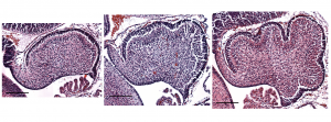
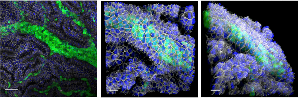
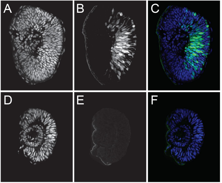
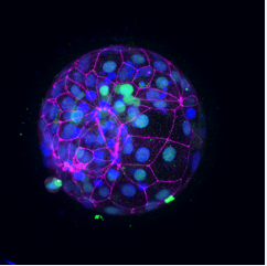
Sagittal midline sections of mouse cerebellum at embryonic day E16.5, E17.5 and E18.5 show the initiation of folding.
The beautiful and robust folds of the human cerebral cortex and the cerebellum increase the synaptic volume and compartmentalize the neural circuits. We have previously shown that the murine cerebellum initiates folding, without a pre-pattern, through differential expansion – between an outer fluid-like layer and an inner solid mass – together with radial and circumferential tension. However, it is not known how the folding amount or pattern is set. Nor is it known how the measured tension is regulated or how the fluidity of the outer layer is maintained. The Lawton Lab uses developmental and mechanical assays, live-imaging, and quantitative analysis to address these issues and to discover the cellular and emergent tissue-level regulation of brain folding.
This project will provide training in classical developmental biology assays, ex vivo slice culture, histology, confocal microscopy, and quantitative analysis.
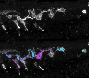
Granule cell precursors labeled with membrane GFP. Cell shapes are masked and quantified
Applying:
Position is to begin fall semester of 2020.
The ideal candidate will have a strong interest in developmental biology, and some experience with microscopy, image analysis, and Matlab. Those with a background in mathematics or physics are also strongly encouraged to apply.
Please send a statement of research interest and a CV with references to alawton@biology.msstate.edu
Apply your developmental biology / molecular biology / neurobiology skills to the problem of brain cancer.
The lab of Dr. Jennifer Chan seeks to recruit a motivated postdoctoral fellow to investigate cell fate decisions during the process of brain tumour development and progression. Research in the lab focuses on growth factor signalling and transcriptional regulation as determinants of neural precursor identity and fate. We use models that include patient-derived glioma cultures, xenografts, and engineered mouse models generated from in utero and postnatal electroporation to address our research questions.
The successful candidate will collaborate with investigators in bioinformatics to apply tools of genomics, epigenomics, and transcriptomics to further define key alterations during neoplastic transformation, and will collaborate with medicinal chemists to determine if identified alterations may be potential therapeutic targets. Early-stage postdocs (within 3 years of receiving PhD or equivalent) with experience in advanced immunohistochemistry, fluorescence microscopy, gene editing, and molecular biology; and/or experience in genomic approaches like RNA-seq, ChIP-seq, ATAC-seq will be preferentially considered.
The position is available immediately.
Other information
Located in Calgary, Alberta, Canada, the Chan Lab is part of the Charbonneau Cancer Institute at the University of Calgary’s Cumming School of Medicine. Within the Charbonneau Institute, we are part vibrant multidisciplinary research groups focused on childhood cancers and brain cancers. Calgary is a very livable and family-friendly city located less than an hour’s drive from the Canadian Rocky Mountains – a haven for outdoor enthusiasts.
Application details
Submit a brief letter of interest, your academic CV, and the names and contact information of at least three references.
Applications should be submitted as a single PDF file and sent as an email with the subject line “Post-Doctoral Fellowship, Glioma Biology” to: jawchan@ucalgary.ca
Welcome to our monthly trawl for developmental biology (and related) preprints.
In recent preprint news, CSHL, which runs bioRxiv, launched Transparent Review in Preprints (TRiP), a new project enabling journals and peer review services to post peer reviews of submitted manuscripts. In linked news EMBO Press and ASAPbio launched Review Commons, a platform that peer-reviews research manuscripts in the life sciences before submission to a journal, and enables authors to publicly post the reviews and their own response to them bioRxiv. Finally, PeerJ Preprints, regular source of preprints for this list, announced it would no longer be accepting new content. So long and thanks for all the preprints!
This month we found a typical wealth and breadth of preprints hosted on bioRxiv, PeerJ, andarXiv. Let us know if we missed anything, and use these links to get to the section you want:
Essential omega-3 fatty acids tune microglial phagocytosis of synaptic elements in the developing brain
C. Madore, Q. Leyrolle, L. Morel, J.C. Delpech, A.D. Greenhalgh, C. Lacabanne, C. Bosch-Bouju, J. Bourel, A. Thomazeau, K.E. Hopperton, S. Beccari, A. Sere, A. Aubert, V. De Smedt-Peyrusse, C. Lecours, K. Bisht, L. Fourgeaud, S. Gregoire, L. Bretillon, N. J. Grant, J. Badaut, P. Gressens, A. Sierra, O. Butovsky, M.E. Tremblay, R.P. Bazinet, C. Joffre, A. Nadjar, S. Layé
STAG2 cohesin is essential for heart morphogenesis
Magali De Koninck, Eleonora Lapi, Claudio Badia-Careaga, Itziar Cossio, Daniel Gimenez-Llorente, Miriam Rodriguez-Corsino, Elena Andrada, Andres Hidalgo, Miguel Manzanares, Francisco X Real, Ana Losada
The enteric nervous system of the human and mouse colon at a single-cell resolution
Eugene Drokhlyansky, Christopher S. Smillie, Nicholas Van Wittenberghe, Maria Ericsson, Gabriel K. Griffin, Danielle Dionne, Michael S. Cuoco, Max N. Goder-Reiser, Tatyana Sharova, Andrew J. Aguirre, Genevieve M. Boland, Daniel Graham, Orit Rozenblatt-Rosen, Ramnik J. Xavier, Aviv Regev
Systematic assessment of regulatory effects of human disease variants in pluripotent cells
Marc Jan Bonder, Craig Smail, Michael J. Gloudemans, Laure Frésard, David Jakubosky, Matteo D’Antonio, Xin Li, Nicole M. Ferraro, Ivan Carcamo-Orive, Bogdan Mirauta, Daniel D. Seaton, Na Cai, Danilo Horta, YoSon Park, HipSci Consortium, iPSCORE Consortium, GENESiPS Consortium, PhLiPS Consortium, Erin N. Smith, Kelly A. Frazer, Stephen B. Montgomery, Oliver Stegle
PI 3-kinase delta enhances axonal PIP3 to support axon regeneration in the adult CNS
Amanda C. Barber, Rachel S. Evans, Bart Nieuwenhuis, Craig S. Pearson, Joachim Fuchs, Amy R. MacQueen, Susan van Erp, Barbara Haenzi, Lianne A. Hulshof, Andrew Osborne, Raquel Conceicao, Sarita S. Deshpande, Joshua Cave, Charles ffrench-Constant, Patrice D. Smith, Klaus Okkenhaug, Britta J. Eickholt, Keith R. Martin, James W. Fawcett, Richard Eva
Evolution of the growth plate into a spatially separated structure allows bone growth on land
Meng Xie, Pavel Gol’din, Anna Nele Herdina, Jordi Estefa, Ekaterina V Medvedeva, Lei Li, Phillip T Newton, Svetlana Kotova, Boris Shavkuta, Aditya Saxena, Lauren T Shumate, Brian Metscher, Karl Großschmidt, Shigeki Nishimori, Anastasia Akovantseva, Irene Linares Arregui, Paul Tafforeau, Kaj Fried, Mattias Carlström, Andras Simon, Christian Gasser, Henry M Kronenberg, Murat Bastepe, Kimberly L. Cooper, Peter Timashev, Sophie Sanchez, Igor Adameyko, Anders Eriksson, Andrei S Chagin
Profiling cellular diversity in sponges informs animal cell type and nervous system evolution
Jacob M. Musser, Klaske J. Schippers, Michael Nickel, Giulia Mizzon, Andrea B. Kohn, Constantin Pape, Jörg U. Hammel, Florian Wolf, Cong Liang, Ana Hernández-Plaza, Kaia Achim, Nicole L. Schieber, Warren R. Francis, Sergio Vargas R., Svenja Kling, Maike Renkert, Roberto Feuda, Imre Gaspar, Pawel Burkhardt, Peer Bork, Martin Beck, Anna Kreshuk, Gert Wörheide, Jaime Huerta-Cepas, Yannick Schwab, Leonid L. Moroz, Detlev Arendt
Universality of clone dynamics during tissue development
Steffen Rulands, Fabienne Lescroart, Samira Chabab, Christopher J. Hindley, Nicole Prior, Magdalena K. Sznurkowska, Meritxell Huch, Anna Philpott, Cedric Blanpain, Benjamin D. Simons
Negligible-Cost and Weekend-Free Chemically Defined Human iPSC Culture
Hui-Hsuan Kuo, Xiaozhi Gao, Jean-Marc DeKeyser, K. Ashley Fetterman, Emily A. Pinheiro, Carly J. Weddle, Hananeh Fonoudi, Michael V. Orman, Marisol Romero-Tejeda, Mariam Jouni, Malorie Blancard, Tarek Magdy, Conrad L. Epting, Alfred L. George Jr., Paul W. Burridge
Cell type specific novel lincRNAs and circRNAs in the BLUEPRINT haematopoietic transcriptomes atlas
Luigi Grassi, Osagie G. Izuogu, Natasha A.N. Jorge, Denis Seyres, Mariona Bustamante, Frances Burden, Samantha Farrow, Neda Farahi, Fergal J. Martin, Adam Frankish, Jonathan M. Mudge, Myrto Kostadima, Romina Petersen, John J. Lambourne, Sophia Rowlston, Enca Martin-Rendon, Laura Clarke, Kate Downes, Xavier Estivill, Paul Flicek, Joost H.A. Martens, Marie-Laure Yaspo, Hendrik G. Stunnenberg, Willem H. Ouwehand, Fabio Passetti, Ernest Turro, Mattia Frontini
Anthony Etuk, Felix Shaw, Alejandra Gonzalez-Beltran, David Johnson, Marie-Angélique Laporte, Philippe Rocca-Serra, Elizabeth Arnaud, Medha Devare, Paul J Kersey, Susanna-Assunta Sansone, Robert P Davey
Location: The Francis Crick Institute, Midland Road, London
Contract: Fixed-term, 4 years, full time
Salary: Competitive with benefits, subject to skills and experience
Vacancy ID: 12287
Short summary
Dr Niakan’s laboratory focuses on understanding the mechanisms of lineage specification in human embryos and the derivation of novel human stem cells. Details of research projects currently being undertaken can be seen at: http://www.crick.ac.uk/kathy-niakan
Research techniques used in the laboratory include: molecular biology, advanced microscopy and image quantification, human and mouse preimplantation embryo culture and micromanipulation, genome modification, genome-wide techniques including single-cell RNA-sequencing, multi-omics analysis and human embryonic and induced pluripotent stem cell derivation.
We seek candidates who are energetic, focused, motivated, productive and collaborative with a desire to work in a congenial, dynamic, and collaborative research environment. Good organisational, analytical, and communication skills are essential.
Project scope
The aim of the project is to characterise early lineage specification in human preimplantation embryos. We aim to generate reporter embryos to perform in vivo lineage tracing to elucidate developmental trajectories of individual cells. The project will explore a range of single cell, imaging and genome editing techniques to understand early lineage specification mechanisms in human embryos. This knowledge will provide fundamental insights into human biology.
About us
The Francis Crick Institute is a biomedical discovery institute dedicated to understanding the fundamental biology underlying health and disease. Its work is helping to understand why disease develops and to translate discoveries into new ways to prevent, diagnose and treat illnesses such as cancer, heart disease, stroke, infections, and neurodegenerative diseases.
An independent organisation, its founding partners are the Medical Research Council (MRC), Cancer Research UK, Wellcome, UCL (University College London), Imperial College London and King’s College London.
The Crick was formed in 2015, and in 2016 it moved into a new state-of-the-art building in central London which brings together 1500 scientists and support staff working collaboratively across disciplines, making it the biggest biomedical research facility under in one building in Europe.
The Francis Crick Institute is world-class with a strong national role. Its distinctive vision for excellence includes commitments to collaboration; developing emerging talent and exporting it the rest of the UK; public engagement; and helping turn discoveries into treatments as quickly as possible to improve lives and strengthen the economy.
If you are interested in applying for this role, please apply via our website.
The closing date for applications is 01 November 2019 at 23:45.
All offers of employment are subject to successful security screening and continuous eligibility to work in the United Kingdom.
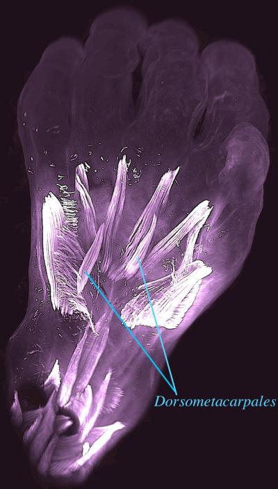
Dorsal view of the left hand of a 10-week old human embryo. The dorsometacarpales are highlighted: these muscles (like others described in this study) are present in adults of many other limbed animals, while in humans they normally disappear or become fused with other muscles before birth. CREDIT: Rui Diogo, Natalia Siomava and Yorick Gitton
A team of evolutionary biologists, led by Dr. Rui Diogo at Howard University, USA, and writing in the journal Development, have demonstrated that numerous atavistic limb muscles – known to be present in many limbed animals but usually absent in adult humans – are actually formed during early human development and then lost prior to birth. Strikingly, some of these muscles, such as the dorsometacarpales shown in the picture, disappeared from our adult ancestors more than 250 million years ago, during the transition from synapsid reptiles to mammals.
Also remarkably, in both the hand and the foot, of the 30 muscles formed at about 7 weeks of gestation one third will become fused or completely absent by about 13 weeks of gestation. This dramatic decrease parallels what happened in evolution and deconstructs the myth that in both our evolution and prenatal development we tend to become more complex, with more anatomical structures such as muscles being continuously formed by the splitting of earlier muscles. These findings offer new insights into how our arms and legs evolved from our ancestors’, and also about human variations and pathologies, as atavistic muscles are often found either as rare variations in the common human population or as anomalies found in humans born with congenital malformations.
Since Darwin proposed his evolutionary theory, scientists have argued that the occurrence of atavistic structures (anatomical structures lost in the evolution of a certain group of organisms that can be present in their embryos or reappear in adults as variations or anomalies) strongly supports the idea that species change over time from a common ancestor through “descent with modification”. For example, ostriches and other flightless birds have vestigial wings, while whales, dolphins and porpoises lack hind limbs but their embryos initiate and then abort hind limb development. Similarly, temporary small tail-like structures are found in human embryos and the remnant of the lost ancestral tail is retained as our coccyx. Researchers have also suggested that atavistic muscles and bones can also be seen in human embryos, but it has been difficult to visualize these structures clearly, and the images that appear in modern textbooks are mainly based on decades old analyses.
This is changing with development of new technology that provides high-quality 3D images of human embryos and fetuses. In the new study published in the journal Development the authors have used these images to produce the first detailed analysis of the development of human arm and leg muscles. The unprecedented resolution offered by the 3D images reveals the transient presence of several of such atavistic muscles. Dr. Diogo said: “It used to be that we had more understanding of the early development of fishes, frogs, chicken and mice than in our own species, but these new techniques allow us to see human development in much greater detail. What is fascinating is that we observed various muscles that have never been described in human prenatal development, and that some of these atavistic muscles were seen even in 11.5-weeks old fetuses, which is strikingly late for developmental atavisms “.
He further added: “Interestingly, some of the atavistic muscles are found on rare occasions in adults, either as anatomical variations without any noticeable effect for the healthy individual, or as the result of congenital malformations. This reinforces the idea that both muscle variations and pathologies can be related to delayed or arrested embryonic development, in this case perhaps a delay or decrease of muscle apoptosis, and helps to explain why these muscles are occasionally found in adult people. It provides a fascinating, powerful example of evolution at play.”
Cells are the building blocks of life. However, in multi-cellular organisms, millions of cells are subject to death due to injury, infection and ordinary cell turnover (Galluzzi et al., 2018). For example, epithelial cells in the small intestine rapidly renew every 2 to 6 days in most mammals, which is crucial to maintain proper function of villus epithelia (Mayhew et al., 1999). Moreover, during embryonic development, cell death serves as a crucial mechanism to remove unnecessary cells, adjust tissue size and shape, as well as correct developmental errors (Arya and White, 2015). In order to maintain homeostasis and avoid unwanted inflammatory responses, cellular debris is usually cleared rapidly by professional phagocytes, such as macrophages and microglia. However, during early neurogenesis when the neural tube develops and large numbers of neurons and glia undergo apoptosis, myeloid-derived professional phagocytes have not yet infiltrated the trunk of developing embryos (Herbomel, Thisse and Thisse, 1999, 2001; McGrath et al., 2003; Bertrand et al., 2013; Stremmel et al., 2018). How dead cells are removed from this region during early development remained largely unknown until recently.
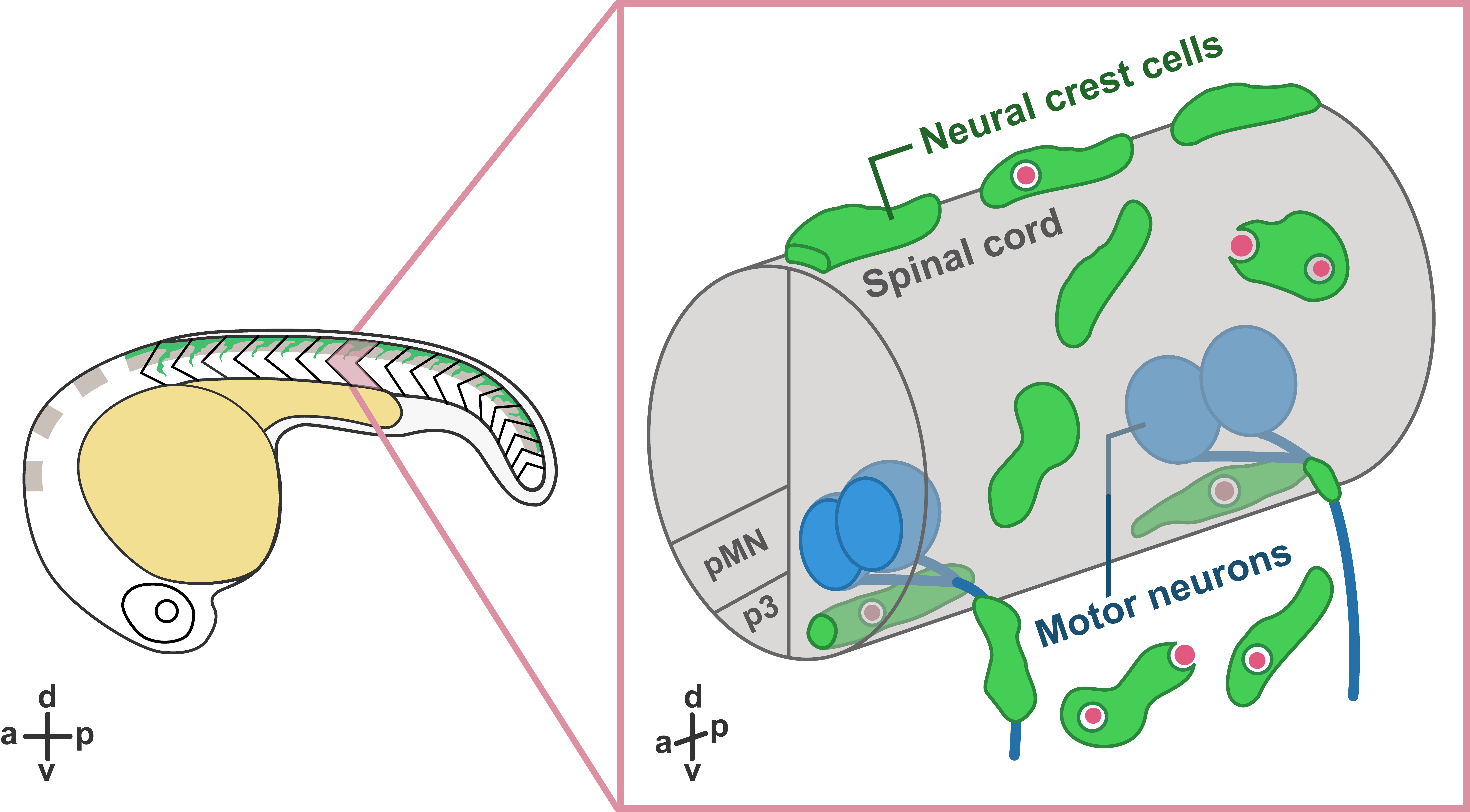
In our recent paper entitled “Migratory Neural Crest Cells Phagocytose Dead Cells in the Developing Nervous System”, we demonstrate an unexpected behavior of neural crest cells (NCCs), which have been studied for more than 150 years, in debris clearance before the colonization of professional phagocytes (Zhu et al., 2019) (Figure 1).
Figure 1. Schematic illustration showing neural crest cells (green) phagocytose dead cells (red) in zebrafish embryos. Right panel shows zoomed view of the trunk.
Interestingly, not only did we not expect this novel function of NCCs, but the discovery was also made by accident! Our original intent was to study how glial populations coordinated their behaviors during early spinal motor nerve development. But early in our studies, Yunlu started to notice NCCs behaving in a manner that didn’t match what the literature described. Some migrated away from their migratory streams, and others appeared to interact with cellular debris. Here, we would like to share the story behind this work.
Neural Crest Cells Clear Debris
What are NCCs and what do we know about them? During the earliest stages of vertebrate nervous system development, the ends of neural plate rise and fold into the neural tube, which further develops into the central nervous system (CNS), including the brain and the spinal cord. During or after the closure of the neural tube, NCCs located at the edge of the neural fold go through an epithelial-to-mesenchymal transition and delaminate from the dorsal neural tube. Multipotent NCCs then migrate through highly conserved pathways and give rise to a variety of cell types, including skeletal tissues, pigment cells, and neurons and glia of the peripheral nervous system (PNS) (Mayor and Theveneau, 2013). Given that NCCs are highly migratory and colonize the entire developing embryo before the infiltration of professional phagocytes, we hypothesized that they may have the ability to clear cellular debris at early developmental stages.
To examine whether NCCs are capable of clearing debris, we performed in vivo, time-lapse imaging in transgenic embryos expressing fluorescent proteins in NCCs. Excitingly, in these movies, we observed that some fluorescently-labeled NCCs migrated towards dead cells located far away from their innate migratory pathways. Then, they reached toward dead cells and engulfed them, resulting in the formation of large engulfment vesicles inside NCCs.
To confirm that the NCC engulfment process was similar to phagocytosis in professional phagocytes, we imaged transgenic lines labeling early endosomes (PI(3)P sensor) and lysosomes (Lamp1-GFP) (Rasmussen et al., 2015). In these movies, we found that, similar to the maturation of phagosomes, NCC engulfment vesicles fused with early endosomes and lysosomes, leading to progressive acidification inside the vacuoles. Interestingly, we observed NCCs phagocytosing a variety of dead cells, including dead NCCs, neuronal debris, and muscle cells.
In these movies, we also observed that PNS NCCs migrated through motor exit point transition zones, where spinal motor neurons send their axons into the PNS, into the ventral neural tube, and phagocytosed CNS debris. Most of these CNS-located NCCs stayed inside the neural tube for 2 to 12 hours and returned to the PNS through motor exit point transition zones. To test whether PNS NCCs were recruited into the spinal cord by cellular debris, we induced CNS cell death by ablating radial glia using nitroreductase-mediated cell death (Smith et al., 2014; Johnson et al., 2016) and observed a significant increase in the number NCCs recruited into the CNS compared to control embryos. Therefore, we conclude that NCCs can migrate toward and phagocytose dead cells in both CNS and PNS.
Are There Subpopulations of Neural Crest Cells?
Interestingly, we noticed dead cells were not always cleared by the nearest NCC. Instead, under most circumstances, they were phagocytosed by NCCs that came from a distance. And that led us to wonder if phagocytic NCCs were a specific subgroup of the neural crest population. To examine whether phagocytic NCCs belonged to a specific lineage, we performed lineage tracing on phagocytic NCCs expressing a photoconvertible protein. Contrary to our expectations, we observed phagocytic NCCs differentiate into a variety of derivatives, including pigment cells, motor axon-associated cells, and dorsal root ganglia cells, suggesting that phagocytic NCCs are not lineage restricted.
Additionally, under physiological conditions, we only observed 5-10% of NCCs that were phagocytic. To examine whether more NCCs could be “activated”, we induced cell death using laser ablation. Surprisingly, immediately after the ablation, the majority of neighboring NCCs started to engulf cellular debris, resulting in the formation of massive numbers of phagocytic vesicles. These results demonstrated that not only could NCC phagocytic abilities be induced by acute cell death, but also that the majority of NCCs have the potential to phagocytose debris. Therefore, these data are consistent with the hypothesis that phagocytic NCCs are not a specialized subpopulation of the neural crest population.
Previous studies show that NCCs migrate along conserved, segmentally restricted pathways. However, we found that phagocytic NCCs move toward cellular debris and sometimes even crossed somite boundaries. So what is the mechanism that directs phagocytic NCCs toward dead cells? To answer this question, we designed an ablation assay to quantify NCC recruitment and inhibited a variety of signaling pathways. We found that NCC recruitment was compromised when we treated embryos with a Caspase-1 inhibitor or a interleukin (IL)-1 receptor antagonist, indicating NCC recruitment was mediated by the IL-1β signaling pathway. Interestingly, we observed IL-1β expression in both cellular debris and phagocytic NCCs. Moreover, the IL-1β level we observed after cell ablation was significantly lower than that in previous studies using spinal cord transection and bacterial infection (Bernut et al., 2014; Nguyen-Chi et al., 2014). Given that our cell ablation is more precise compared to manipulations performed in previous studies, the low level of Il-1β release supports the hypothesis that Il-1β secretion is tightly regulated and dependent upon the strength of the inflammatory stimulus (Lopez-Castejon and Brough, 2011).
Conclusion (Yunlu Zhu)
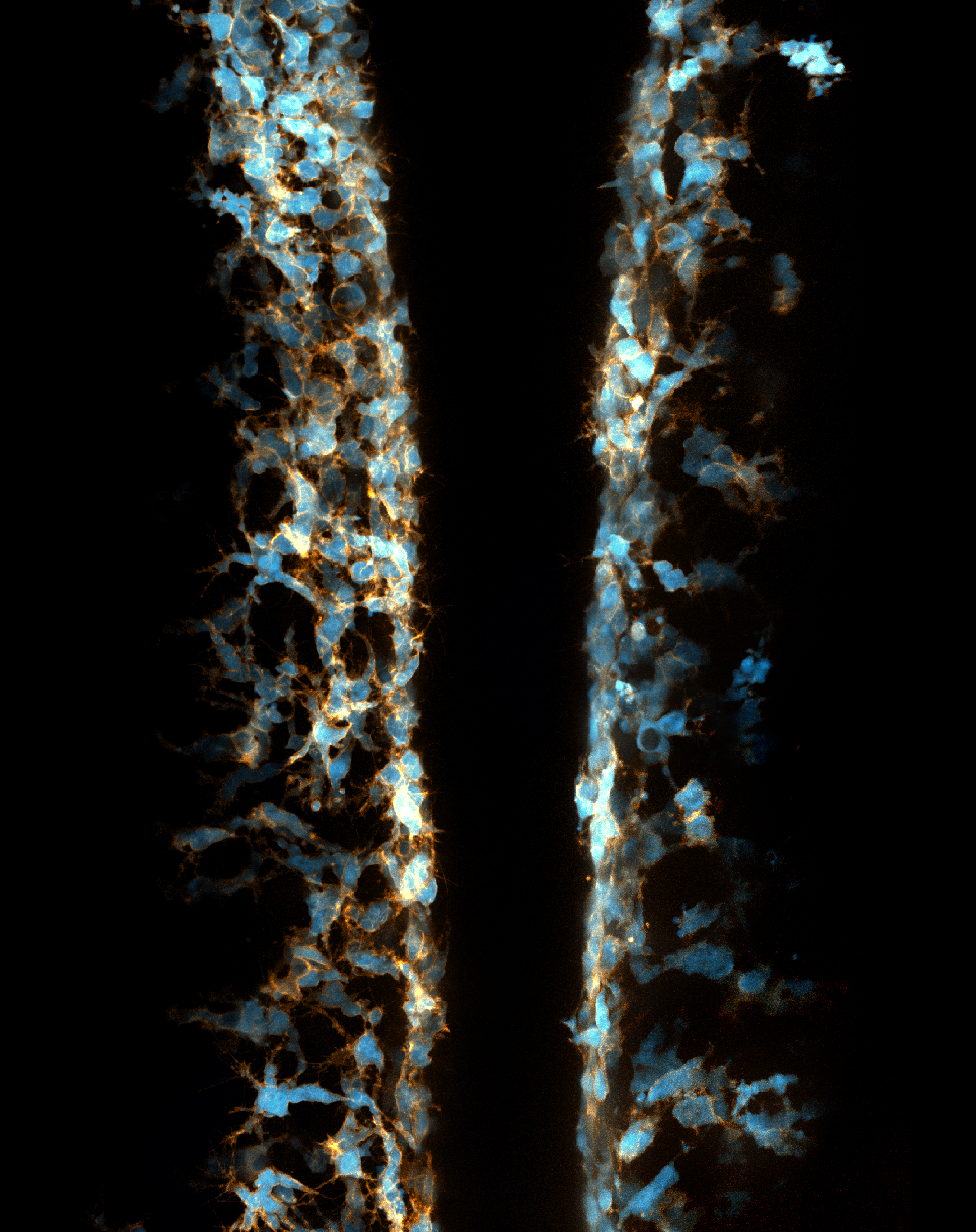
I joined the Kucenas lab at the University of Virginia in early 2014 because I was astonished by the beauty of live imaging in zebrafish embryos (Figure 2).
Figure 2. Two transgenic zebrafish embryos at 22 hour-post-fertilization expressing fluorescent protein in neural crest cells mounted back to back.
My previous focus was the development of perineurial glia and the role of cell-cell interactions in the development of spinal motor nerves. However, while imaging transgenic embryos labeling NCCs, I accidentally observed that some NCCs migrated ectopically away from their innate pathways and had spherical vacuoles with diameters of 3 to 10 µm. I discovered that these weird behaviors of NCCs had never before been described and was deeply attracted by this phenomenon. Therefore, I shifted my focus toward this NCC project.
This novel role of NCCs in debris clearance was quite unexpected but perfectly reasonable. However, the nature of these phagocytic NCCs is not well understood and we have many remaining questions. Given that the majority of NCCs are capable of phagocytosing debris, why don’t they respond to cell death under physiological conditions? What is special about those active NCCs that migrate towards dead cells from a distance? And what happens when professional phagocytes like macrophages colonize the trunk region of the developing embryo? Are their immune-NCC interactions? And what happens if one of these two populations is unable to clear debris? There are so many new questions that have come from this work, and I’m excited to see which ones the next students decide to pursue.
I would like to end this post with a quote from Yogi Berra, which, I think, precisely describes the beauty of live imaging and my experience in this project: “You can observe a lot by just watching”.
References:
Arya, R. and White, K. (2015) ‘Cell death in development: Signaling pathways and core mechanisms’, Seminars in Cell & Developmental Biology. Academic Press, 39, pp. 12–19. doi: 10.1016/J.SEMCDB.2015.02.001.
Bernut, A. et al. (2014) ‘Mycobacterium abscessus cording prevents phagocytosis and promotes abscess formation.’, Proceedings of the National Academy of Sciences of the United States of America. National Academy of Sciences, 111(10), pp. E943-52. doi: 10.1073/pnas.1321390111.
Bertrand, J. Y. et al. (2013) ‘Three pathways to mature macrophages in the early mouse yolk sac Three pathways to mature macrophages in the early mouse yolk sac’, Blood, 106(9), pp. 3004–3011. doi: 10.1182/blood-2005-02-0461.
Galluzzi, L. et al. (2018) ‘Molecular mechanisms of cell death: recommendations of the Nomenclature Committee on Cell Death 2018’, Cell Death & Differentiation. Nature Publishing Group, 25(3), pp. 486–541. doi: 10.1038/s41418-017-0012-4.
Herbomel, P., Thisse, B. and Thisse, C. (1999) ‘Ontogeny and behaviour of early macrophages in the zebrafish embryo.’, Development (Cambridge, England), 126(17), pp. 3735–45.
Herbomel, P., Thisse, B. and Thisse, C. (2001) ‘Zebrafish early macrophages colonize cephalic mesenchyme and developing brain, retina, and epidermis through a M-CSF receptor-dependent invasive process.’, Developmental biology, 238(2), pp. 274–88. doi: 10.1006/dbio.2001.0393.
Johnson, K. et al. (2016) ‘Gfap-positive radial glial cells are an essential progenitor population for later-born neurons and glia in the zebrafish spinal cord’, Glia, 64(7), pp. 1170–1189. doi: 10.1002/glia.22990.
Lopez-Castejon, G. and Brough, D. (2011) ‘Understanding the mechanism of IL-1β secretion’, Cytokine and Growth Factor Reviews. Elsevier Ltd, 22(4), pp. 189–195. doi: 10.1016/j.cytogfr.2011.10.001.
Mayhew, T. M. et al. (1999) ‘Epithelial integrity, cell death and cell loss in mammalian small intestine.’, Histology and histopathology, 14(1), pp. 257–67. doi: 10.14670/HH-14.257.
Mayor, R. and Theveneau, E. (2013) ‘The neural crest’, Development, 140(11), pp. 2247–2251. doi: 10.1242/dev.091751.
McGrath, K. E. et al. (2003) ‘Circulation is established in a stepwise pattern in the mammalian embryo.’, Blood. American Society of Hematology, 101(5), pp. 1669–76. doi: 10.1182/blood-2002-08-2531.
Nguyen-Chi, M. et al. (2014) ‘Transient infection of the zebrafish notochord with E. coli induces chronic inflammation’, Disease Models & Mechanisms, 7(7), pp. 871–882. doi: 10.1242/dmm.014498.
Rasmussen, J. P. et al. (2015) ‘Vertebrate epidermal cells are broad-specificity phagocytes that clear sensory axon debris.’, The Journal of neuroscience : the official journal of the Society for Neuroscience. Society for Neuroscience, 35(2), pp. 559–70. doi: 10.1523/JNEUROSCI.3613-14.2015.
Smith, C. J. et al. (2014) ‘Contact-Mediated Inhibition Between Oligodendrocyte Progenitor Cells and Motor Exit Point Glia Establishes the Spinal Cord Transition Zone’, PLoS Biology. Edited by B. A. Barres, 12(9), p. e1001961. doi: 10.1371/journal.pbio.1001961.
Stremmel, C. et al. (2018) ‘Yolk sac macrophage progenitors traffic to the embryo during defined stages of development’, Nature Communications. Nature Publishing Group, 9(1), p. 75. doi: 10.1038/s41467-017-02492-2.
Zhu, Y. et al. (2019) ‘Migratory Neural Crest Cells Phagocytose Dead Cells in the Developing Nervous System’, Cell, 179(1), pp. 74-89.e10. doi: 10.1016/j.cell.2019.08.001.
One of the biggest open questions in biology is how organisms can form complex patterns (limbs, organs, entire body plans) from initially disordered or very simple states. Every animal does this at the beginning of its life, forming its full complexity from a single cell. Some are capable of similar feats even after their bodies are fully constructed: starfish and flatworms survive when cut in pieces, the zebrafish restores lost fins, the axolotl regrows limbs and organs alike. How does either case work? How are those patterns established so accurately, and how is it that things so rarely go wrong?
I’m trying to approach this question with the help of one of the most impressive regenerators of all: Hydra. As a 1 cm long freshwater cnidarian it’s a lot less impressive than the monster whose name it bears, but what it lacks in scales and fangs it makes up for in raw regenerative ability. The mythological beast grew two heads for every one it lost. The real thing can regenerate its head, can regenerate from a ball of tissue as small as 150 microns across, and can come back from being dissociated down to single cells if enough of those cells are placed together. On top of this, the small size and simplicity of the creature make it very easy to study and manipulate compared to other model organisms. In fact, several theoretical models purporting to describe the patterning process have already been developed.
Of particular interest is the fact that these models propose mechanical forces as drivers of pattern formation. Mechanical forces are well known to be important in embryonic development but their role in regeneration is less clear. As a Hydra regenerates it first forms a hollow sphere, which then undergoes osmotically driven cycles of swelling and rupture. According to the literature, there is a characteristic switch in these oscillations from large amplitude and low frequency to small amplitude and high frequency, and this shift is linked to when the animal sets its body axis. Thus the pattern shift was proposed to represent a link between mechanics and biochemistry. I wanted to determine what the mechanism linking mechanical forces to biochemical axis specification was.
On observing many regenerating animals, one of the first things we noticed was that nearly half of the animals we imaged regenerated without a clear shift in oscillation pattern. As this called into question essentially all previous assumptions about the nature and relevance of the pattern shift, we turned to trying to figure out its exact cause.
It was previously proposed that the change in oscillation behavior might be due to the beginnings of a regenerated mouth. This idea makes logical sense, as adult Hydras open their mouths to relieve internal water pressure as well as to feed, but nobody had experimentally tested it. By using injected fluorescent beads to track the location of successive rupture sites, we determined that the spot where rupture occurs is random during large oscillations but conserved during small ones. These data would be consistent with the theory that the large oscillations are due to the tissue tearing under internal pressure, while small oscillations occur when the mouth is established to act as a vent. As confirmation, a tissue piece containing the mouth of the original animal also produces conserved rupture sites and only shows small oscillations.
To this point we had only shown that oscillation behavior and the mouth are somehow correlated. These experiments do not explain how the mouth might affect oscillation behavior. Thankfully, Hydra’s ease of manipulation offers a direct way to establish causality. It’s possible to eliminate all nerve cells from a Hydra, producing a nerve-free animal that is structurally normal but cannot actively move. The critical experiment was to use mouth-containing tissue pieces cut from such nerve-free animals. These contain a mouth that is fully formed and complete, but cannot open on its own. If the deciding factor for small oscillations to occur is simply that the mouth acts as a weak point and tears more easily, nerve-free samples with a mouth should show small oscillations exactly like their normal counterparts. If on the other hand mouth function is the key, they should show large oscillations.
We find that nerve-free mouth pieces show only large oscillations, indicating a causal link between active control of mouth function and a decrease in oscillation amplitude. This provides a concrete explanation for the oscillation pattern shift in cases where it occurs: it is caused by the animal’s ability to open its mouth at will.
Our study does not answer why some pieces are delayed in developing mouth function or provide exact developmental checkpoints. What it does give us is further constraints and parameters that can be used to improve existing models, and put us one step closer to understanding how Hydra regenerates. As Hydra shares many key biochemical pathways with more familiar animals despite its alien appearance, figuring out patterning here could one day be the basis of a similar understanding in humans.
We are seeking a PhD candidate to join our EvoDevo lab in the University of Barcelona to study our favorite chordate model Oikopleura dioica, in which we are currently interested in heart development, 4D imaging of early embryo cleaving, and early developmental responses to environmental challenges. To meet our unique Oikopleura model -> Click here for a tour “A day in our lab” posted in The Node.
We have also engaged a new EcoEvoDevo line investigating if the developmental mechanisms of marine embryos are ready to respond to climate change, including the effects of biotoxins derived from algal blooms. Click here for a tour on this new EcoEvoDevo adventure.
Our approaches include Single-cell transcriptomics,RNAseq, RNAi Knockdowns, CRISPR and Fluorescent-Microscopy
PhD fellowship Call OPEN (FI Catalan program): October 1st-14th 2019 (contact for enquiries as soon as possible canestro@ub.edu)
REQUIREMENT: to have finished a Master degree
CONTACT: please send an email to Cristian Cañestro (canestro@ub.edu), including a brief letter of interest, and the final scores for the degree and Master (indicating the scale), all together in ONE single pdf file.
Dolly the Sheep via Flickr, Toni Barros (CC BY-SA 2.0)
In this episode from our centenary series exploring 100 ideas in genetics, we’re looking at mergers and acquisitions – but in a biological rather than a financial sense. We find out what happens when two cells decide to move in together, unpack the history of genetic engineering and bleat on about the story of Dolly the Sheep.
If you enjoy the show, please do rate and review and spread the word. And you can always send feedback and suggestions for future episodes and guests to podcast@geneticsunzipped.com
The Department of Biological Sciences at the University of South Carolina (UofSC) invites applications for two tenure-track Assistant Professor positions in neurobiology. The successful candidates will be expected to establish an independent, extramurally funded research programs in: 1) Molecular or Cellular Neurobiology relevant to neural development and/or disease focused on cell-cell interactions; or 2)Molecular or Cellular Neurobiology using animal models to understand the pathophysiology of Autism or neurodevelopmental disorders (see below). Minimum qualifications include a Ph.D. or M.D. and post-doctoral training in a relevant area. The successful candidates will be responsible for teaching courses relevant to their area of expertise, as well as mentoring research training for graduate and undergraduate students.
1) Molecular or Cellular Neurobiology focused on Cell-Cell Interactions (position # 67536): We are interested in applicants focusing on how interactions between different cells, including cellular interactions with axons, contribute to the biology of nervous system development, disease, or response to injury. This individual will closely interact with research groups in the SmartState Center for Childhood Neurotherapeutics, which includes neurobiologists focused on molecular mechanisms of axon growth in development and after neural injury, and the broader UofSC Neuroscience Community. Applications are made online at http://uscjobs.sc.edu/postings/67536. For questions or further information, please contact Dr. Fabienne Poulain (fpoulain@mailbox.sc.edu).
2) Neurobiology of Autism or Neurodevelopmental Disorders (position # 67556): This position is part of a university-wide initiative to enhance research on Autism and Neurodevelopmental Disorders and establish a Center of Excellence (USCAND) to accelerate interdisciplinary efforts in neuroscience. There is a parallel faculty search in the Department of Psychology for this initiative, and several additional USCAND faculty hires planned over the next few years in complimentary disciplines. This individual will closely interact with research groups in USCAND, as well as the SmartState Center for Childhood Neurotherapeutics, Institute for Mind and Brain,andResearch Consortium on Children and Families. Applications are made online at http://uscjobs.sc.edu/postings/67556. For questions or further information, please contact Dr. Jeff Twiss (twiss@mailbox.sc.edu).
Review of applications will begin by November 1, 2019. The review process will continue until the positions are filled. Qualified individuals should submit a curriculum vita, research statement (3 pages), teaching philosophy (1 page), and the names, email addresses and phone numbers of at least three references to http://USCjobs.sc.edu/postings (with position # and links as above).
The Department of Biological Sciences is a multidisciplinary unit of approximately 1,600 undergraduate students, 50 graduate students, and 35 tenure-line faculty representing a broad range of research areas (www.biol.sc.edu). UofSC has a highly interactive neuroscience research community that encourages and precipitates collaborations. UofSC in Columbia (www.sc.edu) is the state’s flagship university (founded in 1801 and currently one of the top 50 “Best Colleges” according to U.S. News and World Report).
Columbia, SC enjoys more than 300 days of sunshine annually and has ready access to pristine beaches, lakes, rivers and mountains.The city hosts historical and cultural attractions, festivals, performing arts and sporting events, parks and outdoor recreation including Congaree National Park and 50,000-acre Lake Murray.
The University of South Carolina is an affirmative action, equal opportunity employer. Minorities and women are encouraged to apply. The University of South Carolina does not discriminate in educational or employment opportunities on the basis of race, gender, age, color, religion, national origin, disability, sexual orientation, genetics, veteran status, pregnancy, childbirth or related medical conditions.
 (No Ratings Yet)


 (1 votes)