The Center for Scientific Collaboration and Community Engagement (CSCCE) has announced its next offering of Scientific Community Engagement Fundamentals (CEF25F), which will run on Mondays and Thursdays beginning Thursday, 4 September until Thursday, 23 October.
The key dates are:
Social hour: Thursday, 4 September at 10am – 11am EDT / 2pm – 3pm UTC
Monday lessons: 8, 15, 22, 29 September; 6*, 20 October at 10am – 11:30am EDT / 2pm – 3:30pm UTC
Thursday Co-Labs: 11, 18, 25 September; 2, 9, 16 October at 10am – 11:30am EDT / 2pm – 3:30pm UTC
Graduation: 23 October at 10am – 12pm EDT / 2pm – 4pm UTC
*CSCCE will be closed on Monday, 13 October 2025 in observance of Indigenous Peoples’ Day. To accommodate this, we have slightly adjusted the schedule for this CEF cohort. “Reading Week” will now only take place during one session, on 2 October 2025, and the Week 7 lesson that would have taken place on Monday, 13 October will be held on Thursday, 9 October.
Scientific Community Engagement Fundamentals is an eight-week course designed to offer new or existing community managers core frameworks and vocabulary to describe their community’s purpose, refine or create strategic programming to engage community members around their shared goals, and identify ways to lower barriers to member participation. While the content is designed for any level of learner, it should not be thought of as a “beginner” course. Rather, it is intended to create common ground so that scientific community managers can converse across disciplines, more efficiently learn from one another, and build successful engagement strategies that are grounded in theory.
Each week, participants will meet virtually (using Zoom) for a 90 minute lesson and a 90 minute Co-Lab. While lessons will involve structured presentations and activities, Co-Lab time is for discussion, reporting out, and seeking feedback from instructors and fellow learners.
Each week will also include approximately 90 minutes of homework, for a total time commitment of 4.5 hours per week.
Pricing
Our pricing structure for CEF reflects the different organizations that community managers work for and the range of available budgets.
Discounted rate: Depending on participants paying the supporting rate, we may be able offer a limited number of discounted tickets for this cohort. If you would like to participate but your organization is unable to cover the whole cost, please complete this course discount request form.
Long range order and topological defects as tissue shapers
My PhD focused on studying the role of CDC42 isoforms during cell polarization and migration1. As CDC42 is a well-known regulator of actin polymerization, I developed extensive training in understanding actin regulation and dynamics in the context of cell polarization and migration.
Towards the final year of my PhD, during my time at the Institut Curie in Paris, I encountered the captivating field of biological active matter. There, I was introduced to the works of several renowned biophysicists working at the intersection of physics and biology. I became deeply intrigued by the emerging studies in this field, which is largely driven by biophysicists exploring long-range order in biological systems and investigating whether singularities in this order—known as topological defects—act as organizational centers that facilitate key biological processes.
Figure 1 Skeleton schematic showing actin long-range order on the Hydra’s body plan
One striking example came from Benoit Ladoux, who demonstrated that cell extrusion events can occur at sites of topological defects in epithelial monolayers2. This was followed by two compelling preprints. The first, from the Roux Lab at UNIGE, showed that myoblasts confined to circular patches organize into nematic order (i.e., long-range, thread-like alignment). At aster-like topological defects, they observed cellular tornado-like 3D bulk cell extrusions, mimicking an in vitro reconstitution of muscle morphogenesis-like events3. The second preprint, from the Keren Lab in 2020, revealed that Hydra exhibits long-range nematic ordering in its supracellular actin organization, with topological defects correlating with head and foot morphogenesis4 (Fig. 1). Circling back to my PhD and my training in actin biology, seeing such single-cell-like highly organized actin structures in a tissue-scale regeneration in Hydra was fascinating and puzzling at the same time. I had so many agitating questions.
Beginning the postdoc: are topological defects shapers of in vivo morphogenesis?
I started in Aurélien Roux’s Roux Lab in September 2021—it was a rocky start, as I was still wrapping up my PhD manuscript and applying for postdoctoral fellowships, with deadlines fast approaching. I was also recovering from medical conditions that had worsened due to the sedentary lifestyle imposed on us during COVID.
In spite of these bottlenecks, I was genuinely excited to take on a new project to study the role of active matter in morphogenesis, especially in light of the recent breakthroughs in the field. Aurélien and I decided to explore the hypothesis of whether—and how—topological defects are required for shaping biological tissues, using various model organisms. We ventured into root morphogenesis in Arabidopsis (in collaboration with Luis Lopez Molina), slug morphogenesis in Dictyostelium (in collaboration with Thierry Soldati), and specifically, head regeneration in Hydra multi-headed mutants (in collaboration with Brigitte Galliot).
While I was juggling these different organisms and studying their fascinating morphogenetic events, I encountered a practical issue: my Hydra were constantly moving during imaging. I was performing these experiments with the help of Matthias Vogg, then a senior postdoc in the Galliot lab. To address the movement issue, I decided to image them using an agarose slab confinement method typically used for imaging plants—and Matthias agreed. Little did I know that this would compress the animal, leading to a mechanical induction of two-headed morphogenesis in Hydra.
Riding on this serendipitous discovery, I began following the phenomenon of mechanically induced morphogenesis in Hydra. During regeneration under compression, I tracked the emergence of new topological defects in the actin organization of the animal, which correlated with the formation of new heads. This observation confirmed the hypothesis proposed by the Keren lab, which suggested that aster topological defects are associated with new head formation during Hydra regeneration.
Topological defects shape animal tissues in a curvature dependent manner
Following this, Aurélien was thrilled and suggested I reach out to a theoretical physicist to explore the physical mechanisms behind how these additional topological defects could influence tissue shaping. He connected me with Daniel Pearce, an active matter theoretical physicist (then a postdoc in Karsten Kruse’s lab), who had already developed a mathematical model describing how long-range organization in Hydra could shape tissue5.
When I showed Daniel my findings, he was extremely excited and immediately came on board to help develop theoretical simulations of tissues under compression. Using his elastic nematic model, he described how +1 aster topological defects organize stresses and generate positive curvature (dome shapes), which is reminiscent of the shape of the Hydra head. In simulations under lateral compression, we observed that placing topological defects at the extremities, as seen in our regeneration experiments, led to the evolution of two dome-shaped structures.
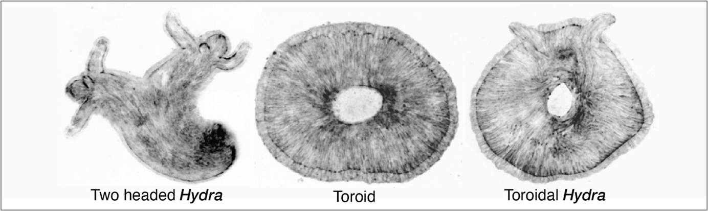
Furthermore, it was Dan who predicted that the orientation of tissue compression could dictate the fate of regeneration. The direction of compression influences how the long-range actin orientation experiences and responds to stress. As he suggested, we observed that when the tissue was oriented parallel to the compressive agarose slab, the sites of +1 aster defects buckled (inverted dome shapes) and underwent tearing, which then healed to form a defect-less toroid (Fig. 2).
This was a ground-breaking observation—it was the first time we observed the abolishment of body axis in an animal tissue that remained viable. I followed the defect-less torus over several days and observed that it failed to regenerate, as it maintained a perfectly symmetric actin configuration in which de novo +1 asters never arose. The tissue continuously attempted to regenerate a head but failed, due to the absence of a +1-aster topological defect.
Lastly, we all collectively thought however we should still be able to generate toroid with a +1 aster. By then we were also in touch with the Kerren lab and she invited me to Israel for a stay in her lab. Everyone suggested that theoretically a torus with defects could be generated. Then I hypothesized that if the compression occurred in a tissue with disordered actin to start with and a simultaneous tear occurred, then while the wound heals to create a 3D hole for the torus the disordered actin will order around it while generating a +1 aster required for a head. That is precisely what happened when we compressed the spheroid tissue that undergoes initial actin disorder and then ordering. This way we generated a torus with a +1 aster therefore a toroidal adult Hydra animal. This was really the cherry on top to see an animal with such a twisted muscle organization and topology.
In conclusion, these experiments established the need for +1 aster actin topological defects as head shapers in the animal. Their presence shapes the head and their absence give rise to no head formation, as observed in the defectless torus. The prospective research is to try to identify such long-range physical morphogens in other organisms, and strengthen the understanding of biological active matter.
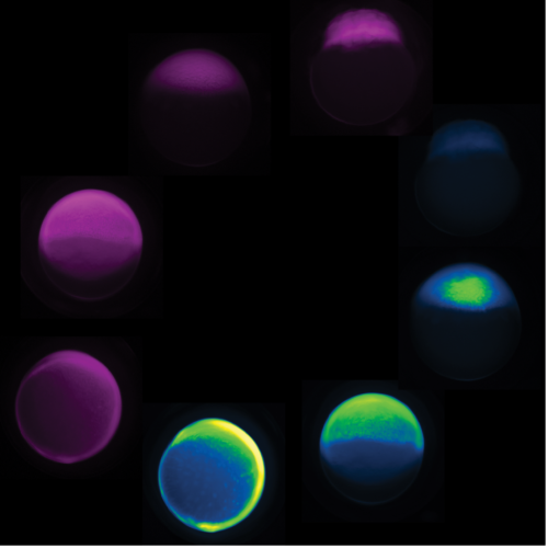
Figure 2 Spinning disk microscopy images of fluorescent actin expressing (GFP-Ecto-LifeAct) Hydra at Day 6 of head regeneration. The three mechanically induced Hydra phenotypes, two-headed, actin-defectless toroid and toroidal Hydra. (2 votes) Loading...
Anyone—regardless of coding skills—should be able to generate a publication-quality plot of their data in minutes. That was the main motivation to develop a series of web apps to make state-of-the-art data visualization more accessible (huygens.science.uva.nl). But who cares, the same result can be achieved with generative AI (genAI) based tools, right?
Before discussing what genAI can bring us for coding a plot, I briefly explain how the web apps work, so we can compare it with genAI later on. The data visualisation produced by the web app (the output) is coded in R and uses the {ggplot2} package. Another R-package, {shiny}, is used to create a graphical user interface (GUI). This GUI enables the user to optimize the data visualization by modifying the (invisible) code, through sliders, buttons, drop-down menus, and text fields. The process of creating a data visualization in a web app is highly interactive. By using a web app, the user can focus on what the data visualization should look like, without dealing with the code.
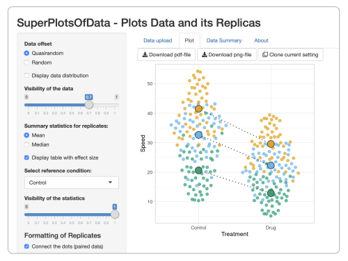
A screenshot of the web app SuperPlotsOfData. Users can optimize the data visualization with sliders, buttons, drop-down menus, and text fields.
By design, the web apps are somewhat limited in their options, so I started an online resource with dataViz protocols as well. My hope was that this resource would lower the barrier for people that wanted more control than what is possible in the web app and therefore would be motivated to learn R&ggplot2. But now, there is genAI. Coding can be done, rapidly and interactively, with websites that spit out code based on Large Language Models. Instead of focusing on the code and the technicalities that are required to build a data visualization, the user can focus on what the data visualization should look like (do you see the parallels with web apps?). This approach is aptly called vibe coding.
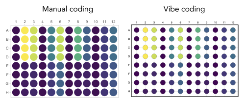
One of the prompts that was used in ChatGPT for vibe coding the data visualization of the output of a 96-wells plate that is shown below.
In a previous blog, I described that vibe coding “felt like I gained some kind of superpower”. But not everything is hunky-dory. It has been nicely documented by Mine Çetinkaya-Rundel that the AI-tool, besides the required changes, makes changes that are not explained and may be difficult to understand. I had exactly the same experience when I tried to vibe code a data valisualization that I had previously manually crafted (protocol 8 in the dataViz protocols book). In the end, the result (see below) is pretty neat, but it took several iterations (prompts), including some debugging of errors. I also noticed that understanding some of the basics (loading packages, knowing where to place the input data, how a plot is built using the {ggplot2} package) is needed to get the code to work. Worse yet, if the code seems to work but actually makes mistakes that are hard to spot things can go really wrong. For instance when doing some calculation for statistics that are difficult to understand or verify.
Graphical representation of readings from a 96-wells plate. The data visualization on the left was manually coded as detailed here and the data visualization on the right was generated by vibe coding in ChatGPT.
Are web apps still relevant when the same result can be obtained with vibe coding? Both the web apps and the genAI tools can be considered as a black box and allow the use to focus on the output. The genAI based tools offer great flexibility, but a strong point of the web apps is their predictable outcome, delivering a fully reproducible data visualization. The underlying code is available and the web apps are documented in (peer-reviewed) papers that can be cited. A practical advantage of web apps is that there is no need to install software or specific packages to run them. So I think there is still a future for the web apps. At the same time, I encourage experimenting with genAI as vibe coding offers new and exciting opportunities for data analysis and visualization. This will require at least a basic understanding of the coding language and sanity checks. Altogether, these are exciting times as the options for generating publication-quality data visualizations are expanding!
This week, we explore the story of Dr. Luis Cedeno-Rosario, a postdoctoral researcher in the Rutter Lab at the University of Utah. Luis’s path into metabolism began with a biochemistry class—an early glimpse into how cells adapt, survive, and respond to their world. His work explores how cancer cells alter their internal wiring to support unchecked growth and resist treatment—uncovering how shifts in metabolism can give tumors a survival advantage. These insights may help identify new ways to target cancer by exploiting its metabolic dependencies. Continue reading to learn how Luis is driven by curiosity, scientific precision, and how having a supportive mentoring environment impacted his journey. Check out his thoughts on how he winds science and music together, and how he views metabolism more than just chemistry— but as a language through which disease reveals its secrets and a window into how life adapts under pressure. Give him a follow over twitter and bluesky.
What’s your first memory of the field of metabolism? Could you share your journey into studying metabolism in disease contexts like cancer and cardiac disorders?
I have always been passionate about understanding how cells adapt to different environments and challenges, with a focus on cancer cell signaling and mitochondrial metabolism. I was taking a biochemistry and cell and molecular biology class as an undergraduate student at the University of Puerto Rico – Humacao and became fascinated by how multiple pathways intersect to regulate this process and their impact on cell behavior. I also had the great opportunity to do summer research internships at UT MD Anderson Cancer Center and at Johns Hopkins University which allowed me to learn more about the cell signaling and metabolism field. This is what led me to pursue a PhD in cell signaling in Dr. Deborah Chadee’s lab at the University of Toledo and a postdoc in metabolism in Dr. Jared Rutter’s lab at the University of Utah.
Introduce us to the field of cancer metabolism – you have worked on different types of cancer cells like ovarian cancer cells and liver cancer cells – tell us about your experiences.
During my first year of graduate school, I knew that I wanted to study cell signaling but I wasn’t sure in what context. I remember listening to Dr. Chadee’s talk in the signal transduction class and I was very fascinated by the complexity of the MAP Kinase signaling pathways and their role in ovarian cancer progression. Therefore, I decided to complete my PhD under the mentoring of Dr. Chadee where I worked on the regulation of the MAP3K MLK3 by CDK1 and CDK2 and their role in controlling cell division and proliferation in ovarian cancer cells (Check out the paper here). For my postdoc in the Rutter lab, I wanted to apply what I learnt during graduate school in the context of mitochondrial metabolism and their signaling pathways that are involved in liver cancer cell proliferation and progression.
How are different cells metabolically heterogeneous within the same tumor? Why is it important to study metabolic heterogeneity in cancer occurrence/progression – in term of both the cells themselves and the microenvironment?
Cells can have different metabolic profiles depending on the metabolites they need or are available in their surroundings. That heterogeneity can also come from where these cells are localized, for example, cells that are in a more hypoxic environment will probably have other metabolic needs than cells that are in a less hypoxic or normal environment. So cells have evolved in a way that they are very smart in choosing or taking what they need to meet their metabolic demands.
Tell us about your current work on metabolic signaling in the context of Wnt/beta-catenin pathway activation in liver cancer cells. How do you link it to the mitochondrial functioning and what future questions are you most excited about?
Our lab has done extensive work in characterizing the importance of the Mitochondrial Pyruvate Carrier (MPC) and its role in proliferation and tumorigenesis. I discovered that activation of beta-catenin represses MPC expression in liver cancer cells, and that this regulation rewires mitochondrial metabolism from glucose oxidation towards fatty acid oxidation. This is particularly interesting in the context of cancers in which MPC is downregulated and fatty acid oxidation is increased. I am very excited for the future since my findings opens up new avenues to explore ways to increase MPC expression in these tumors and increase the quality of life and survival of cancer patients.
Tell us how difficult some of these experiments are – do you have to deal with midnight timepoints or require an army of undergrads/ long hours, has to use some un-conventional/creative tools to overcome experimental challenges etc.
Some of these experiments have been truly a challenge and I have definitely spent many many hours in the lab trying to solve multiple research questions and/or developing new techniques to study the regulation of MPC by beta-catenin. I mentored an amazing summer research student, Nimo Abdi, who helped me a lot in the beginning of this project. I also have excellent collaborators, inside and outside the lab, who have contributed to the development of new ideas and have given me new perspectives on this regulation. I am very grateful to have them as collaborators and truly believe that these efforts will make a great impact in the metabolism field.
Building upon your work in the context of liver cancer metabolism, what are your upcoming plans? What metabolic pathways do you aim to investigate further to understand cancer progression from a cell growth and signaling perspective?
This switch in metabolic profile from glucose towards fatty acid oxidation is very exciting. So we are definitely looking more in depth at the metabolic processes that are changing and at the proteins and enzymes behind that regulation. One of the big questions we are investigating right now is to understand what fatty acids these cells prefer to utilize and their implication in liver cancer progression.
Before this, you studied cell signaling in ovarian cancer cells. How was the transition between fields, and what do you carry over from your previous research? Could you shed some light on your results regarding how MLK3 (Mixed lineage kinase 3) regulates cell cycle of ovarian cancer cells and tell us about your cool findings?
I have always thought about metabolism as another way of cells to sense their environment and metabolites are signaling molecules. These are multiple signaling pathways that are interconnected, and this concept was very similar to what I studied during graduate school. During my PhD, I found that the MAP3K MLK3 (Mixed lineage kinase 3) becomes phosphorylated by CDK1 and CDK2 to control ovarian cancer cell cycle progression. This research was published in the Journal of Biological Chemistry (JBC) doi: 10.1016/j.jbc.2022.102263 and I would encourage everyone to read it. It is a very interesting story that shows how these phosphorylation events act as “on” and “off” switches to control ovarian cancer cell division and proliferation.
How do you think scientific paradigms in the field of cancer metabolism will evolve in the coming decades – in regard to the new upcoming tools? Are we moving toward a more nuanced understanding, or do you see potential pitfalls?
I feel like we have bright future in our metabolism community. We have seen the development of great techniques such as mass spectrometry integrated with equilibrium dialysis for the discovery of allostery systematically (MIDAS) that was developed in our lab to identify novel interactions between metabolites and proteins. This is a fast-growing field and we are opening more doors to understand the complexity of metabolic pathways in multiple contexts, including cancer, cardiac function and neurodegenerative diseases, and in development. I am very excited for our future findings and hope that I can contribute significantly and have a positive impact not only in the research field itself but also in training the next generation of scientists.
What role does curiosity play in your life, both within and outside of science?
I believe that curiosity plays an important role in my scientific career. Understanding what is happening at the cellular level is pivotal in the development of new therapies, and that is what drives my passion for science. I want to be able to use my knowledge from cell and molecular mechanisms to develop new and better ways to treat multiple diseases or to at least increase the quality of life of the people affected by a particular disease.
What are the future research questions you are most excited to ask?
I am very excited about pursing metabolism in the context of cell biology and development. I think it is a field that is also growing very fast and I would like to contribute to it and make new discoveries.
Were there any pivotal moments that shaped your career path —and how have you found ways to build a supportive community in science?
I think that the most pivotal moment in my career path was creating a strong and supportive network of mentors within the cell metabolism and mitochondrial biology field. I have met many of these mentors in conferences and through the Burroughs Wellcome Fund Postdoctoral Diversity Enrichment Program (PDEP) that have been critical in my development as a future independent scientist. I am also very grateful to be part of the biochemistry department at the University of Utah and to receive a lot of internal support as a postdoctoral fellow.
How do you maintain a balance between your rigorous research activities and personal life? Are there hobbies or practices you find particularly rejuvenating?
Music! I have extensive training in classical music and I am actually a member of the Utah Medical Orchestra (UMO) where I play the flute and the piccolo. Music is definitely a big part of who I am.
If you hadn’t embarked on a career in biological research, what other profession might you have pursued, and why?
I would have pursued a law degree or a music degree in flute performance. In the law aspect, I like the complexity of finding new solutions to diverse problems. In the music aspect, I like how we can create art using a universal language and enjoy that art as a whole. Music can bring you different feelings and helps us express ourselves.
Last week we learnt about how viruses rewire and utilize host lipid metabolism using mosquitoes as a host model system with Wolbachia and dengue as viral players, check out the article – Lipids and Labyrinths(Cassandra Koh). Cassandra is a new PI, studying metabolic interactions of symbiosis and virus-virus host interactions. She is seeking motivated students and collaborators.
Madalena M. Reimão Pinto (Schier lab, University of Basel, Switzerland) and Sebastian Castillo Hair (Seelig lab, Washington University, Seattle, USA) joined forces to understand how zebrafish embryos orchestrate protein synthesis during early development. Through this collaboration, their recently published study in Developmental Cell identifies features in mRNA 5′ UTRs that act during early zebrafish development to regulate translation.
Madalena, what brought you to Alex Schier’s lab and how did you embark on this project?
During my PhD at the Vienna BioCenter, I worked on understanding the molecular mechanisms regulating mRNA biogenesis and function in the fruit fly. It so happened that the exonucleolytic enzyme that I was working on at the time had a striking effect on spermatogenesis, and the data pointed to defects resulting from misregulated mRNA translation. By the end of my PhD, I was fascinated by mRNA translational control in the context of organismal development. I quickly realized that we lacked a comprehensive understanding of how mRNA translation initiation – which is rate limiting for protein production – is regulated during the very fast-paced and temporally coordinated stages of early embryogenesis. This motivated me to tackle this question in the context of early vertebrate development. I also realized that my background in RNA biochemistry gave me an edge to start studying developmental biology at a mechanistic level from a different perspective. I chose to address this question using the zebrafish model because it allows me to combine high-throughput approaches with biochemistry and powerful genetic tools in live embryos, as they develop.
I had heard great things about the Schier lab from Andi Pauli (Schier lab Alumna, group leader at the Vienna BioCenter, Austria) and was inspired by Michal Rabani’s work on mRNA stability during zebrafish embryogenesis (Schier lab Alumna, group leader at the Hebrew University of Jerusalem, Israel). On one hand, I was looking to join a lab which had in depth knowledge about developmental biology and zebrafish genetics; on the other hand, I wanted to be part of a multidisciplinary and vibrant group of people who aspired to become group leaders, so the Schier lab was the perfect choice.
The main question motivating my postdoctoral work is: how do embryos know how much protein to make and when? To start dissecting this question systematically, I decided to focus on the 5′ UTR sequence, which is crucial for regulating translation initiation, and developed an approach to interrogate at transcriptome scale how the 5′ UTR contributes to regulating translational dynamics as embryos develop.
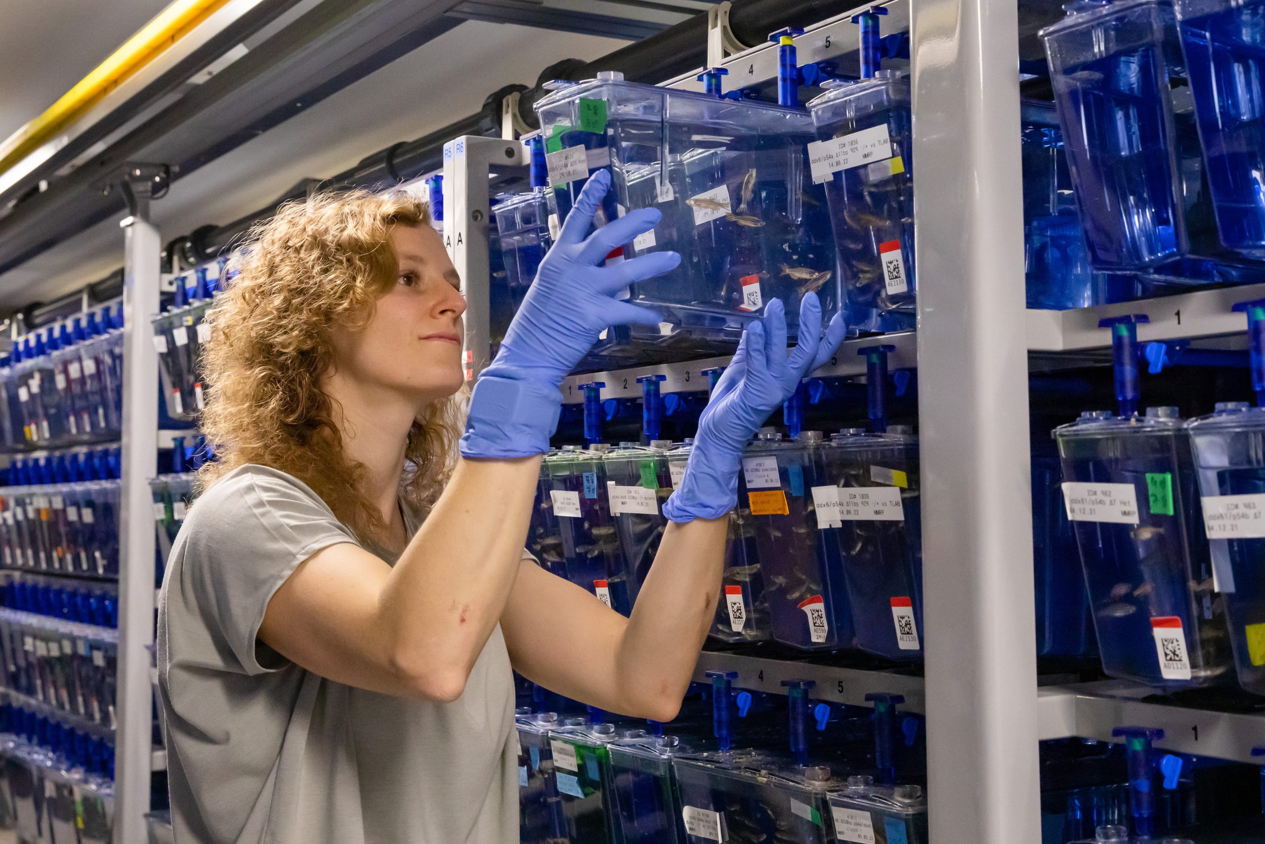
Madalena holding a tank of zebrafish. Photo credit: Annette Roulier, Biozentrum Basel.
How did the collaboration with the Seelig lab start?
Well, it took more than two years to get to a stage when I had designed and generated the 5′ UTR massively parallel reporter assay (MPRA) library, validated it and acquired the in vivo data. To be honest, I also spent quite some time running data analyses before convincing myself that the assay had actually worked and was recovering biologically meaningful information! Once I realized that was the case, I was super excited to try and learn as much from the data as possible. I was familiar with Georg Seelig’s work and had actually met him in person when he gave a talk at the Biozentrum’s Discovery Seminar. At that time, I was still running experiments but I approached Georg and asked if he would be interested in collaborating to explore the data with deep-learning models. Georg was excited about my project and told me to reach out when the right time came, and so I did! He then paired me up with Sebastian, and we started a wonderful collaboration together. Honestly, it was so efficient and so much fun: we would meet every two weeks, and at each meeting discuss our progress, come to an agreement on next analyses and experiments and then execute them. It gave the project a really nice momentum and it was just great to feel part of a team working towards a common goal. At the same time, Georg joined the Schier lab for a sabbatical, so it was great to have the opportunity to discuss and get feedback on a regular basis from both PIs in person. The project would have not been the same without Sebastian’s contributions, who developed the Danio Optimus 5-Prime (DaniO5P) deep-learning model to evaluate and interpret the 5′ UTR MPRA data. And Georg was the one who came up with the model’s name!
What were for you the most exciting findings, or particular moments during the project that stuck with you?
The most exciting moment was running the Kozak sequence and uORF analyses and realizing that the assay had actually technically worked. It also meant that the data likely contained additional sequence information to be uncovered, which was a super exciting prospect. And then of course, seeing motifs emerging from the motif enrichment analysis and the DaniO5P model!
I’d also like to say that when I started this project, I had no experience performing polysome profiling. I was extremely fortunate to meet Sunil Shetty, at the time a postdoc in the Hall group at the Biozentrum (now leading his independent research group at the Tata Memorial Centre in Mumbai, India), who taught me how to prepare sucrose gradients and perform polysome profiling. His happiness and positivity are contagious, and some of the most fun moments of my postdoc were spent next to the polysome profiling machine learning from Sunil. I am deeply grateful to him, and also to Michael Hall for kindly welcoming me in his lab.
And what was most challenging?
For sure it was starting my postdoc and having the COVID-19 pandemic hit a few months later. At the time, my plan was to experimentally define zebrafish 5′ UTRs by long-read sequencing to design the MPRA. There was so much uncertainty about when we could be back in the lab full time, that I decided to instead use an alternative approach to computationally infer 5′ UTR sequences from a public dataset of cap analysis of gene expression data. I don’t have a background in computational biology, so it was definitely a slow and iterative learning process – but also very rewarding in the end, and most importantly it allowed me to move forward with the project.
What’s ahead for you?
I am currently applying for Group Leader positions. Going on interviews and getting to meet faculty members from different research institutions is really an amazing and rewarding experience, but also very time-consuming, so it’s hard to get any experiments done alongside applications and interviews. I am super excited to start my own independent research group soon, and I hope to continue to collaborate with Sebastian on future projects!
Montage of zebrafish embryo images acquired at consecutive stages of development (from 2 to 10 hours post-fertilization) that were injected at the 1-cell stage with a fluorescent dextran dye (magenta) and an mRNA reporter encoding GFP (green), reminiscing of the moon phases. (1 votes) Loading...
At the end of each month, I pick the same month from a random year from the past 15 years of the Node, and take a look at what people were talking about back then.
Previously, I travelled back to February 2011, March 2013 and April 2014 to have a look around the Node. Luckily, I didn’t get lost along the timeline and managed to get back to the present day. But now I’m itching for another adventure. So this time, let’s fasten our seat belts and turn the dial to May 2016…
In this series, we asked prominent researchers to recommend their favourite hidden gems from history – papers that are, for whatever reason, unjustly overlooked today. Read the other Forgotten classics posts.
We love it when you’ve organised a science outreach activity and then tell us how it went. It’s also useful for other researchers to get inspiration about their own outreach events. Check out other ‘outreach’-related posts.
Catarina Vicente was the Node’s Community Manager from 2013 – 2016. What’s she been up to since then? Find out from this conversation with all the past and present Community Managers.
It lives. It lives! What lives, you may ask? Well, somewhere in a lab at Yale University, one young scientist has stuck human brain cells and chimp brain cells together to make a chunk of hybrid brain. A few weeks ago, I met with her to ask more about this research. She admits that it all sounds like “mad science,” but this mad scientist might be taking a big step forward on our path to find out what really makes us human.
Chimpanzees are our closest relatives, and closer than most of us would probably like to think about. We share some 98.8% of our DNA with chimps.1 This means only about 1.2% of our DNA accounts for the uncanny power of our species to build cities, write symphonies, split atoms, and do all the other things we alone do so well. We know that much of this uncanny power resides in our brain, which is massive compared to a chimp’s brain,2 and has a much larger wrinkled region at its front3 that does most of our complex “higher” thinking. This wrinkled region at the front of our brains takes almost twice as long to finish developing in us as it does in chimps,4 and scientists have long thought that its slow development in humans helps to explain our subtle and adaptable “higher” thinking abilities.4 What we do not know is how that meager 1.2% of our DNA goes about making our wrinkly-fronted brains develop so slowly.
For the last two years, a young scientist named Reem Abu-Shamma has been trying to change that. Since graduating summa cum laude from UCLA, Reem has made a career of mutating genes, creating artificial 3D clusters of human intestinal cells (delightfully called “organoids”), and using computer programs to study vast amounts of DNA. These endeavors might sound eerily sci-fi, but have in fact taught us a lot about public health and disease. Her work mutating genes in parasites could shed light, down the line, on how we treat some particularly nasty strands of malaria,5 and her work with human intestinal organoids promises to tell us more about the cellular basis for inflammatory bowel disease. Now, as a PhD student at Yale University, Reem has set her sights on what makes our brains human.
” Slower development means more time to make a big brain…So where in the genetic code is it telling our brains to develop slower? “
To investigate what makes our brains unique, Reem has created something like a hybrid “half-human, half-chimp brain.” This phrase baffled me as much as it’s probably baffling you, so I sat down with Reem to ask her more about what inspired this research. And, as I listened to Reem’s enthusiastic, down-to-Earth explanation for her project, it began to seem less like mad science and more like vital research. “Large brains allowed us to dominate the world for better or for worse,” Reem explains. She wants to find “the underlying code in our cells that has allowed us to do that.” In searching for this code, Reem has focused on the speed at which that wrinkly portion at the front of our brains develops. “Slower development means more time to make a big brain…and we know that the human brain takes a really long time to develop.” This slow pace of human brain development manifests at the cellular level6—individual human brain cells take years to branch out and mature, whereas those of chimps develop much faster. Reem’s research question is simple, then: “Where in the genetic code is it telling our brain cells to develop slower?”
To answer this question, Reem recently joined the Noonan Lab at Yale University, which has a long history of using the best-available gene-editing technology to study human brains. One particular focus of the Noonan Lab has been to find particular bits of DNA that distinguish humans from chimps and other animals. What exactly are these bits of human specific DNA? Well, as Reem explains, “they’re parts of the genome that are not genes,” but “dials” for genes, which make various brain-building genes more or less active as the brain develops.7 These bits of DNA are part of that 1.2% of our genetic code separating us from chimpanzees, and could tell us a lot about how that huge wrinkly portion at the front of our brains develops so slowly, gets so big and complex,5 and makes us so clever. Each one of these bits, once found, “gives us a hint that maybe this part of the genome helped us evolve big brains.” However, that hint alone doesn’t prove the bit’s role in making our brains bigger or tell us how it did. In order to actually verify what these human bits of DNA do, scientists have to mutate them, and see how those mutations affect the development and interaction of different human and chimp brain cells over time. Obviously, no one at any credible research institution wants to mess with the brain of an actual living human—institutional and federal guidelines fortunately forbid that kind of work. But scientists dowant to understand what these human-specific bits of DNA are doing. So how do you mutate realistic human brains without using actual real human brains?
Well, remember those “organoids” I mentioned before? Reem uses those. And they’re a lot less scary than they sound. “We’re not using real animals, or growing real brains” Reem assures me with a laugh. Instead, she’s using what amounts to just a few cells: To create brain “organoids”—again, 3D clusters of living brain cells—she uses cells that other labs have collected from human or chimp skin. These labs treated those skin cells with various molecules to “reprogram” them into stem cells, which can turn into almost any other kind of cell if given the right molecular cues. “In our case,” Reem explains, “we make them turn into neurons.”8 Reem uses this approach to create human and chimp brain cells, and then grows each reprogrammed brain cell into a different 3D cell cluster or “organoid.” The result in each case is a separate ball of brain cells9 for each species that develops much like they would in a real brain.
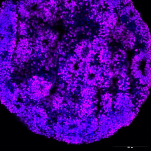
A human brain organoid, 30 days old, made by combining two different human cell lines. Cells are labeled with two overlain molecular markers——a blue one marking all cell nuclei and a violet one that marks “forebrain cortical neuron progenitors” (the kind of cells that end up forming the wrinkly front of our brains). The cells in this organoid have spontaneously arranged themselves into “rosettes”, much like brain cells do in an embryonic brain. Image courtesy of Reem Abu-Shamma.
With each brain organoid, Reem plans to test what our human-specific bits of DNA are doing to make our brains grow slower and larger. She will do this by tweaking or changing10 various human-specific bits of DNA to make them act more like the corresponding regions of chimp DNA, and vice versa. Then she’ll see how these modifications affect the activity levels of various brain cell genes and the “speed” at which those brain cells ultimately develop. “By ‘speed’, we don’t mean absolute time; rather, we have the technology to look at a single cell and figure out how mature it is based on the molecules we observe in it,” Reem clarifies. Then, for each bit of human-specific DNA, she’ll see whether the humanized chimp cells appear to develop more slowly, while the “chimpanized” human cells develop more quickly. Then we would know that this specific bit of our 1.2% unique genetic code is partly responsible for making our brains so weirdly human.
Reem finds the sheer size of this mystery fascinating. “The genome is a really big place,” she explains. “It’s so vast and we don’t know what most of it does. It kind of feels like detective work, because you’re trying to see where in this really big space it’s telling us to be human.” By tweaking little bits of human and chimp DNA so they behave more like their counterparts—a sort of genetic Freaky Friday—Reem can do just that, finding which bits of human-specific DNA tell our brain cells to grow in a human way. This in itself is the stuff of science fiction. However, Reem and her PhD advisor, Dr. James Noonan, are taking this approach one step further.
They aren’t just growing human brain-cell colonies and chimp brain-cell colonies. They’re mixing them together, to make something like a miniature hybrid brain. Despite their different origins, these cells branch out and interconnect much like the cells in our own brains, possibly creating a cellular communication network unseen in nature. “Why would you make a half-human, half-chimp brain?” Reem jokes that her mother and even her colleagues have often asked her this question. But Dr. Noonan initially suggested this approach, and Reem has pursued it, because we can learn a lot from it.
” It kind of feels like detective work, because you’re trying to see where in this really big space it’s telling us to be human. “
Brain cells don’t usually grow on their own. They grow in response to cues from neighboring cells, and these hybrid brains can show us the extent to which human brain cell development is genetically encoded. How much of how our brain cells behave is written in their DNA, and how much is determined by interaction with their cellular neighbors? Specifically, Reem is curious whether the sum of brain cell interactions, and the presence of similar brain cells from other species, together affect how fast that wrinkly portion at the front of the brain develops. Previous studies have found that these external cues (the “cellular environment”) don’t matter much for the development speed of human brain cells.11,12 However, few if any studies have used hybrid chimp-human brain organoids to study that big wrinkly-fronted part of the human brain. By creating hybrid chimp-human organoids with this specific type of brain cell, Reem will finally test whether environmental cues help it grow slower in humans. Reem gives me an example to help me wrap my head around this. So, suppose you “take a human cell and transplant it into a chimp brain organoid,” Reem explains. And then suppose you collect molecular data from human brain cells inside a purely human organoid and then do the same to human brain cells inside a human-chimp hybrid organoid. “If they’re exactly the same, then the environment the cell is in isn’t as important!”
Making and mutating hybrid brains is intense work. To do it right, Reem has to set up hundreds of different brain organoids, each in its own plastic well, and from a variety of different human and chimp donors. She goes into the lab every day. “I check on my cells immediately…first thing. I make sure they’re still alive.” She recounts instances where some of her organoids became cancerous, and others spontaneously collapsed and started dying—both unplanned events that threatened to skew her work and required hours of manual labor to remedy. “So many things can go wrong…it’s a lot of manual labor to make sure they’re alive and happy.” On a daily or weekly basis, she has to feed her many hundreds of brain organoids, and look at each one under a powerful microscope to make sure nothing has gone horribly wrong. She has to modify them all at just the right time, in just the right way. And then, when all of that is done, within the next few months, she’ll have to extract specific molecules from these organoids and analyze the resulting vast amounts of data to see how her mutations changed the approximate speed of brain cell development in her human brain, chimp brain, and hybrid brain organoids.
Reem is eager to find “what inferences we can make about the speed of development using these models”—at what rate the brain cells are likely growing, dividing, branching out, and developing their various special functions. With this approach, Reem wants to pinpoint some of the intrinsic genetic factors responsible for speeding up and slowing down the “molecular rate” of human brain cell development.
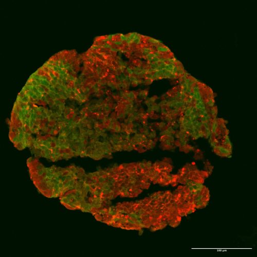
A hybrid human-chimp brain organoid from Reem’s first experiment on this project. Molecular markers tag chimp brain cells red and human ones green. This hybrid chunk of brain is 17 days old. Image courtesy of Reem Abu-Shamma.
When I asked Reem about the benefits of this research, her answer surprised me. Of course, her work does have implications for treating and understanding psychiatric or developmental conditions—autism spectrum disorder, schizophrenia, and other cognitive differences that often relate to brain development. That was the answer I expected. But Reem went on to highlight something else. “This is a very exploratory study,” she explained. “It’s hypothesis-generating,” and “in the history of science, doing fundamental research can sometimes lead you down unexpected paths, just because you’re exploring your curiosity.” This “fundamental research” is done not for its direct societal benefits, but to better understand ourselves and our world, and often has unexpected humanitarian value. For example, Reem points out, CRISPR was discovered by fundamental research projects on a few seemingly random repetitive patterns in microbe DNA. And yet, CRISPR now forms the most promising avenue for therapeutic gene editing and has a variety of other applications for human health and disease worldwide.13,14
Reem’s work on hybrid brains is fundamental research in the same way. Yes, it has biomedical implications. But its potential value is so much broader. It can shed light on the parts of our genetic code that separate us from chimps and other animals. As Dr. Noonan told her when he first suggested making human-chimp hybrids, “no one’s done it before,” and we can hardly begin to predict what it might tell us about what makes us human.
Caleb Gordon is a Postdoctoral Associate at Yale University, where he studies the evolution of reptiles during the time of the dinosaurs.Check out his website to follow his research and popular science writing.
Note from the author: This piece was written as part of a workshop series taught by Carl Zimmer, and organized by Yale’s Graduate Writing Lab, on science reporting intended for a general audience.This workshop challenged us to write a popular science article without any scientific jargon. However, for any scientists missing this jargon, I’ve included more scientific terminology in the References Cited below. This article benefited greatly from feedback by Lauren Gonzalez and Joseph Lee at the Graduate Writing Lab.
[2] For more information about the evolution of human brain size, you can check out this research paper: Smaers, J. B., R. S. Rothman, D. R. Hudson, A. M. Balanoff, B. Beatty, D. K. N. Dechmann, D. De Vries, et al. “The Evolution of Mammalian Brain Size.” Science Advances 7, no. 18 (April 30, 2021): eabe2101. https://doi.org/10.1126/sciadv.abe2101.
[3] This wrinkled region at the front of the brain is called the “prefrontal cortex.” For more information about this remarkable brain region and its implications for our higher executive functioning abilities, you can check out this research paper: Preuss, Todd M., and Steven P. Wise. “Evolution of Prefrontal Cortex.” Neuropsychopharmacology 47, no. 1 (January 2022): 3–19. https://doi.org/10.1038/s41386-021-01076-5.
[4] Additional information on the prefrontal cortex is provided in this research paper from the same journal: Kolk, Sharon M., and Pasko Rakic. “Development of Prefrontal Cortex.” Neuropsychopharmacology 47, no. 1 (January 2022): 41–57. https://doi.org/10.1038/s41386-021-01137-9.
[5] The following research paper summarizes the results from Reem’s collaborative gene-editing work with malarial disease vectors: Subudhi, Amit Kumar, Anne-Lise Bienvenu, Guillaume Bonnot, Reem Abu-Shamma, Faryal Khamis, Hussain Ali Abdulhussain Al Lawati, Stephane Picot, Eskild Petersen, and Arnab Pain. “The First Case of Artemisinin Treatment Failure of Plasmodium Falciparum Imported to Oman from Tanzania.” Journal of Travel Medicine 30, no. 3 (May 18, 2023): taac092. https://doi.org/10.1093/jtm/taac092.
[6] The human brain matures more slowly in part because individual human brain cells take longer to develop. For a great review highlighting the uniquely protracted nature of human brain cell development, check out this recent paper: Lindhout, Feline W., Fenna M. Krienen, Katherine S. Pollard, and Madeline A. Lancaster. “A molecular and cellular perspective on human brain evolution and tempo.” Nature 630 (19 June 2024): 596–608. https://doi.org/10.1038/s41586-024-07521-x.
[7] For more information on what these human-specific bits of DNA are and what they do, check out this recent paper from the Noonan Lab: Pal, Atreyo, Mark A. Noble, Matheo Morales, Richik Pal, Marybeth Baumgartner, Je Won Yang, Kristina M. Yim, Severin Uebbing, and James P. Noonan. “Resolving the Three-Dimensional Interactome of Human Accelerated Regions during Human and Chimpanzee Neurodevelopment.” Cell 188, no. 6 (March 2025): 1504-1523.e27. https://doi.org/10.1016/j.cell.2025.01.007.
[8] For additional information about how these scientists create brain cells from stem cells, check out the following paper: Mariani, Jessica, Maria Vittoria Simonini, Dean Palejev, Livia Tomasini, Gianfilippo Coppola, Anna M. Szekely, Tamas L. Horvath, and Flora M. Vaccarino. “Modeling Human Cortical Development in Vitro Using Induced Pluripotent Stem Cells.” Proceedings of the National Academy of Sciences 109, no. 31 (July 31, 2012): 12770–75. https://doi.org/10.1073/pnas.1202944109.
[9] These colonies of brain cells are called “cortical organoids.” For more information about these remarkable 3D brain cultures, check out the following paper: Pollen, Alex A., Aparna Bhaduri, Madeline G. Andrews, Tomasz J. Nowakowski, Olivia S. Meyerson, Mohammed A. Mostajo-Radji, Elizabeth Di Lullo, et al. “Establishing Cerebral Organoids as Models of Human-Specific Brain Evolution.” Cell 176, no. 4 (February 2019): 743-756.e17. https://doi.org/10.1016/j.cell.2019.01.017.
[10] Reem mutates brain cell colonies using “arrayed CRISPR screens,” which are described in more detail in the following research paper: Bock, Christoph, Paul Datlinger, Florence Chardon, Matthew A. Coelho, Matthew B. Dong, Keith A. Lawson, Tian Lu, et al. “High-Content CRISPR Screening.” Nature Reviews Methods Primers 2, no. 1 (February 10, 2022): 8. https://doi.org/10.1038/s43586-021-00093-4.
[11] This research paper studied pure human and pure chimp brain cell organoids: Otani, Tomoki, Maria C. Marchetto, Fred H. Gage, Benjamin D. Simons, and Frederick J. Livesey. “2D and 3D Stem Cell Models of Primate Cortical Development Identify Species-Specific Differences in Progenitor Behavior Contributing to Brain Size.” Cell Stem Cell 18 (April 7, 2016): 467–480. http://dx.doi.org/10.1016/j.stem.2016.03.003.
[12] This research paper took human brain cells from that wrinkly region at the front of the brain and transplanted them into a live mouse brain: Linaro, Daniele, Ben Vermaercke, Ryohei Iwata, Arjun Ramaswamy, Baptise Libé-Philippot, Leila Boubakar, Brittany A. Davis, Keimpe Wierda, Kristofer Davie, Suresh Poovathingal, Pier-Andrée Penttila, Angéline Bilheu, Lore De Bruyne, David Gall, Karl-Klaus Conzelmann, and Vincent Bonin. Neuron 104 (December 4, 2019): 972–986. https://doi.org/10.1016/j.neuron.2019.10.002.
[13] Doudna, Jennifer A., and Emmanuelle Charpentier. “The New Frontier of Genome Engineering with CRISPR-Cas9.” Science 346, no. 6213 (November 28, 2014): 1258096. https://doi.org/10.1126/science.1258096.
[14] Barrangou, Rodolphe, and Jennifer A Doudna. “Applications of CRISPR Technologies in Research and Beyond.” Nature Biotechnology 34, no. 9 (September 2016): 933–941. https://doi.org/10.1038/nbt.3659.
This year’s venue will be the CRUK Scotland Institute in Glasgow from Tuesday 23rd to Friday 26th of September and will be the 7th since the meeting was first held at the Doherty Institute in Melbourne in 2018, the name having been coined by Prof Elizabeth Vincan at the time. The Glasgow meeting this September will be the first to be held in Europe!
Plenary, Keynote and Symposium sessions, including rapid-fire short talks and poster sessions, make up the programme, which includes many well-known international speakers.
Early bird registration closes on the 15th of June and includes discounted rates for students.
Organisers are very excited to be hosting this meeting, where cutting-edge technologies meet unanswered biological and clinical questions.
Prof Elizabeth Vincan, Clinical Scientist and Medical Researcher in the Department of Infectious Diseases, Melbourne Medical School, University of Melbourne says: “Organoids Are Us brings together delegates from diverse fields and disciplines to show case and learn about the latest advances in organoid technology, and its application to fundamental and clinical research to fast track discovery and translation. This is the seventh conference in this highly successful series, and the first in the UK.”
Prof Ramanuj DasGupta, Professor of Cancer Systems Biology at the School of Cancer Science, University of Glasgow and CRUK Scotland Institute says: “The Organoids Are Us conference aims to bring together the key opinion leaders in the fields of cancer, infectious disease and regenerative biology, who utilise organoid models to address key questions in human disease biology and development of novel therapies.”
This week we will meet Dr Cassandra Koh, who is a new faculty at Institut Pasteur. Cassandra is driven by curiosity and is passionate about decoding the molecular choreography between viruses, their insect hosts, and the lipids that entwine them. Her research traces the intricate ways arboviruses hijack mosquito metabolism to fuel their replication and has delved deep into how symbionts like Wolbachia rewire these metabolic pathways. Her brand-new lab will focus on how virus–virus–host interactions offer a nuanced view of disease transmission and microbial co-evolution. But science for Cassandra is more than experiments — it’s a way of asking questions about the world. She credits strong mentors and surprising inspiration from artists and storytellers for shaping her journey. Whether she’s tracing lipid dynamics in mosquito cells or hosting a long lunch for friends, Cassandra believes in curiosity as a compass. She’s currently welcoming collaborators and postdocs interested in exploring the metabolic intersections of immunity, infection, and symbiosis. Check out her work here ! Give her a follow over Bluesky and Twitter.
What was your first introduction to the fields of metabolism and viral infections?
It started with cholesterol. The fruit fly Drosophilamelanogaster carries a bacterial endosymbiont called Wolbachia that suppresses pathogenic viral infections in its host. This was a very exciting finding that resulted in a biological intervention strategy against mosquito-transmitted viral diseases based on the stable introduction of the Wolbachia wMelstraininto natural populations of a major mosquito vector species, Aedesaegypti. Eric Caragata had showed that Wolbachia-induced viral suppression became weaker when Drosophila flies had more cholesterol in their diets, leading to the conclusion that Wolbachia and Drosophila viruses were competing for host cholesterol (PMID: 24337107).
Naturally, this led to the question of whether Wolbachia and mosquito-transmitted viruses (also called arboviruses) also compete for host lipid resources. During this study, I learned a lot about the role of lipids in viral infections in mosquitoes from a key paper by Rushika Perera (PMID: 22457619). Her work had shown that dengue virus replication in mosquito cells relies on the activity of fatty acid synthase. In addition, as a flavivirus, dengue virus re-organizes the endoplasmic reticulum membranes to form replication complexes, and this is reflected in an enrichment of lipids that promote membrane curvature and permeability.
You use the term “virus-virus-host” interaction for describing the theme of your lab. Could you share your journey into studying this and using mosquitoes as one of your host model systems? Tell us how you got inspired to further branch out into metabolic aspects of viral infections?
The interaction between arboviruses with their mosquito host is a fascinating subject in itself. Mosquitoes have evolved interesting ways to fight off and tolerate viral infections and the virus seeks to complete its transmission cycle while minimizing virulence to its vector host. With the recent appreciation that mosquitoes harbor a multitude of other viruses that constitutes its resident microbiota, virus-mosquito interaction is no longer a two-player game. These resident viruses are called “mosquito-specific viruses” to distinguish them from the ones that infect and cause disease in humans and animals. They have entered the research spotlight as many studies have reported their ability to reduce or enhance arbovirus infection in mosquitoes, which implies that they have a role to play in disease transmission. Many questions have since sprung up about how they interact with arboviruses and the mosquito host. I am curious to see whether these interactions can be observed in the metabolic dimension.
Your work intersects virology, metabolism, immunology and RNA interference – tell us what studying immune-metabolism means for you. How do you integrate these disciplines in your research, and what unique insights have emerged from this approach?
Immune responses cost energy. Viral infections cost energy. A virus infection is therefore a disruption to immune and lipid homeostasis. There is already some evidence of crosstalk between immune signaling and lipid metabolism modulations in bacteria-infected Drosophila (PMID: 30902902, PMID: 33227003). On top of that, viruses hijack host lipid membranes and reconfigure phospholipids to form viral replication complexes (PMID: 33087565), adding to the toll on the metabolic burden of virus-infected cell. Immune and metabolic regulation therefore go hand in hand and would provide a more holistic view of cellular responses to viral infection.
How has Wolbachia evolved in Drosophila and mosquito species – are a lot of mechanisms of symbiosis evolutionarily conserved? How can studying Wolbachia-insect interactions help us understand symbiont-driven metabolic changes? Could you shed more light on these emerging areas of research.
Wolbachia is a common endosymbiont among arthropods including insects like Drosophilamelanogaster and some mosquito species like Aedes albopictus. When it was observed that some Wolbachia strains protect their Drosophila host from viral infection, the notion that the endosymbiont could be introduced into Aedesaegypti mosquitoes, a non-native host, led to the development of Wolbachia-based intervention strategies to limit the spread of viruses transmitted by this major vector species. That Wolbachia is vertically transmitted through the maternal line was a very useful property for a sustainable and self-driving intervention.
Studying how Wolbachia interacts with its hosts would reveal its mechanisms of symbiosis, which would say something about the directions of evolutionary pressures in natural or introduced hosts. These mechanisms might take the shape of metabolic mutualism, immune priming, or something else entirely.
What are the key differences in how Wolbachia and DENV-3 (Dengue virus serotype 3) alter host lipid metabolism?
In our work comparing how Wolbachia and DENV-3 modulate Aedesaegypti lipids, we found that the endosymbiont and the virus individually produce very different lipid alterations. While DENV-3 produced strong elevations, Wolbachia-infected mosquitoes exhibited much milder perturbations of different lipid species, which does not support the Wolbachia-virus competition hypothesis.
Tell us about your findings on association of DENV3 infection with lipid droplet growth and hyperglycemia in mosquitoes? Tell us about your exciting finding of how Cardiolipins are involved in DENV infection.
DENV-3 infection alone strongly elevated levels of triacylglycerols, glycerophospholipids high in polyunsaturated fatty acids, and Amadori-glycated phosphatidylethanolamines in Aedesaegypti mosquitoes. These lipid classes indicate lipid droplet accumulation, cellular membrane remodeling, and viral infection-induced hyperglycemia. The latter is especially interesting as it suggests that this cellular phenomenon, which has previously been observed in human cells infected by dengue viruses, could also occur in mosquito cells.
Cardiolipins were an interesting class of lipids to notice in our dataset because they have not been previously associated with infection. Given its role to maintain mitochondria function, and our finding that cardiolipin depletion disfavors virus replication, we surmised that cardiolipins help to buffer the effects of cellular stresses from viral infection that would otherwise lead to the triggering of the apoptosis regulation pathways.
Building upon your findings in lipid metabolism, what are your upcoming plans? Are there particular metabolic pathways or hormonal regulators you aim to investigate further?
It seems that viruses are strong remodelers of the lipid landscape in mosquitoes. I am keen to see how these modulations take place in different virome backgrounds. The crosstalk between immune and metabolic pathways is something I wish to dive into deeper. It would tell us something about the mechanisms through which host microbiota influence arbovirus infection and transmission.
What role does curiosity play in your life, both within and outside of science? How does studying viral manipulation of host lipid dynamics have implications on evolution of metabolism and its connection to therapies?
Interesting question, because I think I view and approach life in general with curiosity. The fact that I can make a living from finding out stuff is something I am very thankful for.
The insights from host lipids modulation could inform on long-term co-evolutionary dynamics between mosquitoes and their viruses. On the clinical side, understanding what host lipids and metabolites are co-opted by arboviruses, for both the arthropod and vertebrate side of the story, may lead to identification of disease severity risk factors or new therapeutic targets.
Tell us about how you see the future of immune-metabolism evolve with the new upcoming tools – what techniques have you used and which tools are you most excited about?
Single-cell transcriptomics and metabolomics are currently the ‘shiny new things’ in the omics space. As viruses don’t infect every single cell within a tissue, this unprecedented level of resolution would allow us to pinpoint cellular virus-modulated metabolism so much more accurately. I look forward to some paradigm shifting revelations that these techniques will bring about.
What advice would you offer to students and early-career scientists interested in exploring the intersections of metabolism and host-virus interaction? What’s an unexpected place you’ve found inspiration for your work?
Finding the right mentors who showed me what it means to be a scientist has been instrumental in my career path. I have also surprisingly found inspiration from actors and singers, like Hugh Jackman, who (according to a podcast interview I heard) gave himself five years after graduating from drama school to see where his career would take him, or Sabrina Carpenter, who debuted in 2014 and kept going until she produced chart-toppers ten years later.
Science-wise, I think more people should know about this thorough review about virus-vector metabolic interactions (PMID: 37360524).
How do you maintain a balance between your rigorous research activities and personal life? Are there hobbies or practices you find particularly rejuvenating?
Maintaining balance will be always a challenge for me but it helps greatly to know where I draw fulfillment and contentment from. I love my work as a researcher, but personal relationships are what brings me the most joy. I am the grandma friend, so I enjoy hosting long lunches and dinner parties.
If you hadn’t embarked on a career in biological research, what other profession might you have pursued, and why?
I think I would have been a food historian. I like learning about how cultures and geography influence the flavor profiles and methods of diverse cuisines. It is the intersection of anthropology and culinary science. I have questions like: Why do cuisines from warmer climates tend to be rich in spices? And what inspired someone to turn spent brewery yeast into a sandwich spread?
Anything else you’d want to highlight?
If immune-metabolism in vector mosquitoes sounds like your kind of vibe, please do get in touch. I would love to hear from postdoc candidates and/or grant co-writers.
Last week we learnt about treating rare metabolic disorders using Drosophila genetics and basic science research via precision medicine, check out the article – Finding Fruit in Flies (Holly Thorpe)
Prof. Wendy Bickmore elected as an International Member of the US National Academy of Sciences (NAS).
Image of Prof. Wendy Bickmore
Prof. Dr. Andrea Rentmeister receives 2024 Franco-German “Georg Wittig – Victor Grignard” Prize, awarded by the Société Chimique de France.
Image of Prof. Dr. Andrea Rentmeister
Prof. Edan Foley relocating her group from the University of Alberta to the Department of Life Sciences at University of Bath.
Prof. Dr. Ruben Portugues moving to the Department of Neurobiology and Behavior at Cornell University.
Dr. Sumeet Pal Singh joins the School of Natural Sciences at Shiv Nadar University as an Associate Professor.
Dr. Shawn Burgess named Associate Editor for GENETICS Journal.
Dr. Laurie Nemoz Billet awarded Accessit 2025 from SFBD for her thesis on the role of the ECM in motor nerve development and regeneration in zebrafish.
Zebrafish Rock! Slack tankspace surpassed 500 active monthly users. Register your interest, if you are keen to join at https://linktr.ee/zebrafishrock
Dr. Luís Hernández-Huertas of Pablo de Olavide Lab
Drs. Briana Davis & Maggie Morash of John Rawls Lab
Now you can submit news, jobs and research to the #DanioDigest without a social media account! Get direct access to the #zebrafish community and beyond by filling out our Google Form with the details: https://forms.gle/H4nFUYqY5feMhBgQ8
Special thanks to Maddie Ryan, Charli Corcoran & Michaela Noskova Fairley for putting this digest together! If you would like to thank the Zebrafish Rock! team for their time & effort, you can buy us a strong cuppa at the link below. Every little bit keeps us caffeinated and motivated! We appreciate your support 🙂
 (No Ratings Yet)
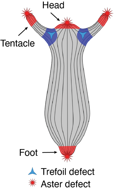
 (2 votes)