A deep-dive into the many cool (and free) resources available to zebrafish researchers!
Disclaimer:This is not a comprehensive list but a list of useful websites that are free (or have free versions that are useful)
Featured Resources
ZFIN (https://zfin.org/): “The Mothership” – Contains vital resources such as The Zebrafish Book, Thisse’s expression data & curated publications.
Alliance of Genome Resources (https://www.alliancegenome.org/): “The Central Hub of Model Organism Databases” – The future of MODs.
MARRVEL (https://marrvel.org/ ): “Disease Modeling on Steroids” – Super powerful resource including links to OMIM & ClinVar.
Ensembl (https://www.ensembl.org/Danio_rerio/Info/Index): “Ol’ Reliable Genome Browsing” – Contains current & past genome assemblies in a easy(-ish) format. BLAST/BLAT is a powerful tool for queries.
Integrative Genomics Viewer (https://igv.org/): “Simplified Genome Browsing Software” – Better as a desktop app. Lovely interface.
UCSC Genome Browser (https://genome.ucsc.edu/): “Genome Browsing With Bells and Whistles” – Contains custom tracks such as ‘DANIO-CODE’ & ‘ZebrafishGenomics’ with super helpful CRISPR/Cas9 guide predictions via CRISPRscan.
GEO DataSets (https://www.ncbi.nlm.nih.gov/gds): “One Repository to Rule Them All” – Shared resource for the life sciences & beyond.
DANIO-CODE (https://danio-code.zfin.org/): “The Exclusive Fishy Repository” – Contains multiple stages across lifespan.
DAVID (https://davidbioinformatics.nih.gov/): “Work-(sea)horse of Bioinformatics” – A beast of a website. A free alternative to Ingenuity Pathway Analysis.
CHOPCHOP (https://chopchop.cbu.uib.no/): “The Controversial (sgRNA Predictions)” – Fantastic for selecting guides for generating F0 crispants and CRISPR knock-ins.
Daniocell (https://daniocell.nichd.nih.gov/): “All Hail the UMAP!” – Turning into the gold-standard for zebrafish transcriptomics during early development.
Zebrahub (https://zebrahub.sf.czbiohub.org/): “Into the Multi-Ome” – The future is here! Dynamic repository of development. And you must check out the videos!
CiteAb (https://www.citeab.com/): “Never-ending Search for Antibodies” – If a zebrafish antibody does exist, it exists here.
ZF REG DATABASE (http://zfregeneration.org/ ): “Regeneration Rules!” – See if your gene-of-interest has a role in regeneration across tissues.
STRING (https://string-db.org/ ): “Missed Connection” – Finds the common link (e.g. expression data or literature reference) between your gene-of-interest & other potential hits in your datasets.
FaceBase (https://facebase.org/): “Trusted Resource for Craniofacial Researchers” – Comparative dataset of musculoskeletal tissues across species.
The 2025 Developmental Biology Gordon Research Seminar (GRS) was held on March 29-30 2025. This year, the GRS co-chairs Ana R. Hernandez Rodriguez and Anastasia Repouliou decided to organise an image competition by asking the GRS participants to submit their images and vote for their favourite.
“I have always believed that one of the great strengths of developmental biology is its remarkable visual appeal. The Developmental Biology GRC was a wonderfully diverse meeting with a variety of established and emerging model organisms, and we wanted to provide a platform for researchers to share captivating images of their work. These images were displayed throughout the week during breaks and poster sessions, creating an inspiring backdrop for cutting edge scientific discussions and highlighting the diversity and creativity within our community.”
Ana Hernandez (Co-Chair GRS Developmental Biology 2025)
Among the great selection of images, Paul Bump’s “Life as a drop in the ocean” won the most votes – congratulations!
You can browse through all the great entries to the competition in the image gallery below.
Browse through the image gallery (click to expand)
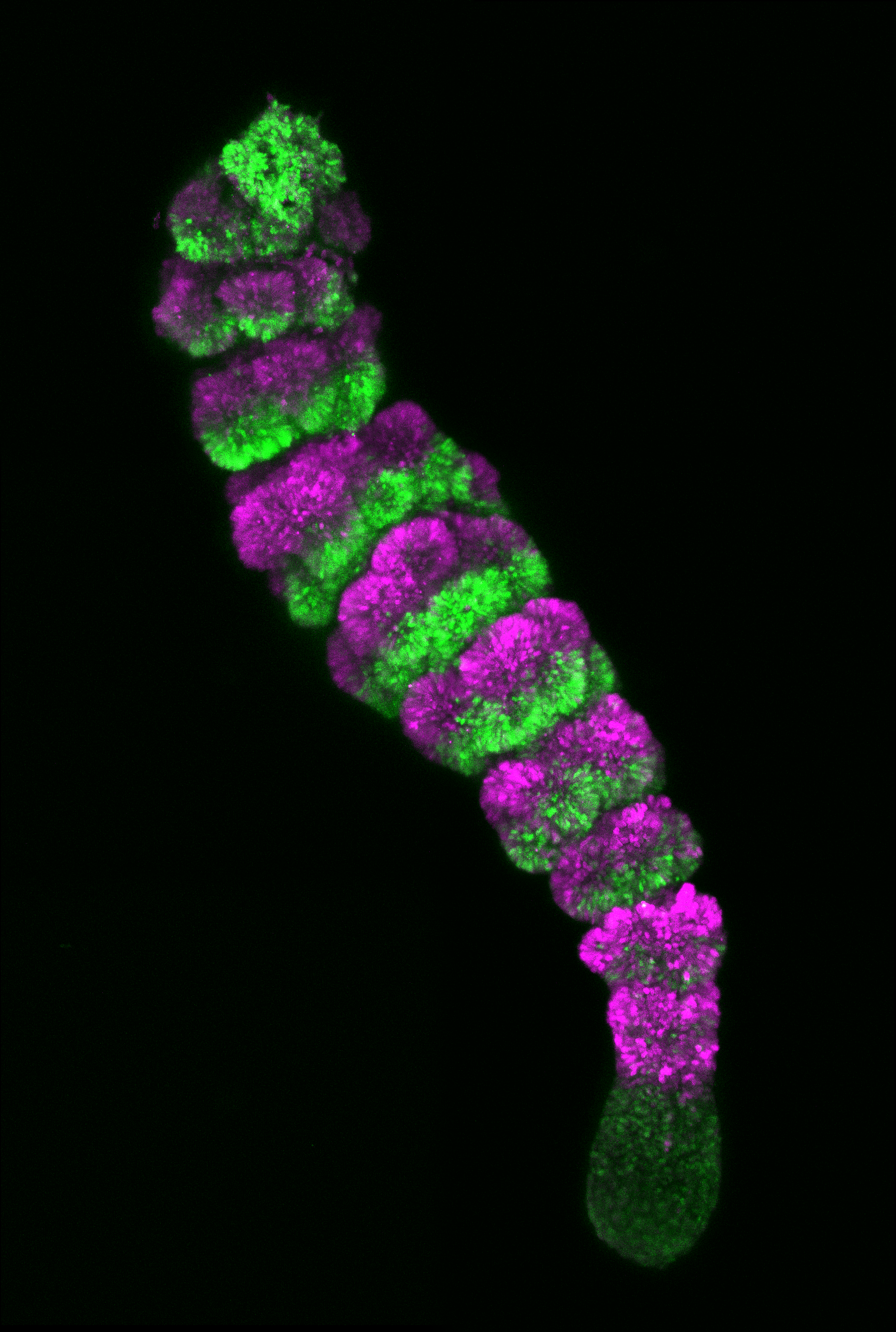
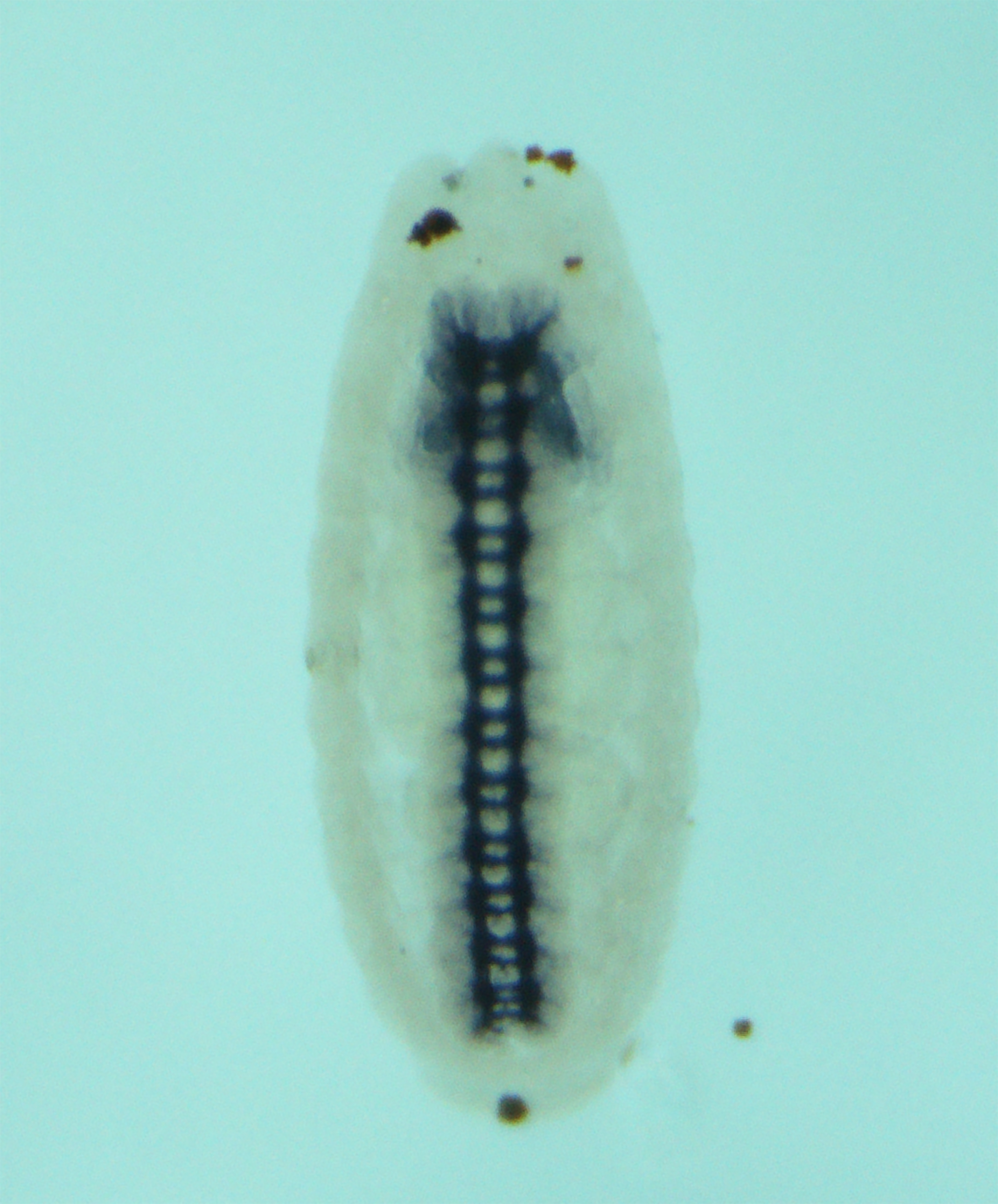
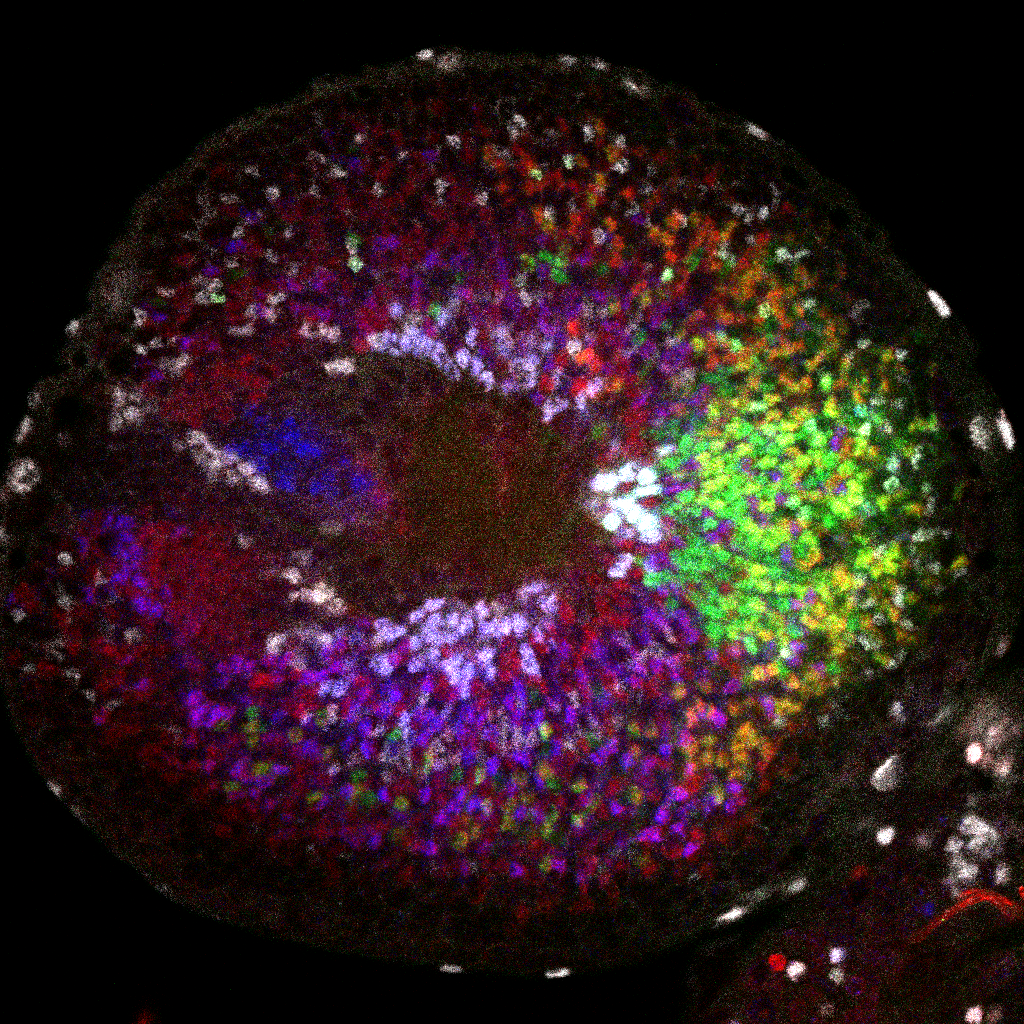
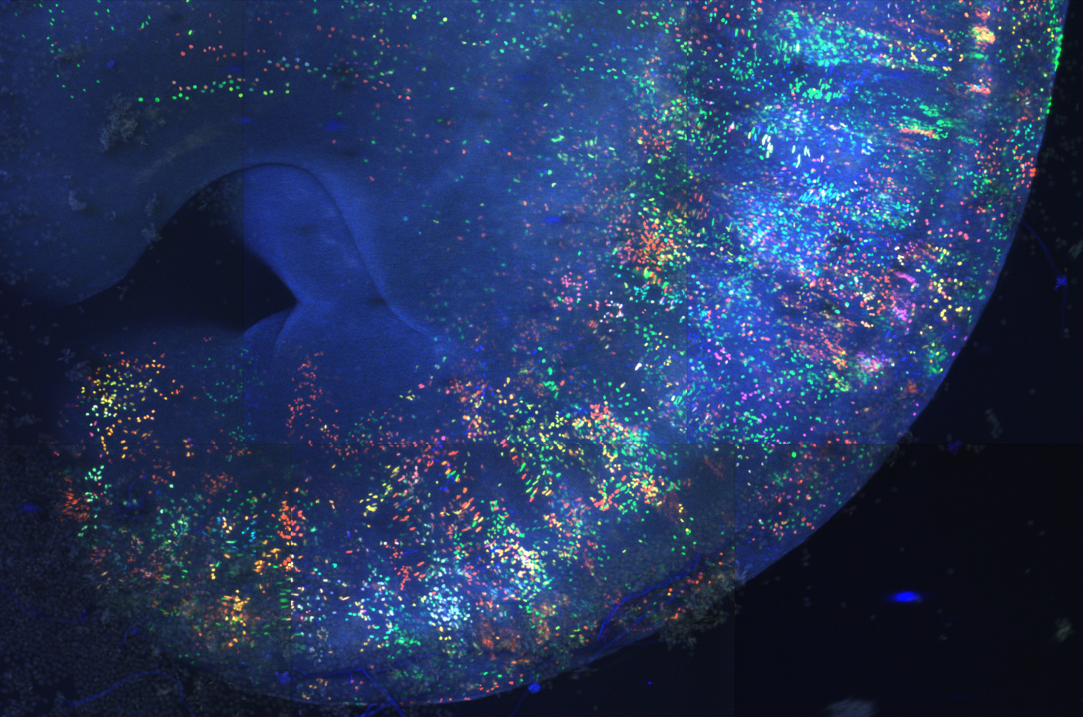
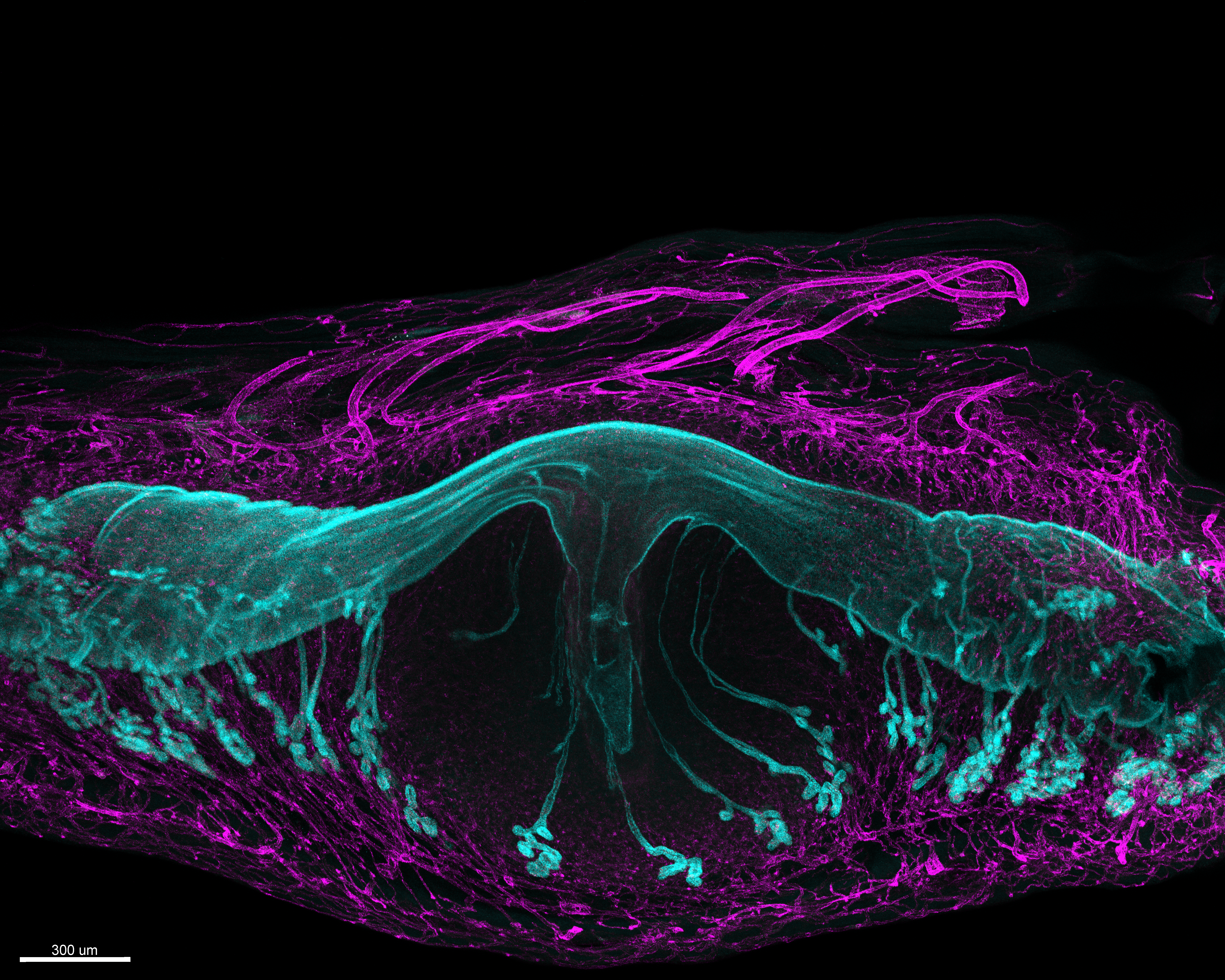
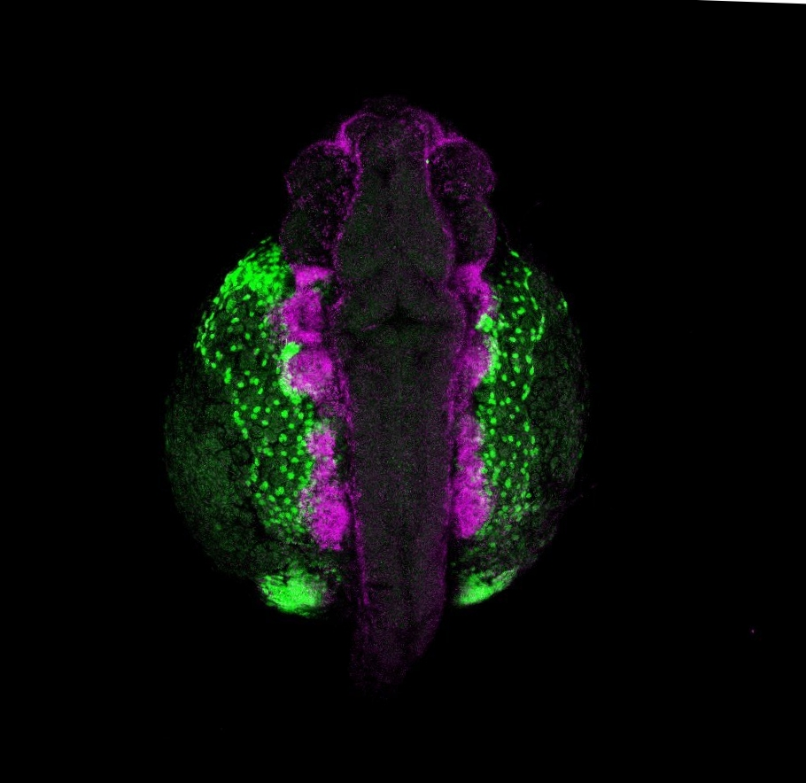
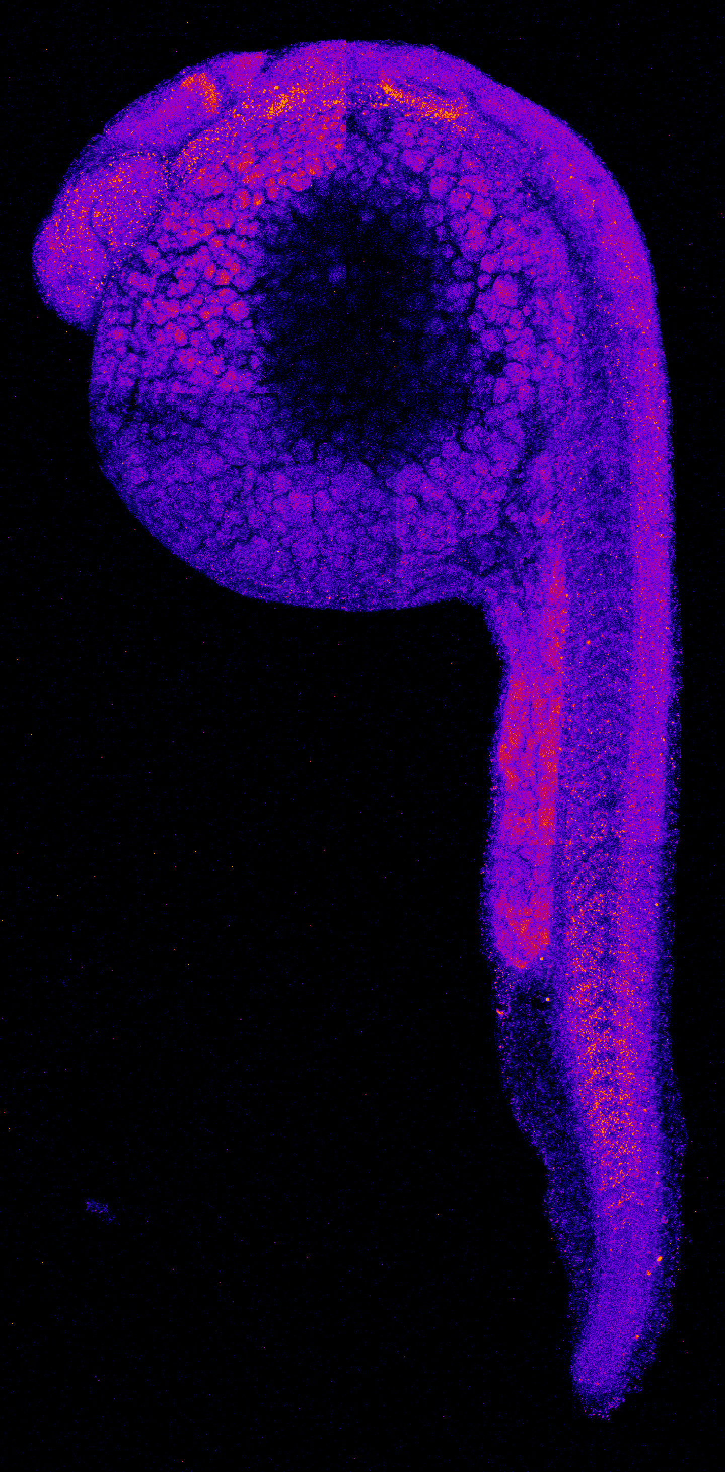
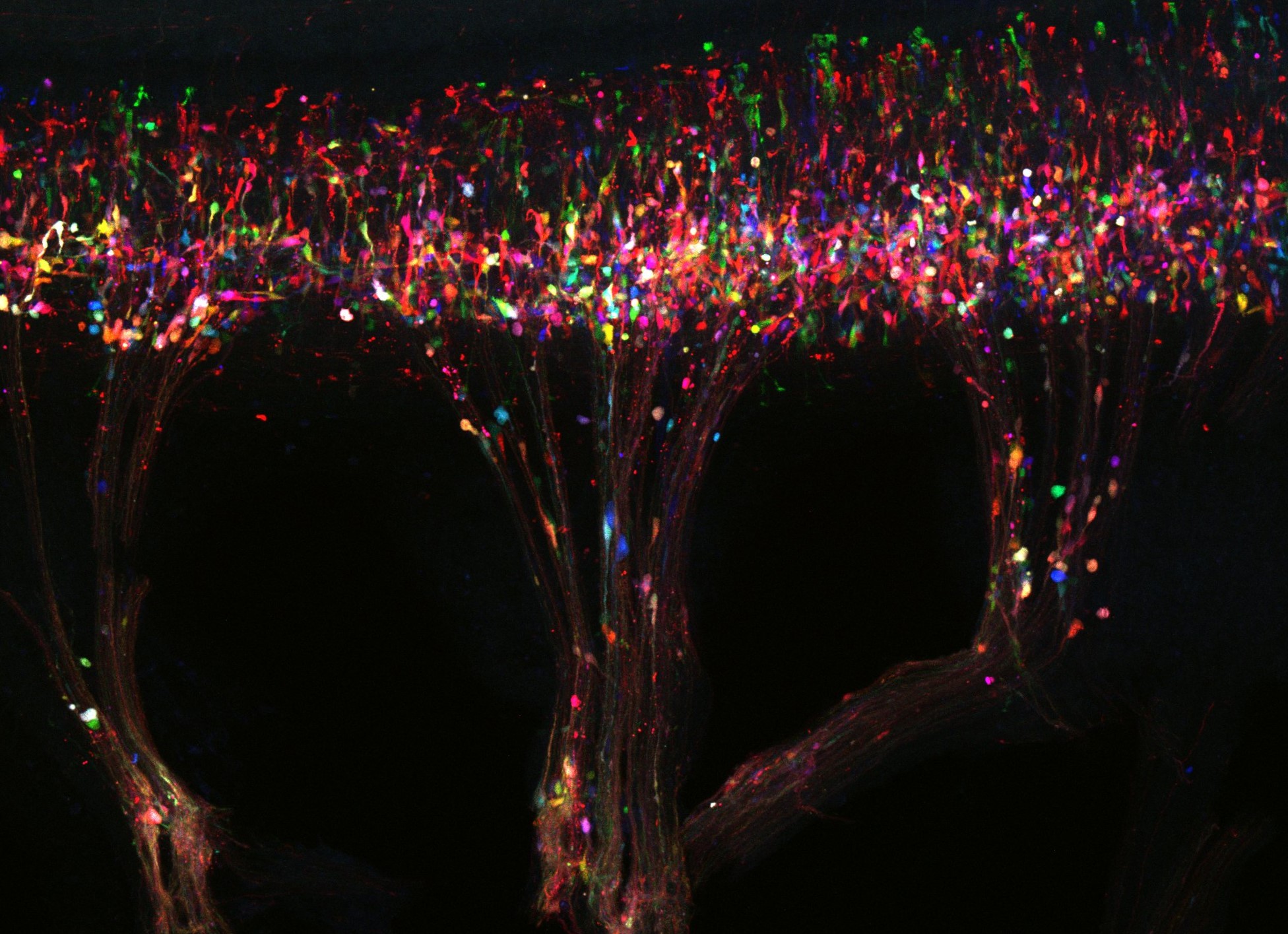
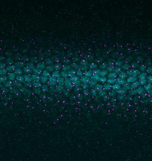
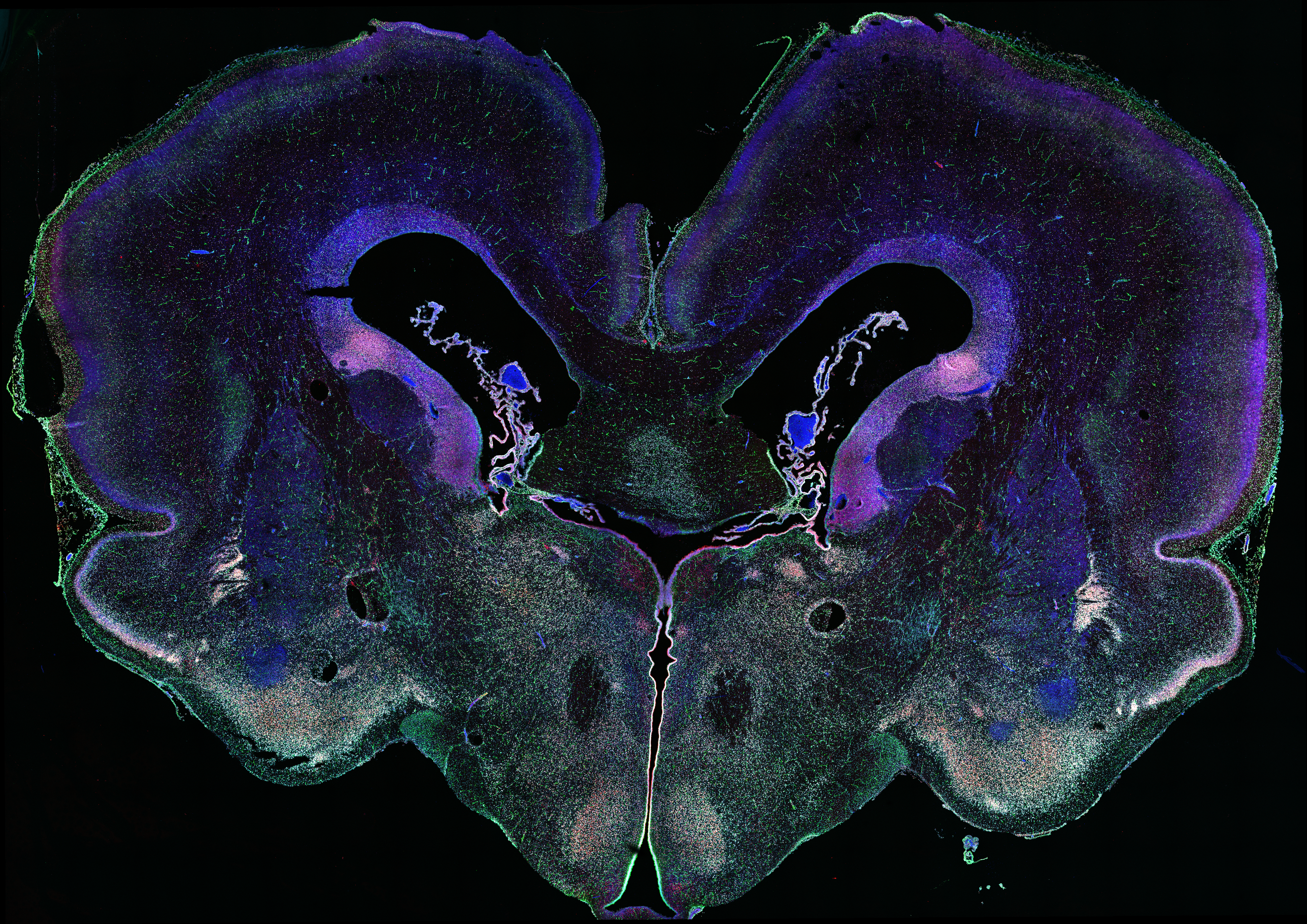
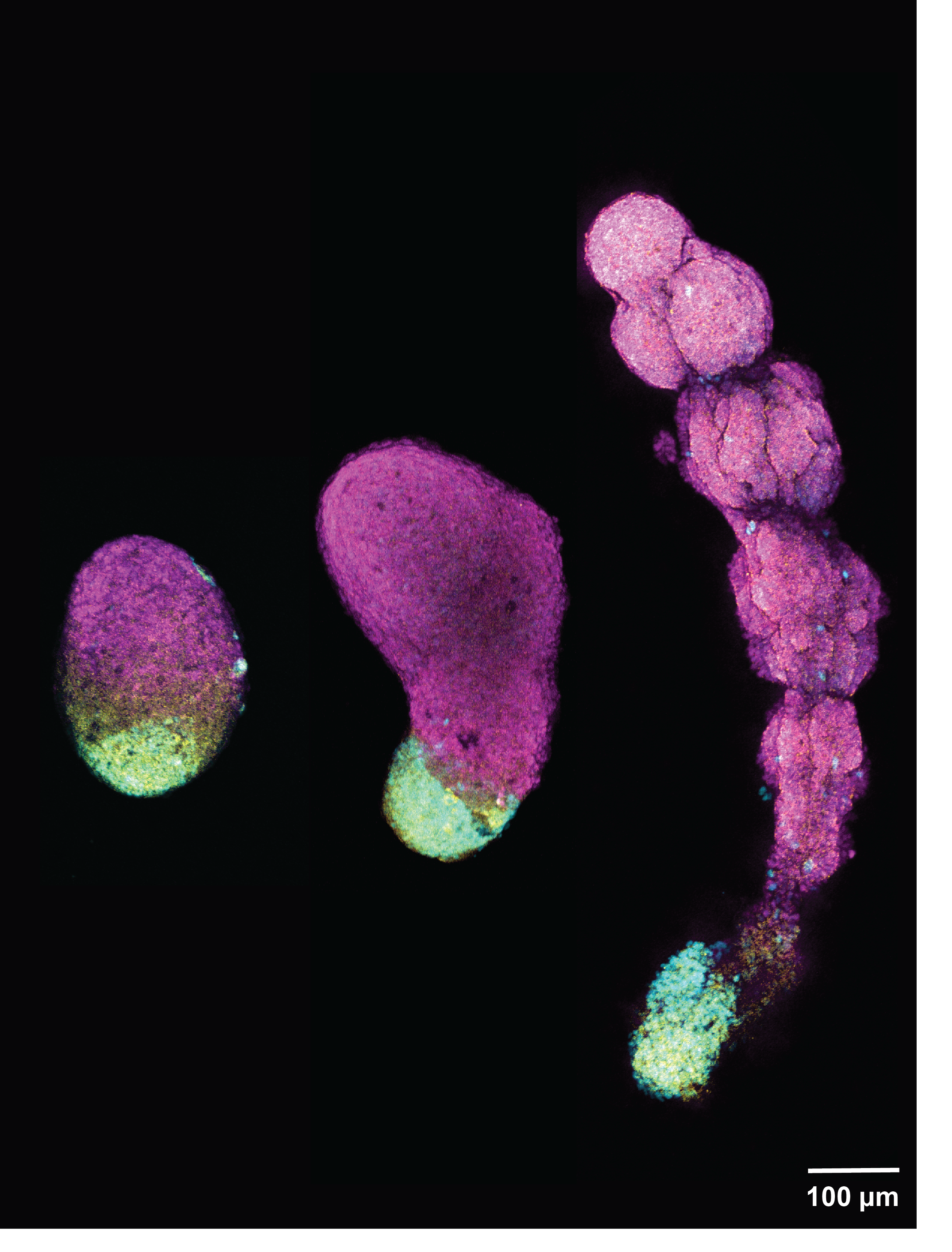
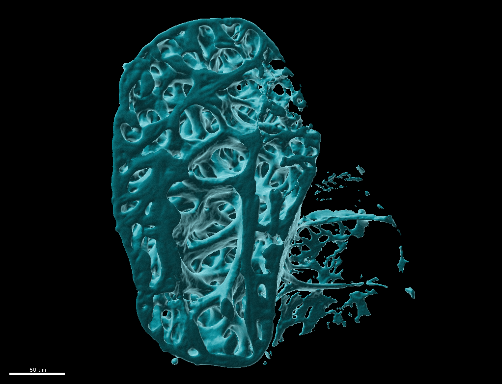
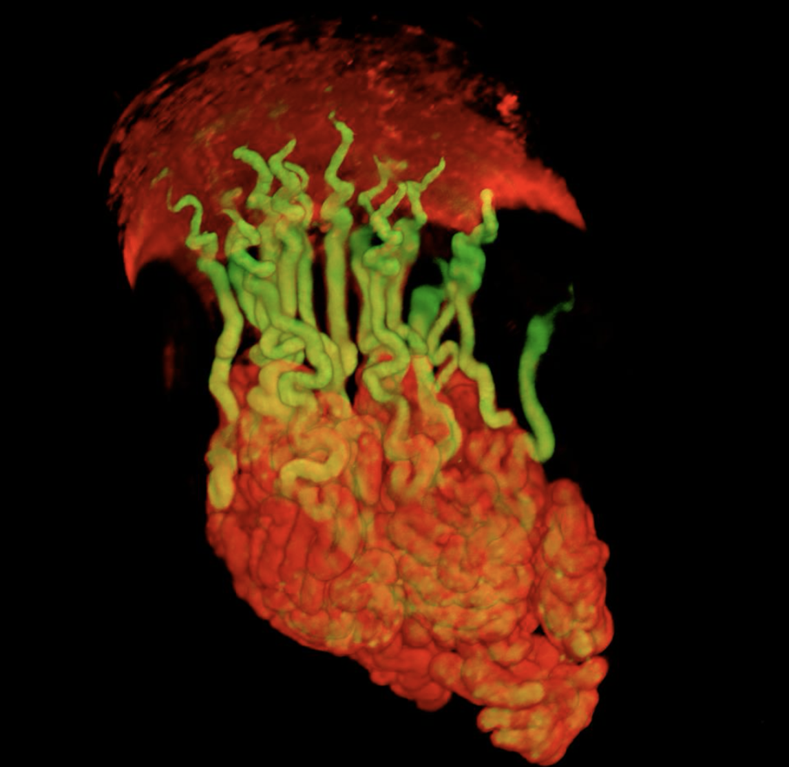
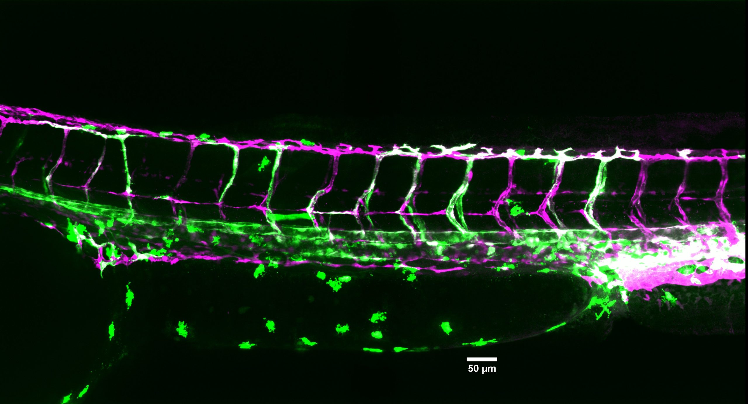
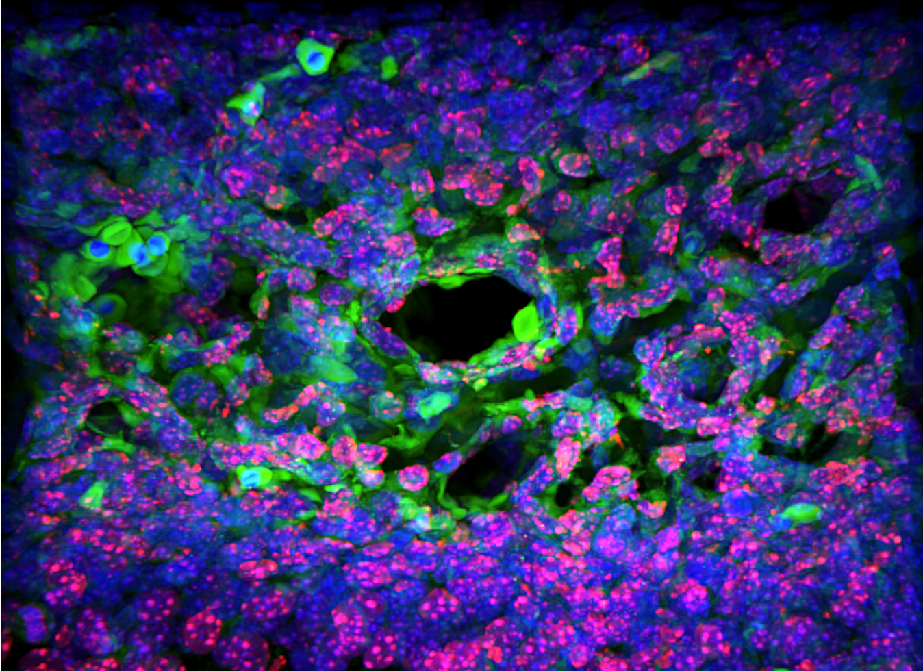
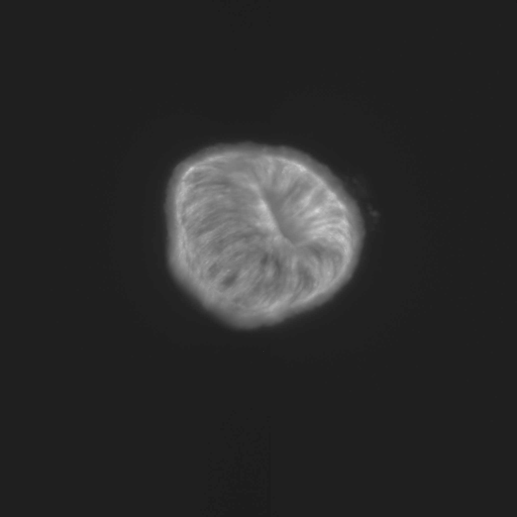
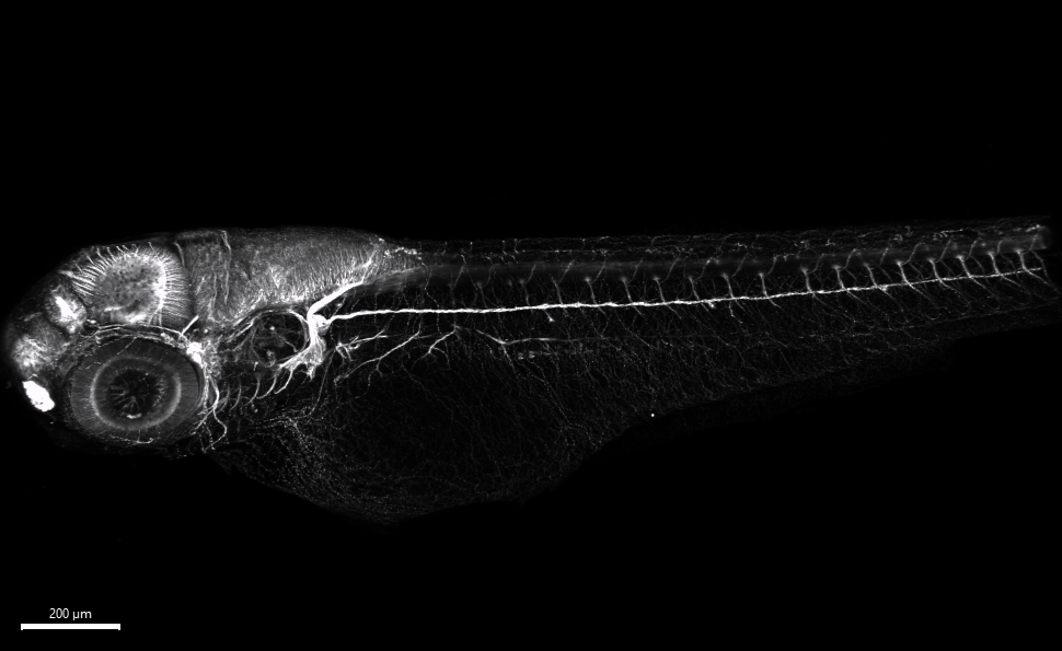
“Life as a drop in the ocean” This image is from an embryo of the marine acoel worm Hofstenia miamia, with cell membranes and nuclei highlighted. Early in development nuclei are shaped as rosettes but reasons or consequences for this remain mysterious. – Paul Bump“Human segmentoid” Organoids recapitulating human axial elongation and segmentation derived from induced pluripotent stem cells – Yuchuan Miao“Drosophila embryo Zipper” The Drosophila embryo central nervous system (CNS) stained with anti-body BP102 – Huangping An“Cycling through the cell cycle” Somite formation in chicken embryos. The pre-somitic mesoderm, the somites and the notochord are clearly visible, as the embryo grows towards the posterior, the anterior tissues mature. FUCCHI chicken line developed at the NARF and the Roslin Institute. Green marks cells in G2/M, Red marks cells in G1. – Ana R. Hernandez Rodriguez“Developing Neural Kaleidoscope in Drosophila melanogaster” Neuronal markers (TfAP-2 (red); Vsx1 (green); Ap (blue)) in the larval optic lobe of Drosophila melanogaster. Imaging was done with a Leica SP8 confocal microscope and processed with the LAS X and Fiji imaging software. – Yen-Chung Chen“Chicken Aurora” Tail of chicken embryo electroporated with Brainbow plasmids – Prikshit“Implantation Infrastructure: Uterine Glands and Vessels around the mouse embryo implantation chamber” Uterine Glands (Blue branched structures) and Blood Vessels (Pink) surround the elongating V-shape embryo implantation chamber (in the center) and are critical for supporting early pregnancy in the mouse. – Harini Raghu Kumar (Arora Lab)“A detour on the path to reproductive system morphogenesis in C. elegans” A young adult hermaphrodite C. elegans expressing markers for the somatic gonad (green) and germ cells (magenta) and carrying a mutation causing turning defects in gonad morphogenesis. – Noor Singh“Lateral neighbors” Developing zebrafish embryo showing the expression patterns of twist1 (magenta) and hand2 (green), dorsal view, anterior to the top. – Amanda Garfield“Fiery fish” A fire filter applied to a gene expressed in neurons and muscles along with DAPI to stain nuclei in a 24 hours old zebrafish embryo. – Zainab Afzal“Multichromatic Acacia” E6 chick embryo spinal cord electroporated with next generation Brainbow transgenes. This whole-mount acquisition allows tracing of neural progenitors to migrating neurons, following with axonal projections, showcasing lineage dynamics during development. – Thea Chrysostomou“Gradients” This image reveals the spatial coordination of BMP signaling and gene expression during early embryonic development. Dorsal view of a Drosophila melanogaster embryo at nuclear cycle 14, stained with anti-pSMAD antibody and smFISH for a BMP target gene. Captured using a Leica Stellaris confocal microscope and post-processed in Fiji. – Susanna Brantley“Symmetry on the Brain” This is a coronal section of a mid gestational embryonic piglet brain stained with DAPI in dark blue, phosphorlated s6 kinase in green, PROX1 in red, and NR2F2 in cyan to label different neuronal progenitor classes. Image aquired on a Leica Steallaris laser scanning confocal microscope then stitched and processed using ImageJ. – Emma Horton“Shaping it up like clockwork” Somite-forming organoids derived from human pluripotent stem cells allow the modelling of early vertebrate development and congenital disorders affecting the spine. Somites are transient block-like structures formed periodically, like clockwork, stacked on top of one another during early development, later developing into the vertebral column. The image shows a somitoid throughout its development from an oval spheroid to a stack of somites. Somite cells (magenta) develop on the anterior end, and progenitor populations (co-stained by cyan and yellow, which appear green) necessary for somite formation mark the posterior end. – Pranav S. Ramesh“Salivary cauliflower” Mouse embryonic salivary gland stained for Hes1 (blue) and p63 (yellow). Imaged using a 40x objective on a Zeiss LSM780 on the PICT Core facility at Institut Curie, Paris. – Robin Journot“Fishing for sex chromosomes” The X chromosomes (green) and Y chromosomes (magenta) painted by DNA FISH in a E3.5 mouse blastocyst; DNA stained with Hoechst (blue). – Aurélien Courtois“Radially migrating cortical neurons” After being born, the cerebral cortical projection neurons migrate from the proliferative zone of developing cortex to destined cortical layers. This extended depth of focus image shows these radially migrating cortical neurons at embryonic day 17 which are labelled with GFP expressing plasmid through in-utero electroporation. The staining in red shows EdU positive cells. This image has been taken using AXR Nikon microscope at CEAF Confocal facility, IIT Kanpur, India. – Nitin Agnihotri“Craniofacial mesenchyme”Craniofacial mesenchyme of HH16 chick embryo stained for ALX1 (magenta), ALX4 (green), PITX2 (red) and DAPI (blue). Image acquired with Nikon confocal microscope and processed with NIS element and FIJI. – Shirley Ee Shan Liau“Neural connections in the developing frog head” Stage 40 Xenopus laevis head labeled with an anti-neurofilament antibody, highlighting several neuronal connections. Antibody used: DHSB 3A10 (AB_531874). – Sudipta Mukherjee“Coming Together” This image captures the process during which the embryonic foregut forms an epithelial septum to give rise to the esophagus and trachea. Cells coming together in the septum remodel their Golgi apparatus (magenta), losing the tubular morphology and apical localization to the nucleus (cyan) characteristic of other epithelial cells, and resemble the surrounding mesenchymal cells. – Rui Yan“An Ewing Fish” Ewing sarcoma development through NCCs reprogramming. a massive green tumor driven by the expression of human oncofusion in neural crest cells. This fish also has an ectopic extra-dorsal fin, demonstrating an intriguing connection between normal development and tumorigenesis. – Elena Vasileva“Love on the Brain” Maximum intensity projection of an adult D. melanogaster brain from a fly carrying a GAL4 insertion crossed to a UAS-mCD8::GFP fluorescent reporter, expressing membrane-targeted GFP (in green) stained against phalloidin (in magenta) showing that the GAL4 insertion drives expression in the mushroom body of the fly brain. – Anastasia Repouliou“Web of Life” 20 days old zebrafish heart ventricle, membranes of cardiomyocytes are marked by fluorescent protein, imaged at 40x zeiss 980 and image rendered on Imaris. – Kirti Gupta“C. elegans digestive tract” – Lauren Cote– Catherine Pei-Ju Lu“The Developing Vasculature in Zebrafish” – Elithabeth Jones“The saga of bone genes and proteins” – Pragati Shekhar“Toroidal Hydra” – Daniel Pearce– Lucia RivasSomites and PSM eletroporated in chicken embryo (Gallus gallus). – Nisia MartinsFast green image of collagen in yellow and nuclei stained with DAPI in the suture mesenchyme of the mouse E15.5 skull. – Sasha Degtyareva Actin cytoskeleton in a developing Drosophila embryo – Nilay Taneja (No Ratings Yet) Loading...
About 6.5 years ago now, I wrote my first post for the Node, ‘Hello from Alex‘, introducing myself as a new Reviews Editor for Development. I was thrilled by the opportunity to stay in touch with the science I first came across at university and with which I instantly fell in love. In the years that followed, I’ve enjoyed helping authors publish their review-type articles, interviewing leaders of the field from all career stages and meeting the community at conferences. Six years is a relatively short time, but I’ve seen single-cell sequencing go from state-of-the-art technology to an almost routine approach, an explosion in the development of stem cell-based systems and a renaissance of fundamental aspects of biology, such as metabolism, genetics and mechanics, interrogated from new perspectives and with new tools. My hairline may have receded over these years, but my love for developmental biology, and indeed for the journal, has only grown. It’s been a journey of continuous learning and constant discovery where I’ve been able to get to grips with the wonderful world of plant biology, finally understand the complex anatomy of an early mammalian embryo and even grasp some mathematics.
It was – is – the perfect job and although I thought that, one day, I might attempt the leap to Executive Editor, I was fully prepared to wait another 20 years for the chance. So, when it was announced that Development’s previous Executive Editor, Katherine Brown, was moving on to a new post within The Company of Biologists, it came as a surprise. For over a decade, Katherine has been a pillar in the colosseum that is our community and the centre pole in the tent that is Development. It is, indeed, the end of an era. As much as I loved being a Reviews Editor, I’ve learned that personal and professional growth can only come through change and challenge – by venturing from the comfortable. I had hestitations about whether I was ready, but I’m fortunate in that, throughout my career (in addition to the privileges afforded to me as a cis white man), I’ve had supervisors and mentors like Katherine willing to take a risk with me, and I’ve tried my best to live up to that potential. I can’t hope to emulate Katherine, nor will I try, but the best news for me is that, as the Publishing Director at the Company, she will remain a source of guidance, expertise and superhuman capability.
What does this change mean for the journal? In the short term, my focus will be on keeping business as usual during this transition and supporting our excellent team of academic Editors in handling your papers. Real change, in whatever form that might be, occurs on a longer timescale. I bring with me a few ideas (and one or two of those, I hope, are actually sensible), but the main purpose of my role is to bring about the vision of James Briscoe, Editor-in-Chief; I’ve quoted below an article published earlier this year about the history of Development in which James talks about the future.
Over the next few years, we can expect continued change in the business models supporting scientific publishing, whether this is in new forms of OA or further innovation in publishing pathways. We are committed to being as inclusive as possible and giving all authors, wherever you are in the world, options that allow you to publish in Development. Preprints present new ways to enhance scientific communication and new approaches to peer review, and we will continue to develop new ways to help researchers navigate and use the preprint literature effectively. Over the next few years, we also expect to see innovative use of artificial intelligence in scientific publishing, complementing human experts. These technologies are emerging as valuable tools for information and literature discovery, manuscript processing, and journal production, offering possibilities to improve scientific publishing while maintaining rigorous standards.
Development has a long-held reputation for being respected and trusted, but the journal has not rested on its laurels. The journal has regularly introduced practices and polices to make publication easier and more transparent for authors, such as format-free submission, cross-referee commenting and publishing referee reports. It is my plan to continue these efforts, to turn avid readers of Development into frequent authors and to tackle the misconception that publishing in Development is ‘hard’. I’ve been overwhelmed by the response from the community about my appointment, and it’s a priority of mine to meet even more of you. I look forward to hearing your ideas, suggestions, queries and indeed your grievances so that the journal might continue to develop.
Finally, if you love developmental and stem cell biology, wish to remain in the community but not in the lab, and want to join our new, enthusiastic and supportive team on this new journey, we are currently recruiting a Community Manager for the Node.
I was excited to be in Liverpool for the Biologists @ 100 meeting this past March. Not only is a meeting organised by The Company of Biologists always guaranteed to be friendly and exciting, this was also my first science-related activity back in the UK since I started a postdoc in Boston last November, so I was doubly happy to be there.
Sunny views on the River Mersey, Liverpool’s aquatic pride and joy.
The most intriguing aspect of the meeting for me was the fact that it drew together an unusually broad range of scientists – this wasn’t your average BSDB/BSCB joint meeting! It was a celebration of the 100 years since the founding of The Company of Biologists and the 5 journals that are published under its umbrella. Accordingly, in addition to the cell and developmental biology track, there were three more included in the meeting, each in reality a mini symposium or satellite meeting: sensory perception (the Journal of Experimental Biology’s symposium), interdisciplinary approaches to combatting antimicrobial resistance (Disease Models & Mechanisms’ symposium), and experimental biology and impact: solutions to climate change and biodiversity loss, organised by the Society of Experimental Biology.
The ACC, venue of the conference.Talks in the ACC.
Bringing together many different groups of scientists that might not typically encounter each other at meetings and pooling resources has some practical perks: firstly, the meeting was held in the ACC, a big, comfortable, and supremely well-located venue right on the River Mersey. We were also very close to the Museum of Liverpool, where a welcome reception was held at the end of the first day, and St George’s Hall, where the conference’s gala dinner was held on the penultimate day of the conference.
St George’s Hall, where the gala dinner was held.
However, what I appreciated most was getting to hear about many different types of science that I would not have normally expected to hear at a conference (and especially at the same conference!). The biodiversity and climate change plenary lectures by Jane Francis of the British Antarctic Survey, and Hans-Otto Pörtner from the Alfred Wegener Institute for Polar and Marine Research in Germany were on the one hand fascinating, and on the other hand greatly sobering. They highlighted the essential role every scientist (not just climate scientists) should play in disseminating accurate information about the climate catastrophe our planet is suffering, and what we should be doing to combat it.
The parallel session format of the conference meant that you could hop between tracks several times throughout the day and take in all the different types of science that converged at the meeting. For example, the sessions on sensory perception were very comprehensive, with talks on vision, light pollution, thermosensation, sound perception, magnetic and chemical senses. There were also talks and posters on insect behaviour and insect-plant parallel evolution – this is a very subjective opinion, but butterflies always make a conference more fun. I was also excited to attend one of the sessions on drug discovery and ‘omics. Talks ranged from large-scale efforts to combat the development of antimicrobial resistance, to systematically exploring the breadth of organic chemistry to discover new drugs. This might not be news to some readers, but I was not aware of the tendency of microbes that develop drug resistance to concomitantly become more sensitive to other classes of drugs. This is something that researchers are actively investigating in their efforts to combat antibiotic resistance.
There was also a handful of plant-themed posters and a couple of talks on plant biology. There I heard the best description of leaves from Dr Chris Whitewoods from the Sainsbury Lab in Cambridge: “kind of green, kind of flat, sort of pretty in the sunshine.” For someone who gave a talk on leaf air space development, it was hilarious to get such a fun, entry-level description of leaves. Speaking to the (relatively few) plant scientists that were present at the meeting, I wondered why there tend to be such few plant researchers at developmental and cell biology conferences. They shared with me that they tend to congregate at plant science conferences instead, so this is my personal plea to plant scientists and developmental biology conferences: please sign up for dev bio conferences / invite plant scientists to speak at them! It is so refreshing to hear about research in non-animal models (and this is coming from someone with a passion for animal development).
Overall, the Biologists @ 100 conference was a big success. It was fantastic to see old colleagues and former lecturers, meet new people and discuss potential collaborations, and above all be exposed to an unusually broad suite of scientific investigation. There is nothing quite as inspiring as sharing one’s excitement about research with others!
All the world’s a metabolic dance, early career scientists are leading the way!
Emerging perspectives in metabolism
Dr. Lautaro Gandara, Postdoctoral researcher, EMBL, Heidelberg.
This week we will get to know insights from Dr. Lautaro Gandara, who is a postdoctoral researcher in the Crocker lab at EMBL Heidelberg. Lautaro’s work delves into the profound interplay between metabolism, toxicology, and development. As he prepares to establish his own lab, Lautaro is driven by a deep curiosity about how life adapts to environmental challenges, and how metabolic shifts shape the very essence of biological resilience. From his studies of Drosophila melanogaster to the impact of environmental stressors on insect populations, his research questions the fundamental nature of life’s response to stress and transformation. For Lautaro, science is not just a pursuit of answers but a journey of discovery, where each question unfolds new dimensions of understanding. He believes that the study of metabolism and development is not merely academic—it’s a window into the intricate ways life connects, adapts, and evolves. Follow his journey as he continues to explore these deep questions and check out his work here.
Could you share your journey into studying metabolism and what inspired you to specialize in metabolic studies using Drosophila melanogaster.
When I took an introductory course in chemical biology as an undergrad, I remember being less than enthusiastic about the field. Metabolism was presented as a completed research program—a field in which human metabolic maps had already been established, flux control theory had provided all the relevant dynamical information, and the only open questions were clinical ones. It was only after I started my PhD work in Pablo Wappner’s lab that I got access to the then-new research showing how metabolism, far from being a housekeeping process, can actually transmit information by regulating gene expression, signaling pathways, and so on. At the time, I was studying the response to oxygen deprivation (hypoxia) in flies, and we soon realized that many metabolic facets of this process remained unexplored. Drosophila larvae can perform lactic acid fermentation in hypoxic environments, but at that time there was little information on the spatial and temporal properties of this metabolic switch. These questions became the focus of my work as a grad student.
How did you get interested in the field of toxicology and impact of chemicals on insect health and metabolism? How do you think toxicology and metabolism fields overlap and how they regulate each other or how they are connected?
I have always been fascinated by the way in which life actively responds to environmental change—its intrinsic ability to preserve itself by regulating its own activities and structures. When you look at these processes more closely, all the reductionist metaphors of “life as mere machines” start to crumble, right? This is where my interest in the hypoxia response originally came from. So as a postdoc in Justin Crocker’s lab at EMBL, I wanted to expand on the approaches I had used as an undergraduate to further explore these phenomena. Instead of focusing on just one environmental perturbation (hypoxia) and one phenotype (metabolism), we decided to test more than 1000 different chemical stressors and measure how the effects induced by these molecules propagate across the different scales of biological organization.
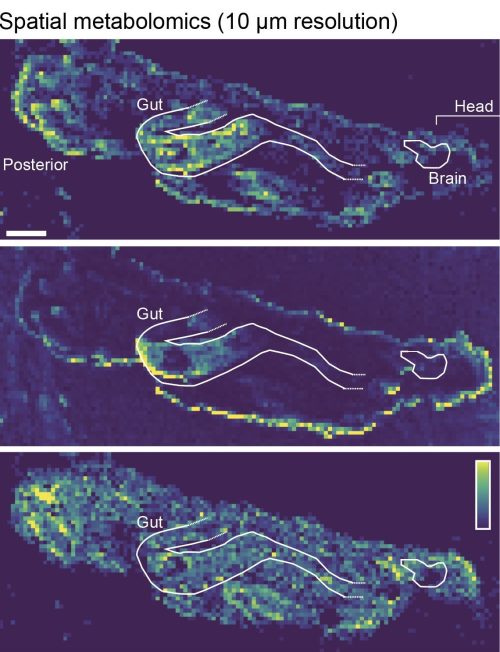
High spatial resolution MALDI-imaging analysis of a Drosophila larva (scale bar = 100 μm). The images show relative intensities of individual lipid species, each for an individual m/z value: upper panel = 544.3373 (C26H52NO7P); middle panel = 177.0158 (C7H6O4); lower panel =744.5537 (C41H78NO8P). Image source: LG.
Tell us about how you see the future of metabolism evolve with the new upcoming tools like the FRET biosensors you worked on. What changes have you seen in the community regarding exploring metabolic aspects of development ?
I consider the development of tools to be an essential driver of science, and I think that the interplay between development and metabolism is a paradigmatic case of this phenomenon. In the first half of the 20th century, embryologists such as Joseph Needham studied the metabolic facets of development using the technology available at the time (calorimetry, respirometry, etc.). However, as embryology was transformed into developmental biology, and the focus shifted from organismal-level processes to gene- and cell-level phenomena, this set of tools proved ill-suited for exploring metabolism at this scale. As a result, questions about the role of metabolism during development were put on hold. Recently, the combination of metabolic FRET sensors, spatial omics techniques (especially spatial metabolomics), and flux analysis by isotope tracing has reignited interest in these old questions and revitalized the field. Ongoing research by many different groups working on different model systems around the world is providing priceless information about the precise role of metabolism in the development of multicellular organisms.
One of your reviews is titled “Metabo-Devo: A metabolic perspective of development”. Could you elaborate on the key findings and their implications for the field? How do you integrate different disciplines – metabolism, development, and evolution in your research, and what unique insights have emerged from this approach?
In that review article, we proposed a conceptual framework to discuss how metabolism interacts with developmental processes. We classified these interactions as either 1) bioenergetic functions, 2) regulation of gene expression through changes in the epigenome, or 3) signaling functions.
Bioenergetic processes are those that provide energy or building blocks to developing tissues. Many cell populations that proliferate at high rates acquire a particular metabolic state, called aerobic glycolysis, that allows them to synthesize macromolecules at the right pace. This metabolic switch was reported ~100 years ago in the context of cancer biology (i.e. the Warburg effect), but it is now becoming clear that aerobic glycolysis is also required for cell proliferation in developing organisms. Ongoing research efforts aim to elucidate the developmental processes during which this metabolic transition occurs, and how it is regulated.
In addition to this bioenergetic role, specific metabolic pathways can directly regulate gene expression. Certain metabolites have been shown to act in developmental contexts as rate-limiting substrates for histone and DNA modification. And this metabolic control of gene expression has been shown to play an essential role in key developmental processes, such as the zygotic genome activation. Similarly, many metabolites are directly involved in the post-translational modification of signaling-related proteins, while several metabolic enzymes have been reported to act as multifunctional “moonlighting” proteins that can perform alternative functions not necessarily related to metabolism. Thus, metabolites and metabolic enzymes have the potential to modulate signaling pathways that are essential for development.
Although we think that the classification described above provides a useful conceptual framework for designing and discussing experiments, dissecting the actual role of metabolism in specific processes remains challenging, especially because the same metabolites and enzymes may simultaneously play bioenergetic and signaling roles that affect the same developmental phenomenon. In any case, the emerging view is that metabolism and development are deeply intertwined processes that cannot be disentangled. Thus, this observation highlights the need for a discipline or research area—developmental metabolism or “metabo-devo”—that directly addresses these issues.
You are currently studying the impact of toxic substances from genotypic to phenotypic perspectives and your work suggests the use of agrochemicals is the root cause of insect decline. Can you briefly discuss this work ?
We decided to focus on agrochemicals (i.e. insecticides, fungicides, plant growth regulators, etc.). Increased use of pesticides has been proposed as a potential cause of the widespread declines in insect populations, but studies investigating the effects of these molecules on insects are often limited to a few chemicals and a single insect species. We started with a screen that tested the effects of 1024 agrochemicals on the behavior of Drosophila larvae. Behavioral changes often have a mechanistic basis at simpler phenotypic levels, and thus monitoring behavior can provide important information about the state of the biological system as a whole. Surprisingly, we found that ~60% of the molecules in our library significantly alter larval behavior! And these effects are not limited to flies—we also detected similar sublethal behavioral changes in mosquitoes and butterflies, suggesting a generalizable effect on insect populations.
By exploring some of the screen hits further, we found that the effects go well beyond behavior. Exposure to sublethal doses triggered widespread phosphoproteomic changes, revealing how these chemicals affect many different physiological processes. And chronic exposure led to delayed development and reduced reproductive output, potentially contributing to the decline in insect populations. Thus, the study showed that even non-insecticidal pesticides at field-realistic sublethal concentrations can have profound ecological consequences, highlighting the need for better safety assessments that take sublethal effects into account.
How difficult some of those experiments work – did you have to deal with midnight timepoints or require an army of undergrads/ long hours ?
The behavioral screen was very time consuming. We ended up testing 3072 different conditions (different molecules and concentrations), so including replicates and controls, we ran more than 10000 individual assays… It was a lot of work, but I got a lot of help from everyone in the lab, and fortunately we managed to get it done in just a few months. It was truly a collaborative effort!
Can you shed light on the big picture of the field, what are you most excited about and how does it all connect to impacting insect/human health.
Understanding how animal systems respond to stress is becoming increasingly important in the current context of human-induced global environmental change. Going back to my fascination with the resilience of biological systems, I think our previous work has opened up an exciting opportunity to test some of the open questions in the field of stress response. There is this idea that multicellular organisms need to activate a system-level stress response to restore homeostasis. This process would be based on the well-known “integrated stress response” pathway—a process that occurs at the cellular level—but would also involve organismal level stress defense systems involving cell differentiation processes, metabolic switches, physiological changes, and even behavioral effects. Testing this hypothesis, however, is not straightforward because it would require measuring the degree of interconnectedness among all these different processes across a wide range of environmental perturbations. By performing the behavioral screen I mentioned earlier, we have defined a panel of chemicals that induce widespread systemic changes in fly larvae, but at sublethal concentrations, meaning that these animals can orchestrate a successful response to these stressors and recover from these injuries. Thus, this panel of chemicals can be used next to explore the level of integration between the different stress defense systems operating at the organismal level. I hope to start my own group soon, and this is one of the first problems I’d like to tackle.
What advice would you offer to students and early-career scientists interested in exploring the intersections of metabolism, development, and evolution?
I’d tell them to have fun! These are indeed exciting times to be doing metabolic research in developmental systems. New technologies are allowing us to explore the various ways in which metabolism transfers information not only across space (inter-organ communication, metabolic coupling between cell types, etc.), but also across time (developmental and cell differentiation processes). I think this is the time to be bold and creative in finding ways to make the most of this technological advantage.
What role does curiosity play in your life, both within and outside of science? How important is it for you to answer basic science questions about behavioral and metabolic aspects of toxicology and how are you planning to use insect models to bridge basic science and applied research ?
Curiosity does play an essential role in my life and in my research. The project I mentioned earlier, in which we studied the sublethal effects of agrochemicals on insects, has some facets that are obviously relevant to the community as a whole. But I strongly disagree with the idea that it’s only worth studying certain natural phenomena if they affect us directly, or if we can use them for our direct benefit in the short term. Basic and applied research aren’t in opposition to each other—on the contrary, they are involved in a dialectical feedback in which the former feeds the latter with information about how fundamental processes work, while the latter not only highlights which questions are most pressing, but also drives the development of new tools and methods that can then be applied to basic research. Hypoxia research is a good example. The oxygen sensor—the molecule that allows cells to determine oxygen availability and trigger an appropriate response when oxygen levels become too low—was identified more than 20 years ago in fundamental work on C. elegans. Years later, this molecular machinery was found to have enormous clinical relevance, as it could be manipulated to induce angiogenesis and treat the symptoms of cardiovascular disease or prevent it and limit tumor growth. But this useful knowledge first came from curiosity-driven research on basic genetics.
Were there any pivotal moments that shaped your career path? What’s an unexpected place you’ve found inspiration for your work?
It may sound simple, but for me the most important source of scientific inspiration is talking to other people. I can think about some problems for hours, but in my case, ideas really take shape when I express them, either by writing them down or by discussing them with someone else. It’s as if, by trying to communicate my thoughts, I organize them into a coherent narrative—a logical structure—from which new ideas can occasionally emerge. And then different people, with different backgrounds and different opinions, will steer your train of thought in completely different directions, certainly leading to unexpected places… But I’m not talking about big meetings full of people here—it’s the one-on-one discussions that force you to interact longer with a single concept and exhaust all its multiple possibilities that are often more productive for me.
If you hadn’t embarked on a career in biological research, what other profession might you have pursued, and why?
I don’t really see myself doing anything other than biological research. But if it weren’t for biology, I think I would have pursued a career in the social sciences. Human societies are incredibly complex entities, but understanding how they work is not just a matter of academic curiosity. Especially now, looking at the current times, explaining how society, economics, or history actually works has become a pressing issue. I think that on an individual level—and as citizens —we can’t afford to ignore these problems any longer.
Anything you’d want to highlight for the future.
I’m currently looking for a place to set up my own lab. Aside from the global uncertainty we discussed earlier, I think these are exciting times to start a group. New technologies are making old questions experimentally tractable, new species are being proposed all the time as novel model systems, and AI promises to change the way we approach data analysis. There’s no doubt that the way we do science is going to change dramatically in the coming years, and that’s an idea I find particularly appealing.
Last week we learnt about how males and females are metabolically differently rewired – from the perspective of lipid storage and utilization – The Fat of the Matter (Lianna Wat)
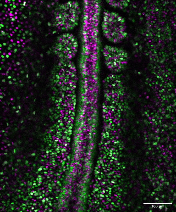
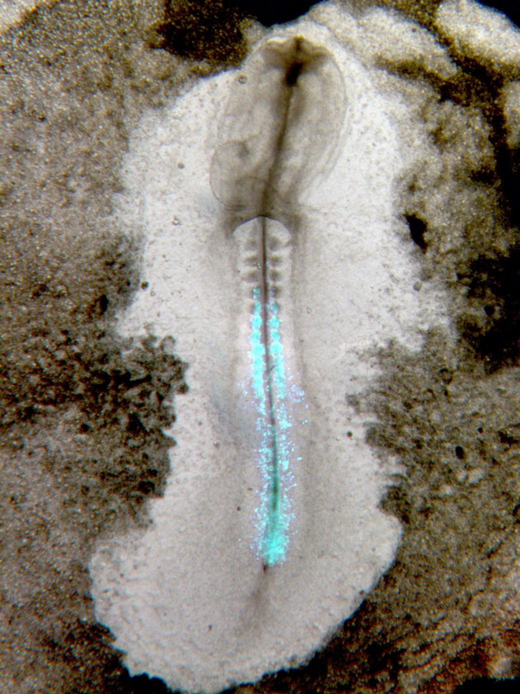
In our recently published paper ‘Ectoderm barcoding reveals neural and cochlear compartmentalization‘, we utilized ultrasound-guided in utero nano injections to deliver heritable DNA barcodes to cells exposed to the amniotic fluid, performing the first high-throughput single cell lineage tracing study of the developing nervous system and inner ear. Our results led to the reclassification of cell lineages in the cochlea and provided a comprehensive single-cell atlas of neural and cochlear clonal relationships.
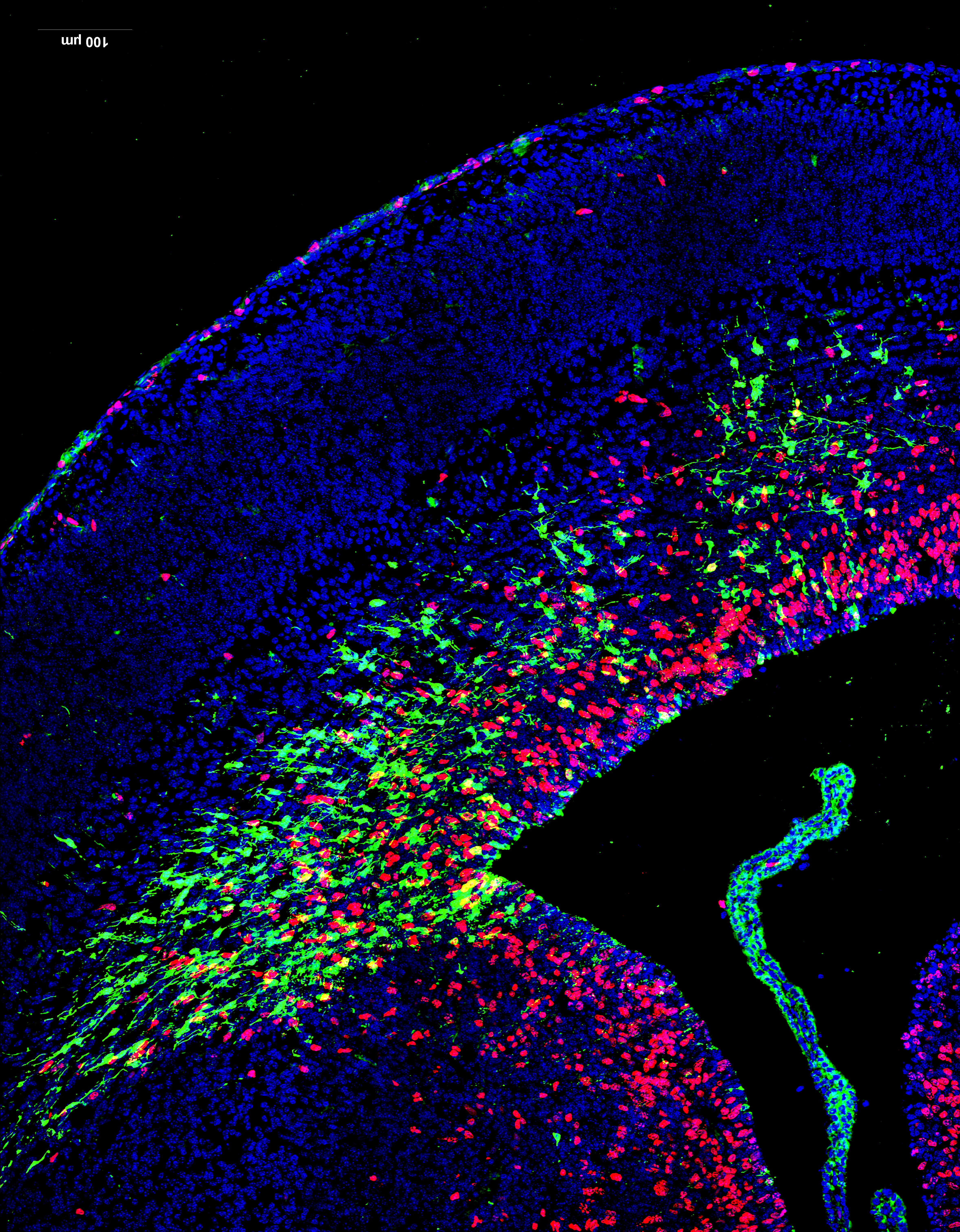
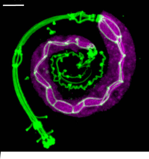
Sandra’s perspective: Work related to this publication already started a long time before I (Sandra) joined Emma R. Andersson’s lab back in 2019 to pursue my PhD studies. Work done by Katrin Mangold and Jingyan He, PhD students in the lab at the time, laid the foundation for successful in utero injections into the amniotic cavity to target the neural plate (Mangold et al, 2021). Like the neural plate, the progenitors of the inner ear are exposed to the amniotic fluid during an early time window of development (~E7 – E9) and we therefore hypothesized that we would be able to target the otic placode and manipulate progenitors of the inner ear using this technique. In fact, preliminary data from a Master’s student, Sanne Stokman, showed some targeting of the vestibular system of the inner ear. This data hinted that we should be able to target the cochlea as well – and laid the foundation to further explore this during my PhD studies. Drawing on expertise from two laboratories—the Andersson lab at the Karolinska Institute, specializing in developmental biology, Notch signaling, and in utero injection techniques, and Matt Kelley’s lab at the National Institutes of Health, experts in inner ear developmental biology—the first experiments targeting the cochlea were conducted in 2019. I vividly recall witnessing the targeting of the cochlea, for the first time, using low-titer H2B-GFP lentivirus injections performed by Jingyan. The mosaic-like targeting of hair cells along the cochlear spiral was truly remarkable (Fig. 1). I remember sharing these initial positive results with Emma via text while at the confocal microscope. Together with Jingyan, who truly mastered the injection technique, and together with the Infinigene core facility (established by Emma), we optimized our injection strategy to target the inner ear, including the injection volume, embryonic stage and viral titer. This journey was, of course, not without its challenges. At times, we faced difficulties with mouse breeding, low viral titers, and, not to forget, a pandemic that occurred along the way. Finally, in 2022, injections using high-titer virus yielded high-targeting efficiencies – of over 90% of the hair cells and supporting cells in the Organ of Corti. I remember observing the high efficiency targeting, quantifying targeting efficiencies late at night the same day, and presenting the results the next day over Zoom during the Kelley lab meeting – excited to share the new results.
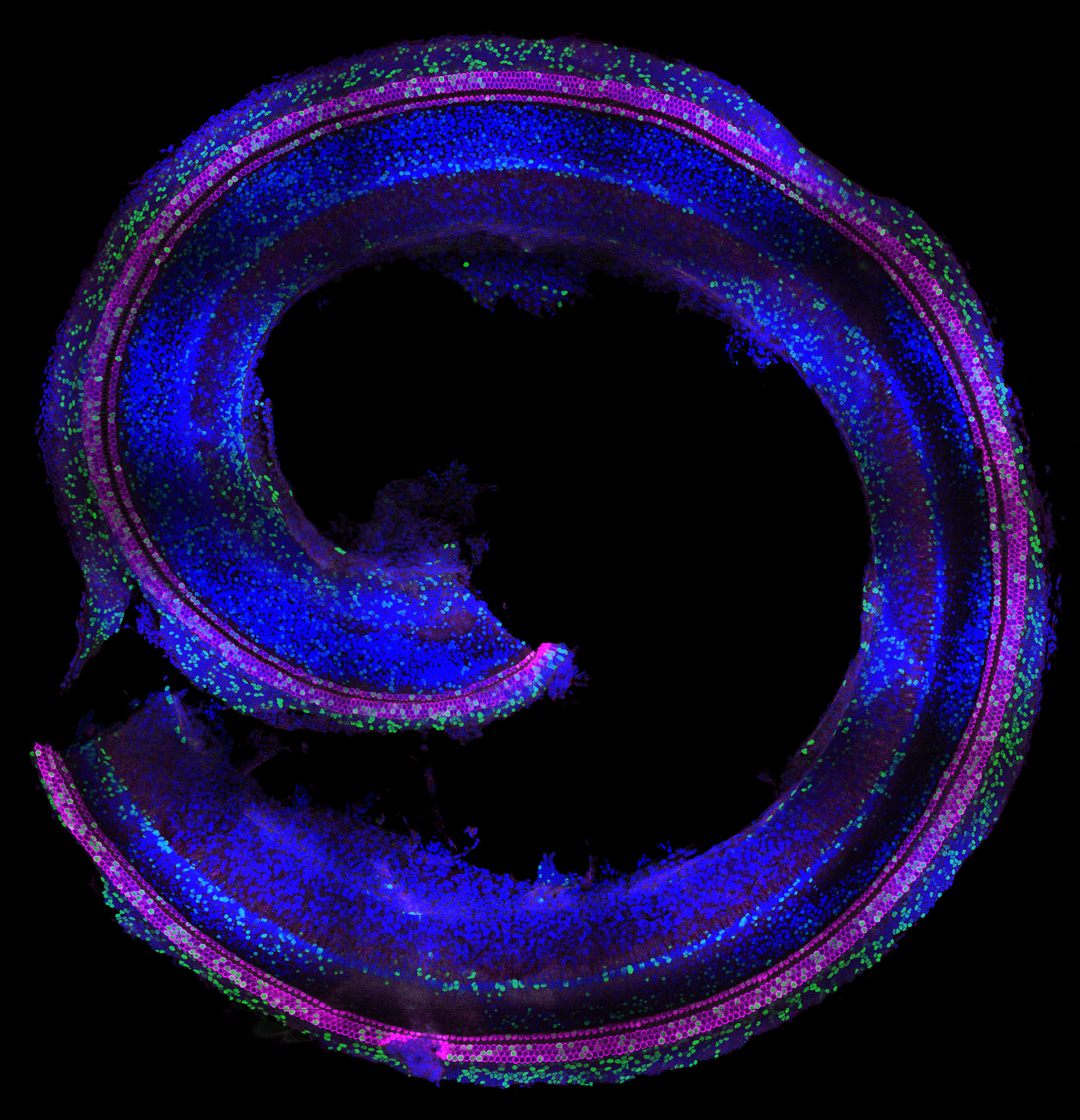
Fig1: First cochlear targeting results using low-titer H2B-GFP lentivirus. Injections performed at embryonic day 8, collection at postnatal day 0. Hair cells shown in magenta, GFP-labelled cells shown in green. DAPI (nuclei) in blue
Using viral delivery of heritable DNA barcodes, we were able to perform high-throughput lineage trace studies in the inner ear for the first time, answering fundamental questions about inner ear development. I am still thrilled that we could apply this advanced method to explore lineage relationships within the inner ear. Although we initially focused on the divergence of medial and lateral cochlear cells, we soon realized that the most intriguing findings came from cells outside the Organ of Corti, including Hensen’s cells and populations within the stria vascularis. One of my favorite insights is the classification of Hensen’s cells. At the time, more research was performed on this specific population of cells in the cochlea, but no consensus existed on whether this cell type should be considered a supporting cell subtype or grouped with cells lateral to the Organ of Corti. Our data indicated that Hensen’s cells should be classified as lateral to the Organ of Corti, rather than being a supporting cell subtype of the Organ of Corti – if basing this definition on lineages. This classification might be relevant for future strategies focusing on regeneration and differentiation of cells within the cochlea. These findings also nicely aligned with results from another project of my PhD studies, in which we showed that Hensen’s cells respond differently to the loss of Jag1-mediated Notch activation compared to lateral supporting cells (De Haan et al 2024, Development).
As dissociation of cochlear cells relies on physical dislocation through microdissections, we inadvertently included cell types in our analysis that were not initially the focus of the study, including spiral ganglion neurons and glia. The analysis of barcode sharing between these populations proved to be quite complex. Contamination between spiral ganglion neurons and glia cells often occurs in single-cell preparations, so we investigated the barcode sharing between these populations to determine whether it was due to contamination or if subtypes of neurons might share a common origin with glia cells, which would challenge the current view. Ultimately, this experience taught me the importance of remaining open to new discoveries while ensuring that data collection and experimental design are well-suited to address the research questions. It highlighted the need for careful experimental planning, robust data collection and validation to draw accurate conclusions from complex datasets.
Jingyan’s perspective: While contributing to the inner ear lineage tracing project, I (Jingyan) was also focused on the overarching goal of my PhD projects: advancing the in utero injection technique to label non-ectodermal cells, building upon previous success in ectoderm targeting. By exploring different injection approaches, I successfully established a technique to label diverse cell types with other embryonic origins (ongoing work). As part of this highly ambitious project, we lineage traced tall ectoderm-derived cells using amniotic cavity injections at E7.5. We collected whole embryos at E9.5 and E10.5 after barcode labeling at E7.5 with amniotic cavity injection. This approach allowed us to study the lineage relations of central nervous system, neural crest-derived cells, as well as various epithelial lineages, including the otic lineage. This part of data was later incorporated into the inner ear lineage tracing paper to add a more comprehensive understanding of neurodevelopment and illustrate the potential clonal relations between otic epithelial lineages and other cell types.
One of the key challenges in this work was balancing the viral transduction efficiencies across different collection time points to ensure an optimal number of labeled cells for the single cell RNA sequencing and clonal analyses. Although E9.5 and E10.5 are just one day apart, the difference in total cell number is substantial. We had to pool a few E9.5 embryos to obtain sufficient cell numbers for clonal analyses. However, for E10.5, if we used the same amount of viral particle and reached the same transduction efficiency as E9.5 collection, a single E10.5 embryo yielded so many labeled cells that we needed to split them into multiple reactions when preparing the sequencing libraries.
Viral transduction efficiency itself was influenced by a variety of factors, such as the subtle differences of the embryos’ stages when injecting, viral storage time, freeze-thaw cycles and the variability between different virus production batches, making it difficult to consistently control the number of transduced cells recovered from each injection.
Collection days were always highly intense and stressful, involving a full workflow from the setting up of the cell sorter, embryo collection, dissection, dissociation, cell sorting, to library preparation, all within a tight window to preserve cell viability and RNA quality. Sandra and I always teamed up to streamline the workflow, assisting each other with reagent preparation, cell counting, and other time-sensitive steps.
Joint perspective: The manuscript, initially focused on cochlea only, was submitted shortly before Sandra’s successful PhD defense in August 2024. The E9.5/E10.5 ectodermal lineage tracing data was initially intended for a separate publication, but in response to reviewer and editorial feedback, we decided to incorporate it into the current paper during the revision process. This addition significantly enriched the manuscript and provided a more comprehensive understanding of neurodevelopment. As a result, Jingyan and Sandra shared the first authorship of the paper, highlighting the collaborative nature of our research and the importance of these findings.
Now that the paper has been published and new projects are underway in the Andersson lab applying this technique to different tissue systems, we are both excited to see how the technology will be used, further developed, and what biological insights it will uncover. We’re proud that our work also laid the foundation for an ERC Consolidator Grant to the Andersson lab, who will continue to push the frontiers of the technology – and the lab is looking for post docs!
An important new resource for researchers, reviewers, and funders has been developed with major contributions from the Mary Lyon Centre at MRC Harwell.
A group of researchers and policy leaders from organisations involved in in vivo and ex vivo research have unveiled a transformative tool to assist scientists, policy makers, funders and reviewers in eliminating persistent sex bias in biomedical research. Published today in Nature Communications, the Sex Inclusive Research Framework (SIRF) introduces a structured and interactive approach to ensure equitable inclusion of male and female samples in preclinical studies.
Preclinical research has long favoured male animals and cell lines, leading to a skewed knowledge base that does not represent the human population. Consequently, it leads to non-optimal use of animals and resources as it ultimately results in less reliable data and less successful therapeutic interventions. Despite mandates by funding bodies to include female and male samples (funders such as the Medical Research Council have introduced this requirement since 2022), many research proposals fall short due to ingrained misconceptions and inconsistent evaluation standards. Recent media attention on this subject was reflected by this Guardian article https://www.theguardian.com/science/article/2024/jul/22/sex-bias-labs-women-losers-research-ageing?CMP=Share_iOSApp_Other, highlighting some of the negative impacts of sex-biased research.
SIRF addresses these gaps with an intuitive, traffic light–based decision tree that evaluates the appropriateness of sex inclusion in experimental design. Developed through collaboration across academia, industry, funding agencies, and animal welfare organisations, the framework assesses whether proposals include balanced sex representation and appropriate analysis plans, and whether single-sex studies are scientifically justified.
“Sex bias in research isn’t just a scientific issue, it’s an equity issue,” said Natasha Karp, lead author, “SIRF provides the structure, rationale, and accessibility needed to make inclusive design the standard, not the exception.”
SIRF offers:
A clear, reproducible evaluation method for use by researchers, ethics boards, and funders.
Detailed guidance to debunk myths that hinder sex-inclusive practices.
An open-access web interface with interactive features and educational resources.
The framework complements and improves upon prior initiatives, such as NIH and SAGER guidelines, by emphasising transparency, scientific justification, and practical implementation.
Available now as an interactive tool or downloadable resource, SIRF is set to reshape how research proposals are developed, reviewed, and funded—ultimately enhancing the validity and impact of biomedical discoveries for all sexes.
Our Director, Sara Wells, was part of the original working group that set out the scope and direction of this work and contributed with the wealth of data and experience from years of in vivo preclinical work at the Mary Lyon Centre. Commenting on this milestone and the resource now available to the community, she said: “The translatability of in vivo experiments is a key element of the success of preclinical studies. The analysis of every variable affecting experimental outcomes is a major objective as we work towards achieving this goal. Sex has now been shown to be a fundamental piece of the data variability puzzle, and the SIRF framework supports the community in assessing where its inclusion is essential, as well as providing resources for adequate data analysis. The implementation of the framework will contribute to the community’s drive for more relevant, translatable data and effective uses of both animals and financial resources as we strive for more successful therapeutics.”
Lilian Hunt, Lead Advisor to the Executive Director of Equity, Wellcome said:
“We’re pleased to see the publication of this vital tool for research. Ensuring sex inclusion in in vivo and ex vivo research is key to ensuring excellent, reproducible, and translatable research that benefits health equity. Wellcome will be exploring how best to integrate this guidance into our expectations of inclusive research.”
As you may have seen in an earlier post, I’m moving on from Development after 13+ years as the journal’s Executive Editor. The good news (for me at least!) is that I’m not going far – I’m taking up a new position here at The Company of Biologists as the Publishing Director, overseeing our editorial programme and supporting the activities of all five of our journals. This means I’ll still be very involved with Development, though much more on the publishing side of things than the science – and this is definitely a real wrench for me. I love developmental biology, I love the community and I love working with such a fantastic and dedicated team of academic editors and in-house staff. But it was time for me to move on – after all this time, I’m ready for a new challenge. And I’m excited to announce that we’ve appointed Alex Eve, who’s been with the journal as a Reviews and then Senior Editor since late 2018, as the new Executive Editor. His knowledge of, enthusiasm for and dedication to the field mean that the journal’s going to be in great hands. You can expect to hear a bit more from Alex and his plans in the coming weeks.
As I turn my attention to my new role, I’ve also been thinking back over the past 13 years and thought I’d share some (perhaps a slightly random selection of!) particularly memorable activities and moments (big and small) from my time here at Development…
When I first joined the journal in late 2011, Olivier Pourquié had been in place as Editor-in-Chief for almost two years, and one of his major focusses was on attracting stem cell scientists to Development as a journal. Olivier, earlier than most, recognised the potential in the synergy between the established field of in vivo embryology and the burgeoning in vitro stem cell field, but also saw a divide between the two communities. I’ll admit that – at first – I was sceptical about the hype surrounding stem cell biology, but Olivier’s viewpoint was persuasive and he rapidly won me around. One of my early tasks was therefore to think about ways in which we could bring the stem cell and developmental biology communities closer together – to benefit both the journal and the fields more broadly. One major initiative in this area was the ‘From stem cells to human development’ meeting – which Olivier and I initially conceived in 2013, and which first ran in September 2014. I’ll be writing more extensively about these meetings for Development later in the year, but suffice to say that I’m super-proud of how this first meeting, and the biennial series it spawned, panned out in supporting and promoting the growing field of human developmental biology. The meeting is still going strong, and I’m delighted that – next year – the journal is partnering with the Wellcome-funded Human Developmental Biology Initiative (HDBI) to run the next edition of this conference.
Another highlight from my early years was visiting the Woods Hole Embryology course back in 2013. Nipam Patel, then the course director and an Editor at Development (and now Director of the Marine Biological Laboratory; MBL), invited me to the MBL to give a talk to the students and find out more about the course – which The Company of Biologists has been supporting for many years (look out for more on the relationship between the Company and the course in an upcoming issue of the journal). I was able to tag this visit on to an already-planned trip to Boston for the International Society for Stem Cell Research (ISSCR) Annual Meeting (incidentally leading to a hotel booking error that left me homeless for a night in Boston!), and visiting Woods Hole – albeit for just 24 hours – reinforced my regret at never having taken the course myself. As well as giving a talk, Nipam roped me in to helping the students with fly imaginal disc dissections – something I’d not done for around a decade at the time but somehow still retained the muscle memory for – and with some imaging experiments. While I’ve never regretted leaving lab science behind, it’s the dissections, injections, and transplantations that I missed – so having just a brief opportunity to do some of this again was a real pleasure for me! As well as the ISSCR meeting, that trip also took in the International Society for Developmental Biology (ISDB) conference in Cancun (and the added bonus of watching a turtle crawling up on to the beach at night to lay eggs) – making it a really memorable, if hectic, couple of weeks.
Another ISSCR meeting makes my list of conferences never to be forgotten, though for a very different reason. 23 June 2016 – I was in San Francisco while, back here in the UK, the country was voting in the Brexit referendum. As votes were being counted, I was at the President’s Reception at the ISSCR meeting, where I should have been chatting about the latest stem cell research, but was actually watching my phone, incredulous, as the ‘leave’ vote mounted – with a similarly shocked group of academics around me. I remember going to bed with the radio on, listening to David Cameron resign and thinking that I might just stay in the US (we were still in the Obama days back then…!). The UK is still feeling the negative ramifications of that vote, though I’d definitely rather be here than Stateside right now…
Back in the office, one of the contributions I feel I’ve personally made to the Company and its journals has been in pushing the preprint agenda. Again, Olivier was prescient on this front and James Briscoe – the journal’s current Editor-in-Chief – has also been very active in this area. When bioRxiv launched in late 2013, Olivier and I really felt this was a game-changer in the publishing ecosystem, so we rapidly changed journal policy to allow preprinting and initiated discussions with bioRxiv to facilitate co-submission to the journal and the preprint server. Since then, I’ve been very involved in various initiatives related to preprints – from the launch of preLights to cross-publisher discussions around preprint peer review – and it’s great to see how the uptake of preprint posting has grown in our community over the years. Moving forwards, this is something that I’ll continue to work on: the value in early sharing of manuscripts, both for the individual researcher and the broader research community, is undeniable and I see the preprint ecosystem as a benefit not a threat to journals.
Over the past couple of years, I’ve also really enjoyed setting up the Pathway to Independence programme. Kudos for this one goes to James – the idea of setting up a scheme to support postdocs going on the job market was all his, but I’m delighted to have played my part in putting this into action. We’ve just selected our third cohort of PI Fellows, and we’re looking forward to starting to work with them. It’s a privilege to help support the next generation of leaders of our field, and I do hope that – in a small way at least – we’ve helped some of them get their foot on the ladder. I hope that, going forwards, we can continue to grow a supportive network of PIs from across the breadth of our field.
From a scientific perspective, the past decade-and-a-bit has been hugely exciting too. It seems bizarre, but when I started at Development, we had no CRISPR-based genome editing, no single-cell RNAseq, and organoids were very much in their infancy (one of the earliest Review articles I comissioned and edited at the journal on was this piece from Yoshiki Sasai and colleagues; his early work on recapitulating optic cup development in vitro will forever be one of the most mindblowing papers I’ve read). To name just a few areas, we’ve also seen tremendous advances in 4D imaging of developmental processes, in our appreciation of the contribution of biophysical forces to development and in our understanding of how genomic elements interact to direct the complex and dynamic patterns of gene expression required to orchestrate development. But we’ve still got so much more to learn – and while I know that there has been a lot of angst in the developmental biology community regarding its place in the broader scientific enterprise and how the field is prioritised for funding, I maintain that (current political circumstances aside) there is no more exciting time to be a developmental biologist than now.
Finally, though, what I’m perhaps most proud of from my time at Development is the people I’ve worked with – both the academic editors, whose dedication to this role never fails to impress me, and the in-house team. When I first arrived at the Company – with zero management experience, limited knowledge of how publishing works and feeling very apprehensive about the new challenge – I was told by the then Company Secretary “you’ll be fine – Development’s got a great team”. They were right, and this continues to be true to this day. I’ve been lucky to work with a wonderful group of in-house staff, including four individuals who were part of my team when I joined and still work for the Company to this day, and several who’ve left and gone on to do fabulous things elsewhere. I hope that I’ve helped them succeed with their career aspirations, whatever they may have been, and I celebrate their ongoing successes. I’m super-lucky that two of them – Seema Grewal (now Executive Editor of Journal of Cell Science) and Alex Eve – are a part of my new team so I can continue to work with them in the months and years to come.
Right now, I’m still figuring out exactly what my new job involves – it’s a new position here at the Company – and for the time being, I’ll be focussing on learning about the other journals and their communities and really getting my head around what matters most to our authors, reviewers and readers. These are interesting times in academic publishing: there’s a fair bit of (understandable) discontent out there about how the whole process works which, combined with things like changing business models and the rise of AI-based technologies, means that there’s an awful lot for me to think about!
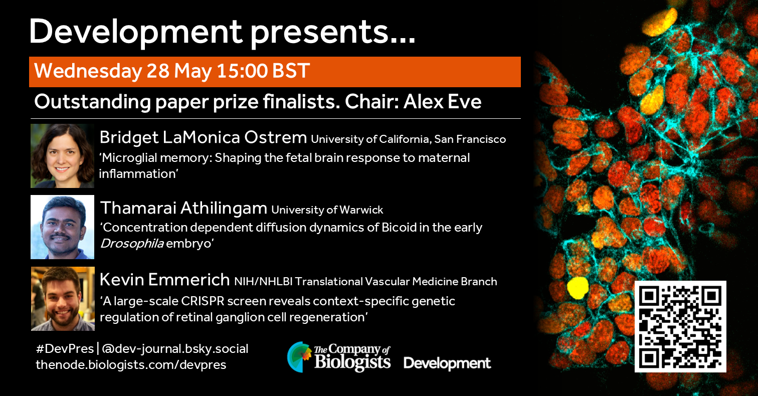
Join us to celebrate the finalists of Development’s 2024 outstanding paper prize, where three first authors will present their work. The webinar will be chaired by Development’s Executive Editor, Alex Eve.
Wednesday 28 May – 15:00 BST
Bridget LaMonica Ostrem (University of California, San Francisco) ‘Microglial memory: Shaping the fetal brain response to maternal inflammation’
Thamarai Athilingam (University of Warwick) ‘Concentration dependent diffusion dynamics of Bicoid in the early Drosophila embryo’
Kevin Emmerich (NIH/NHLBI Translational Vascular Medicine Branch) ‘A large-scale CRISPR screen reveals context-specific genetic regulation of retinal ganglion cell regeneration’
At the speakers’ discretion, the webinar will be recorded to view on demand. To see the other webinars scheduled in our series, and to catch up on previous talks, please visit: thenode.biologists.com/devpres
Spotted a preprint in this list that you love? If you’re keen to gain some science writing experience and be part of a friendly, diverse and international community, consider joining preLights and writing a preprint highlight article.
Sophie Wiszniak, Dimuthu Alankarage, Iman Lohraseb, Ceilidh Marchant, Genevieve Secker, Wendy Parker, John Toubia, Melissa White, Sandra Piltz, Markus Tondl, Eleni Giannoulatou, David Winlaw, Gillian M. Blue, Congenital Heart Disease Synergy Group, Patrick P. L. Tam, Paul Thomas, Natasha Harvey, Sally L. Dunwoodie, Quenten Schwarz
Jens Bager Christensen, Alex P.A. Donovan, Marzieh Moradi, Giada Vanacore, Mohab Helmy, Adam J. Reid, Jimmy Tsz Hang Lee, Omer Ali Bayraktar, Andrea H. Brand, N. Sumru Bayin
Jade A. Phillips, Jessica Perochon, Cai T. Johnson, Matthew Walker, Colin Nixon, Mark Hughes, André Barros-Carvalho, Yachuan Yu, Louise Mitchell, Karen Blyth, Massimo Vassalli, Julia B. Cordero
Gabriel Baonza, Tatiana Alfonso-Pérez, Gonzalo Herranz, Carlos Quintana-Quintana, Carmen Gordillo-Vázquez, Yara El Mazjoub, L.M. Escudero, David G. Míguez, Elisa Martí, Nuria Martínez-Martín, Fernando Martín-Belmonte
Aishwarya Ramamurthy, Masha D Bandouil, Likhita Aluru, Esther Yoon, Nicholas Bodkin, Jennifer Z Cheng, Carina G Biar, Jeffrey D Calhoun, Gemma L Carvill
Ruben I Calderon, Nirvay Sah, Molly Huang, Ryan H. Kittle, Walee Shaik, Jennifer N Chousal, Sampada Kallol, Tony Bui, Robert Morey, Alejandra Mitre, Norah M.E. Fogarty, Claudia Gerri, Claire Zheng, Peter ME DeHoff, Pratik Home, Kathy Niakan, Heidi Cook-Andersen, Kathleen M. Fisch, Soumen Paul, Francesca Soncin
Rashi Agarwal, Joergen Benjaminsen, Katharina Lust, Clara Becker, Natalia Fuchs, Eva Hasel de Carvalho, Fanny Eggeler, Omnia El Said Ibrahim, Narges Aghaallaei, Baubak Bajoghli, Joachim Wittbrodt
Damián García-García, Dunja Knapp, Minjoo Kim, Katelyn Jamwal, Heath Fuqua, Ryan P. Seaman, Riley E. Grindle, Sergej Nowoshilow, Maria Novatchkova, Fred W. Kolling, Joel H. Graber, Prayag Murawala
Adrien Franchet, Yuhong Jin, Clare L. Newell, Victor Girard, Gérard Manière, Yaël Grosjean, Christopher Barrington, James I. MacRae, Ian S. Gilmore, Alex P. Gould
Jennifer Falconi, Miriam Rodríguez-Vázquez, Katrin Strobel, Céline Jahannault-Talignani, Lisa Heron-Milhavet, Patrice Lassus, Charles Géminard, Alexandre Djiane
Zhenzhen Peng, Thitinee Vanichapol, Phong Dang Nguyen, Hao-Han George Chang, Katrinka M. Kocha, Lori L. O’Brien, Peter D. Currie, Peng Huang, Alan J. Davidson
Declan L. Turner, Hannah Baric, Katelyn Patatsos, Sahel Amoozadeh, Michael See, Kathleen A. Strumila, Jack T. Murphy, Liam Gubbels, Elizabeth Ng, Andrew Elefanty, Melanie Neeland, Shivanthan Shanthikumar, Sarah L. Londrigan, Mirana Ramialison, Fernando J. Rossello, Ed Stanley, Rhiannon B. Werder
Alexander Walker, Paula Olaizola, Euan Brennan, Edward J Jarman, Yuelin Yao, Elizabeth Carmichael, Andreea Gradinaru, Alexander EP Loftus, David H Wilson, Anabel Martinez Lyons, Laura Charlton, Kimberley Ober-Vliegen, Wunan Mi, Amy Broeders, Kyle Davies, Neil O. Carragher, Asier Unciti-Broceta, Timothy J Kendall, Luc van der Laan, Monique MA Verstegen, Margaret C Frame, Scott H Waddell, Luke Boulter
Esra Sengul, Helen G. Potts, William T. Stockdale, Ryan D. Carter, Laura Bevan, Maria Nozdrina, Rita Alonaizan, Zhilian Hu, Abigail Goodship, Jun Ying, Konstantinos Lekkos, Lucy O’Byrne, Madeleine E. Lemieux, Rebecca Richardson, Mathilda T.M. Mommersteeg
Jennifer Saile, Hannah Walter, Moritz Denecke, Patrick Lederer, Laura Schütz, Andreas Hiltbrunner, Katharina Lepp, Sofia Lobato-Gil, Petra Beli, Andreas Wachter
Patrycja Sokolowska, Matthias Jost, Wolfram Buss, Brett Ford, Peter Michael Chandler, Wolfgang Spielmeyer, Andy Phillips, Alison K. Huttly, Danuše Tarkowská, Rocío Alarcón-Reverte, Suzanne J. Clark, Stephen Pearce, Peter Hedden, Stephen G. Thomas
Juan Zapata-Muñoz, Juan Ignacio Jiménez-Loygorri, Michael Stumpe, Beatriz Villarejo-Zori, Sandra Alonso-Gil, Petra Terešak, Benan J. Mathai, Ian G Ganley, Anne Simonsen, Jörn Dengjel, Patricia Boya
Cerys E. Currie, Aleksandra Byrska, Deborah M. Taylor, Muriel Erent, Daniela Bakalova, Xuhui Sun, Constandina Koki, Nigel J. Burroughs, Richard. A. Anderson, Adele L. Marston, Geraldine M. Hartshorne, Andrew D. McAinsh
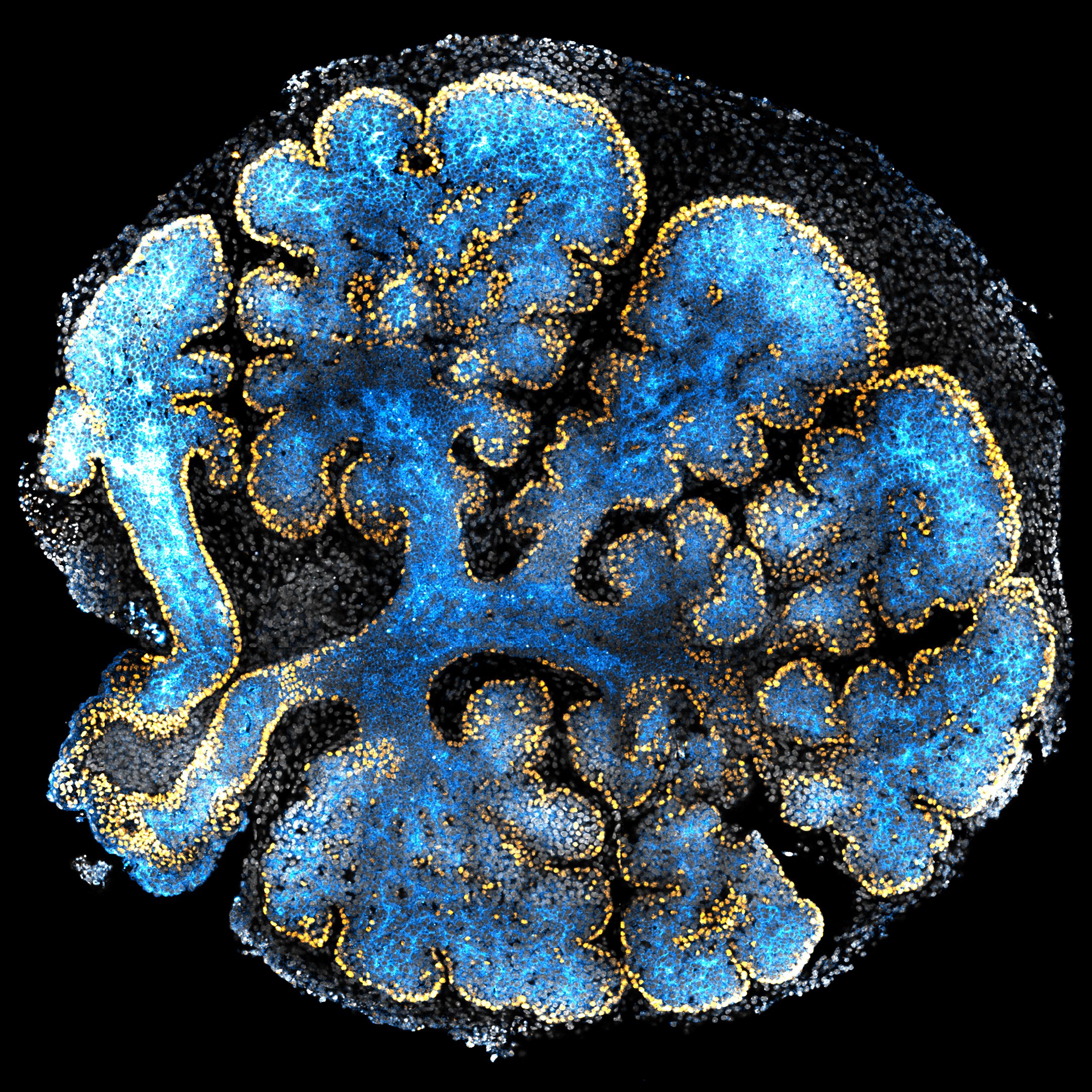
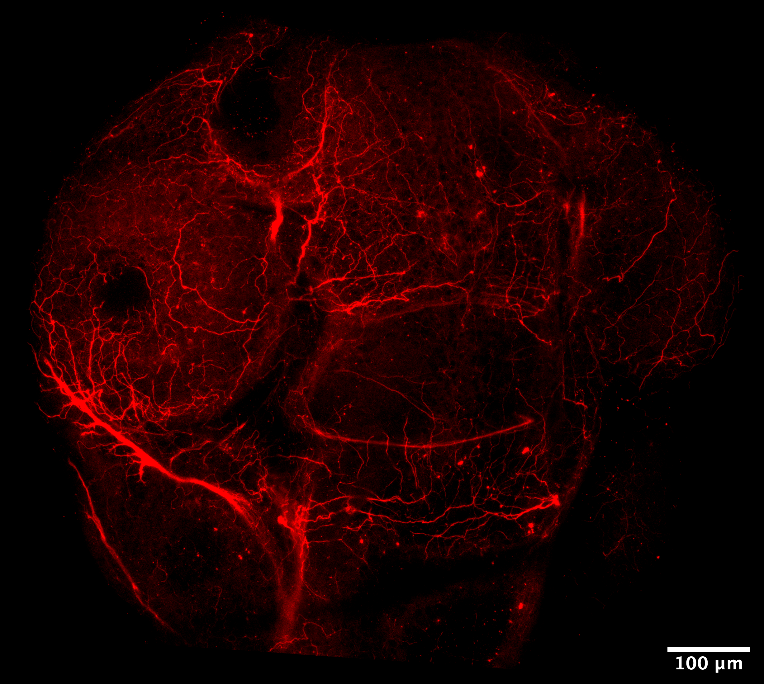
Marina Venero Galanternik, Daniel Castranova, Ryan D. Gober, Tuyet Nguyen, Madeleine Kenton, Gennady Margolin, Aurora Kraus, Abhinav Sur, Louis E. Dye 3rd, Van Pham, Adilenne Maese, Melanie Holmgren, Aniket V. Gore, Bakary Samasa, Allison Goldstein, Andrew E. Davis, Avery A. Swearer, James Iben, Tianwei Li, Steven L. Coon, Ryan K. Dale, Jeffrey A. Farrell, Brant M. Weinstein
From Galanternik et al. This image is made available for use under a CC0 license.
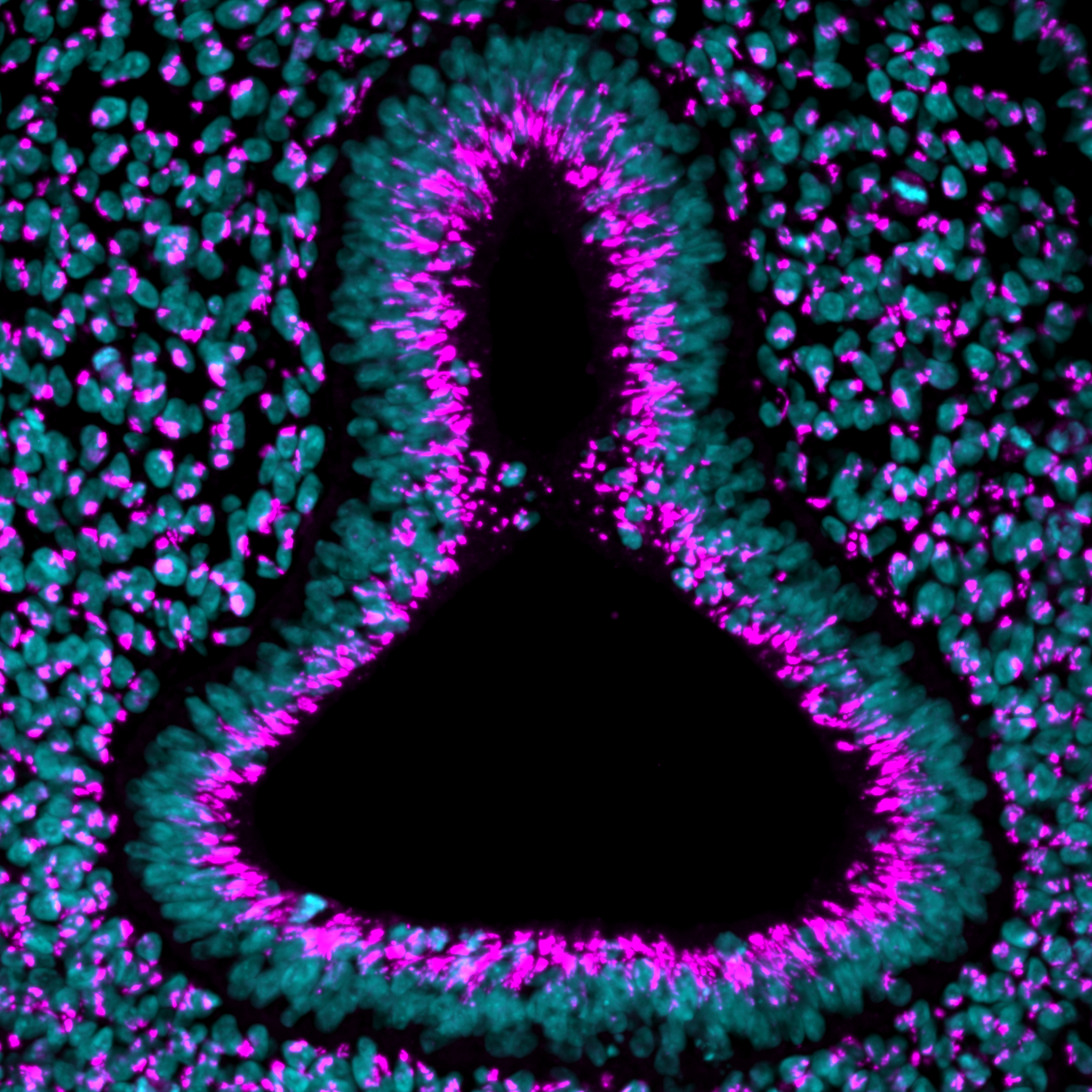
Leanne E. Iannucci, Velanganni Selvaraj Maria Thomas, Micaela R. Murphy, Caitlin E.T. Donahue, William K. Anderson, Catherine E. Rogers, Allison J. Saul, Katherine W. Rogers
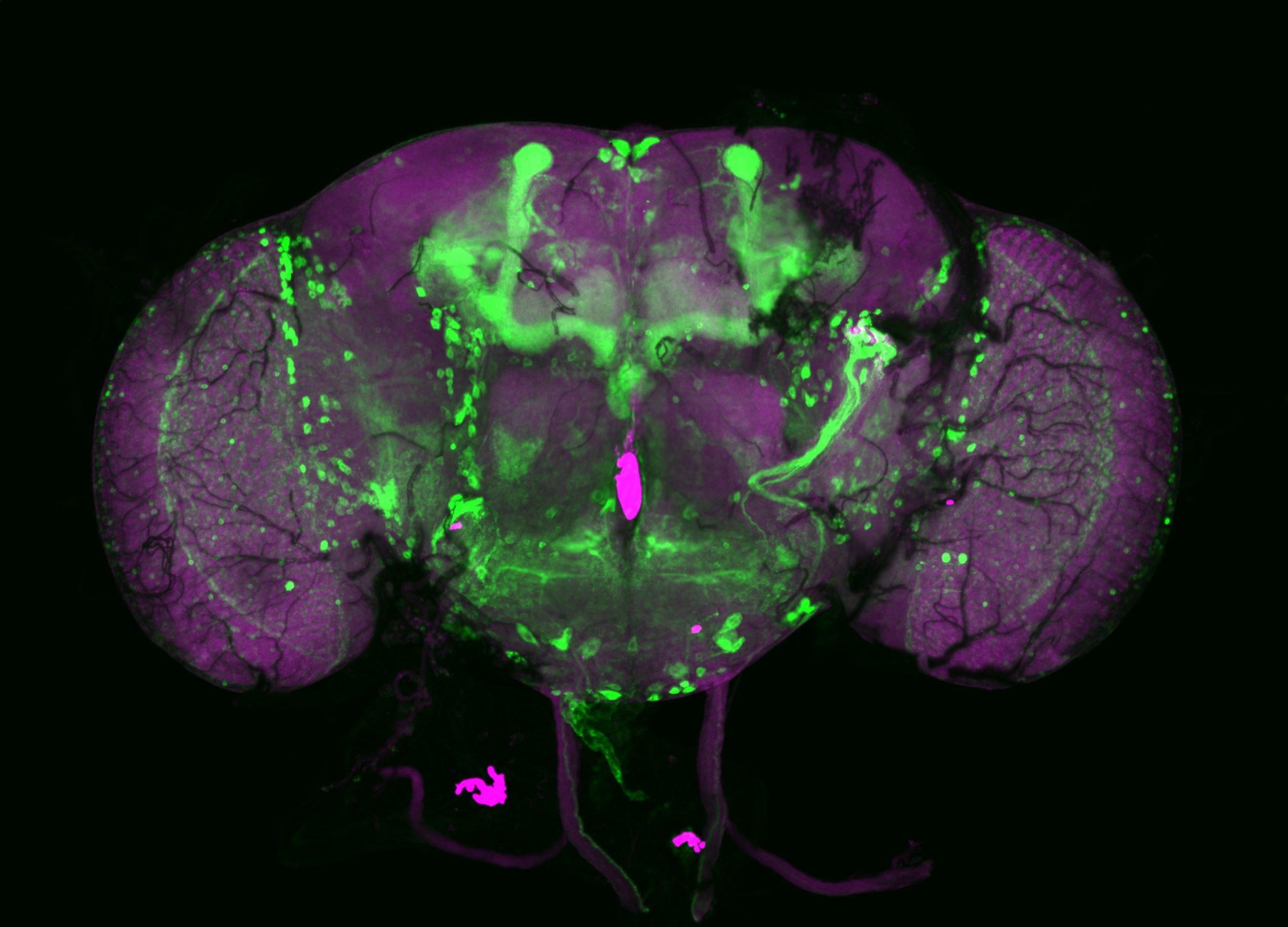
Dominic W.H. Shayler, Kevin Stachelek, Linda Cambier, Sunhye Lee, Jinlun Bai, Bhavana Bhat, Mark W. Reid, Daniel J. Weisenberger, Jennifer G. Aparicio, Yeha Kim, Mitali Singh, Maxwell Bay, Matthew E. Thornton, Eamon K. Doyle, Zachary Fouladian, Stephan G. Erberich, Brendan H. Grubbs, Michael A. Bonaguidi, Cheryl Mae Craft, Hardeep P. Singh, David Cobrinik
 (8 votes)
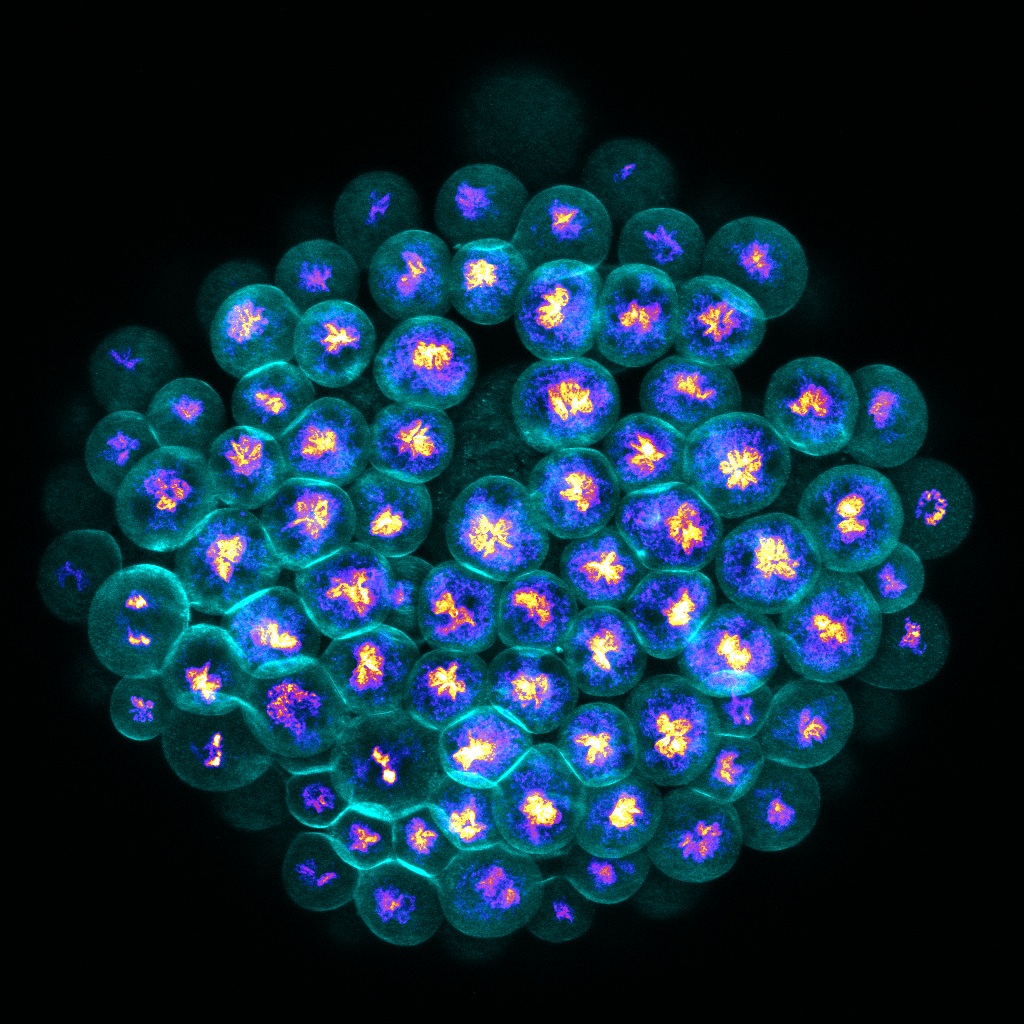

 (No Ratings Yet)