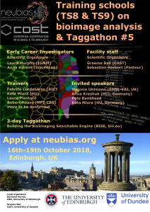
The focus of training will be on construction and automation of BioImage Analysis workflows, using as examples more than one toolbox and different exercises. The schools will be held in Edinburgh 16-19th of October 2018, hosted by the MRC Center for Regenerative Medicine/Imaging facility and co-organized by the University of Edinburgh and University of Dundee.
NEUBIAS schools are an excellent opportunity to learn from many experts in Bioimage Analysis (we are expecting >30 specialists at the event) and “….a great mix of intensive learning and community networking” (former trainee testimonial!). The schools will include practical sessions and seminars on ImageJ for analysis and publication, scripting/macros in ImageJ and Matlab, Omero, CellProfiler, QuPath, Ilastik, Ethics in Image Analysis and work on own data.
Applications are now open (each school has 20 available seats and ~7 trainers)
Applicants are highly encouraged to bring their own laptops and data.
Within the COST framework (funders of NEUBIAS), 7 travel grants per school are offered to applicants who qualify.
Registration requires a “letter of motivation” (filled in the application form) and later a confirmation of status.
Application deadline: May 11th, 2018
Selection notification: 1st week of June 2018.
More information about schools (programme & trainers) and venue, travel & lodge available at our website (see the linked pages for each school in above)
We kindly ask that you help us reach all potentially interested applicants.
Stem cells are defined by the dual capacity to self-renew and to differentiate. These properties sustain homeostatic cell turnover in adult tissues and enable repair and regeneration throughout the lifetime of the organism. In contrast, pluripotent stem cells are generated in the laboratory from early embryos or by molecular reprogramming. They have the capacity to make any somatic cell type, including tissue stem cells.
Stem cell biology aims to identify and characterise which cells are true stem cells, and to elucidate the physiological, cellular and molecular mechanisms that govern self-renewal, fate specification and differentiation. This research should provide new foundations for biomedical discovery, biotechnological and biopharmaceutical exploitation, and clinical applications in regenerative medicine.
CAMBRIDGE STEM CELL COMMUNITY
The University of Cambridge is exceptional in the depth and diversity of its research in Stem Cell Biology, and has a dynamic and interactive research community that is ranked amongst the foremost in the world. By bringing together members of both the Schools of Biology and Medicine, this studentship will enable you to take advantage of the strength and breadth of stem cell research available in Cambridge. Choose from over 50 participating host laboratories using a range of experimental approaches and organisms.
PROGRAMME OUTLINE
Students are expected to have chosen a laboratory for their thesis research prior to application, and to have obtained the support of the PI. A list of eligible supervisors can be found here.
Students will have access to our Discussion course during the first year, where they will:
Study fundamental aspects of Stem Cell Biology through a series of teaching modules led by leaders in the field.
Learn a variety of techniques, such as advanced imaging, flow cytometry, and management of complex data sets.
ELIGIBILITY
We welcome applications from those who hold a relevant first degree at the highest level (minimum of a UK II.i Honours Degreeor equivalent) as well as a Master’s Degree in a relevant discipline. You must have a passion for scientific research.
Wellcome provide full funding at the ‘Home/EU’ rate. Funding does not include overseas fees, so non-EU applicants will need to find alternative funding sources to cover these.
Another bumper month for developmental biology and related preprints, this time featuring spiders, lampreys, amphiouxus and plenty of zebrafish, worm and fly research. Also a big month for fans of Sox genes, which turned up in the title of five preprints! My visual highlight is the preprint from David Parichy’s group on zebrafish scale development – well worth downloading the PDF to gawp at the imaging.
The preprints were hosted on bioRxiv, PeerJ, andarXiv. Use these links to get to the section you want:
Discovery of a new path for red blood cell generation in the mouse embryo
Irina Pinheiro, Ozge Vargel Bolukbasi, Kerstin Ganter, Laura A. Sabou, Vick Key Tew, Giulia Bolasco, Maya Shvartsman, Polina V. Pavlovich, Andreas Buness, Christina Nikolakopoulou, Isabelle Bergiers, Valerie Kouskoff, Georges Lacaud, Christophe Lancrin
Yap regulates glucose utilization and sustains nucleotide synthesis to enable organ growth
Andrew Cox, Allison Tsomides, Dean Yimlamai, Katie Hwang, Joel Miesfeld, Giorgio Galli, Brendan Fowl, Michael Fort, Kimberly Ma, Mark Sullivan, Aaron Hosios, Erin Snay, Min Yuan, Kristin Brown, Evan Lien, Sagar Chhangawala, Matthew Steinhauser, John Asara, Yariv Houvras, Brian Link, Matthew Vander Heiden, Fernando Camargo, Wolfram Goessling
Tracking mandibular arch cell rearrangements in Tao, et al.’s preprint
Oscillatory cortical forces promote three dimensional cell intercalations that shape the mandibular arch
Hirotaka Tao, Min Zhu, Kimberly Lau, Owen Whitley, Mohammad Samani, Xiao Xiao, Xiao Xiao Chen, Noah A. Hahn, Weifan Liu, Megan Valencia, Min Wu, Xian Wang, Kelli D. Fenelon, Clarissa C. Pasiliao, Di Hu, Jinchun Wu, Shoshana Spring, James Ferguson, Edith P. Karuana, R. Mark Henkelman, Alexander Dunn, Huaxiong Huang, Hsin-Yi Henry Ho, Radhika Atit, Sidhartha Goyal, Yu Sun, Sevan Hopyan
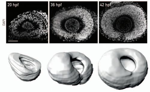
Tracking development of the zebrafish retina, from Matejcic, et al.’s preprint
Single-cell analysis identifies EpCAM+/CDH6+/TROP-2- cells as human liver progenitors.
Joe M Segal, Daniel J Wesche, Maria Paola Serra, Bénédicte Oulés, Deniz Kent, Soon Seng Ng, Gozde Kar, Guy Emerton, Samuel Blackford, Spyros Darmanis, Rosa Miquel, Tu Vinh, Ryo Yamamoto, Andrew Bonham, Alessandra Vigilante, Sarah Teichmann, Stephen R Quake, Hiromitsu Nakauchi, S Tamir Rashid
Randal Burns, Eric Perlman, Alex Baden, William Gray Roncal, Ben Falk, Vikram Chandrashekhar, Forrest Collman, Sharmishtaa Seshamani, Jesse Patsolic, Kunal Lillaney, Michael Kazhdan, Robert Hider Jr., Derek Pryor, Jordan Matelsky, Timothy Gion, Priya Manavalan, Brock Wester, Mark Chevillet, Eric T. Trautman, Khaled Khairy, Eric Bridgeford, Dean M. Kleissas, Daniel J. Tward, Ailey K. Crow, Matthew A. Wright, Michael I. Miller, Stephen J Smith, R. Jacob Vogelstein, Karl Deisseroth, Joshua T. Vogelstein
| Genome tools
An optimized toolkit for precision base editing
Maria Paz Zafra, Emma M Schatoff, Alyna Katti, Miguel Foronda, Marco Breinig, Anabel Y Schweitzer, Amber Simon, Teng Han, Sukanya Goswami, Emma Montgomery, Jordana Thibado, Francisco J Sanchez-Rivera, Junwei Shi, Christopher R Vakoc, Scott W Lowe, Darjus F Tschaharganeh, Lukas E Dow
Robust single-cell DNA methylome profiling with snmC-seq2
Chongyuan Luo, Angeline Rivkin, Jingtian Zhou, Justin P Sandoval, Laurie Kurihara, Jacinta Lucero, Rosa Castanon, Joseph R Nery, Antonio Pinto-Duarte, Brian Bui, Conor Fitzpatrick, Carolyn O’Connor, Seth Ruga, Marc E Van Eden, David A Davis, Deborah C Mash, M. Margarita Behrens, Joseph R Ecker
Community-driven data analysis training for biology
Bérénice Batut, Saskia Hiltemann, Andrea Bagnacani, Dannon Baker, Vivek Bhardwaj, Clemens Blank, Anthony Bretaudeau, Loraine Guéguen, Martin Čech, John Chilton, Dave Clements, Olivia Doppelt-Azeroual, Anika Erxleben, Mallory Freeberg, Simon Gladman, Youri Hoogstrate, Hans-Rudolf Hotz, Torsten Houwaart, Pratik Jagtap, Delphine Lariviere, Gildas Le Corguillé, Thomas Manke, Fabien Mareuil, Fidel Ramírez, Devon Ryan, Florian Sigloch, Nicola Soranzo, Joachim Wolff, Pavankumar Videm, Markus Wolfien, Aisanjiang Wubuli, Dilmurat Yusuf, Rolf Backofen, James Taylor, Anton Nekrutenko, Björn Grüning
Seán I O’Donoghue, Benedetta F Baldi2, Susan J Clark, Aaron E Darling, James M Hogan, Sandeep Kaur, Lena Maier-Hein, Davis J McCarthy, William J Moore, Esther Stenau, Jason R Swedlow, Jenny Vuong, James B Procter
David Trafimow, Valentin Amrhein, Corson N. Areshenkoff, Carlos Barrera-Causil, Eric J. Beh, Yusuf Bilgiç, Roser Bono, Michael T. Bradley, William M. Briggs, Héctor A. Cepeda-Freyre, Sergio E. Chaigneau, Daniel R. Ciocca, Juan Carlos Correa, Denis Cousineau, Michiel R. de Boer, Subhra Sankar Dhar, Igor Dolgov, Juana Gómez-Benito, Marian Grendar, James Grice, Martin E. Guerrero-Gimenez, Andrés Gutiérrez, Tania B. Huedo-Medina, Klaus Jaffe, Armina Janyan, Ali Karimnezhad, Fränzi Korner-Nievergelt, Koji Kosugi, Martin Lachmair, Rubén Ledesma, Roberto Limongi, Marco Tullio Liuzza, Rosaria Lombardo, Michael Marks, Gunther Meinlschmidt, Ladislas Nalborczyk, Hung T. Nguyen, Raydonal Ospina, Jose D. Perezgonzalez, Roland Pfister, Juan José Rahona, David A. Rodríguez-Medina, Xavier Romão, Susana Ruiz-Fernández, Isabel Suarez, Marion Tegethoff, Mauricio Tejo, Rens van de Schoot, Ivan Vankov, Santiago Velasco-Forero, Tonghui Wang, Yuki Yamada, Felipe C. Zoppino, Fernando Marmolejo-Ramos
Christos D Arvanitidis, Richard M Warwick, Paul J Somerfield, Christina Pavloudi, Evangelos Pafilis, Anastassis Oulas, Giorgos Chatzigeorgiou, Vasilis Gerovasileiou, Theodoros Patkos, Nicolas Bailly, Francisco Hernandez, Bart Vanhoorne, Leen Vandepitte, Ward Appeltans, Robert Adlard, Peter Adriaens, Ahn Kee-Jeong, Ahyong Shane, Akkari Nesrine, Gary Anderson, Angel Martin, Claudia Arango, Tom Artois, Stephen Atkinson, Ruud Bank, Anthony D Barber, Joao P Barbosa, Ilse Bartsch, Denise Bellan-Santini, Jimmy Bernot, Annalisa Berta, Rüdiger Bieler, Magda Błażewicz, Phil Bock, Ruth Böttger-Schnack, Philippe Bouchet, Nicole Boury-Esnault, Geoff Boxshall, Christopher B Boyko, Simone Nunes Brandão, Rod Bray, Niel L Bruce, Stephen Cairns, Tania N Campinas Bezerra, Paco Cárdenas, Benny KK Chan, Tin-Yam Chan, Lanna Cheng, Morgan Churchill, Laure Corbari, Ralf Cordeiro, Astrid Cornils, Keith A Crandall, Thomas Cribb, Jean-Loup D’hondt, Meg Daly, Mikhail Daneliya, Jean-Claude Dauvin, Peter Davie, Claude De Broyer, Valentin De Mazancourt, Nicole De Voogd, Peter Decker, Danielle Defaye, Henk Dijkstra, Martin Dohrmann, Daryl Domning, Rachel Downey, Inna Drapun, Ursula Eisendle-Flöckner, Christine Ewers-Saucedo, Marien Faber, Diego Figueroa, Julian Finn, Gustavo Fonseca, Ewan Fordyce, William Foster, Hidetaka Furuya, Horia Galea, Oscar Garcia-Alvarez, Rade Garic, Rebeca Gasca, Santiago Gaviria-Melo, Sarah Gerken, David Gibson, João Gil, Arjan Gittenberger, Chris Glasby, Serge Gofas, Samuel E Gómez-Noguera, David González-Solís, Dennis Gordon, Michal Grabowski, Cinzia Gravili, José M Guerra-García, Roberto Guidetti, Katja Guilini, Kerry A Hadfield, Ed Hendrycks, Bachiller Herrera, Ju-Shey Ho, Jens Høeg, Oleksandr Holovachov, Matthew D Hooge, John Hooper, Tammy Horton, Lauren Hughes, Matús Hyžný, Luiz IF Moretti, Tohru Iseto, Viatcheslav N Ivanenko, Gerhard Jarms, Damià Jaume, Krzysztof Jazdzewski, Ivana Karanovic, Young-Hyo Kim, Rachael King, Michelle Klautau, Jürgen Kolb, Alexey Kotov, Traudl Krapp-Schickel, Antonina Kremenetskaia, Reinhardt Kristensen, Andreas Kroh, Sven Kullander, Rafael La Perna, Sara LeCroy, Daniel Leduc, Rafael Lemaitre, Anne-Nina Lörz, Jim Lowry, Enrique Macpherson, Larry Madin, Tomasz Mamos, Renata Manconi, Bruce Marshall, David J Marshall, Patrick Martin, Sandra McInnes, Jan Mees, Tõnu Meidla, Kelly Merrin, Dmitry Miljutin, Claudia Mills, Vadim Mokievsky, Tina Molodtsova, Rich Mooi, André C Morandini, Rosana Moreira Da Rocha, Fabio Moretzsohn, Jonas Mortelmans, Jeanne Mortimer, Luigi Musco, Thomas A Neubauer, Eike Neubert, Peter NG Neuhaus, Anh D Nguyen, Claus Nielsen, Jon Norenburg, Tim O’Hara, Hisayo Okahashi, Dennis Opresko, Masayuki Osawa, Yuzo Ota, Gustav Paulay, Vincent Perrier, William Perrin, Iorgu Petrescu, Bernard Picton, John F Pilger, Andrzej Pisera, Dan Polhemus, Gary Poore, James D Reimer, Hans Reip, Michael Reuscher, Pilar Rios Lopez, Marc Rius, Klaus Rützler, Alexander Rzhavsky, José Saiz-Salinas, André F Sartori, Heinrich Schatz, Bernd Schierwater, Andreas Schmidt-Rhaesa, Simon Schneider, Christine Schönberg, André R Senna, Cristiana Serejo, Shabuddin Shaik, Shokoofeh Shamsi, Jyotsna Sharma, Noa Shenkar, Andrew Shinn, Jacek Sicinski, Volker Siegel, Petra Sierwald, Elizabeth Simmons, Frederic Sinniger, Duncan Sivell, Boris Sket, Harry Smit, Nicole Smol, Jesser F Souza-Filho, Jörg Spelda, Sérgio N Stampar, Eric Stienen, Pavel Stoev, Sabine Stöhr, Malin Strand, Eduardo Suárez-Morales, Mindi Summers, Billie J Swalla, Stefano Taiti, Masaatsu Tanaka, Anne H Tandberg, Danny Tang, Mark Tasker, Harry ten Hove, Jan J ter Poorten, Jim Thomas, Erik V Thuesen, Ben Thuy, Juan T Timi, Antonio Todaro, Xavier Turon, Peter Uetz, Sergiy Utevsky, Jean Vacelet, Risto Väinölä, Sancia ET van der Meij, Ton van Haaren, Virág Venekey, Chris Vos, Genefor Walker-Smith, Chad T Walter, Les Watling, Matthew Wayland, Christopher Whipps, Gary Williams, Robin Wilson, Moriaki Yasuhara, Joana Zanol, Wolfgang Zeidler
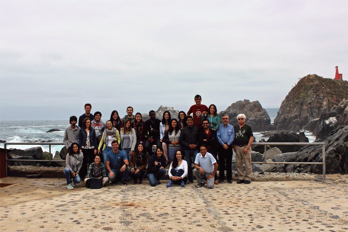
In January 2018, twenty-one graduate students and early career researchers from across South and North America participated in the International Course on Developmental Biology, an EMBO Practical Course held at the Marine Biology Station of Quintay-Chile (CIMARQ). This two-week course was led by eight world-renowned researchers, Drs. Roberto Mayor, Nipam Patel, John Ewer, Raymond Keller, Alejandro Sánchez-Alvarado, Kathleen Whitlock, Claudio Stern and Andrea Streit, many of whom started or currently base their careers in Latin American countries. First offered in 1999, the creation of this course reflected the enthusiasm of Latin American researchers to promote and enhance the field of Developmental Biology in this region by providing an opportunity for young scientific investigators to learn current research techniques and become familiar with model organisms used in this field. A successful outcome from the course was the creation of the Latin American Society of Developmental Biology (LASDB) in 2003, which holds biannual meetings for researchers to disseminate their findings and network with colleagues throughout Latin America. Although the changing political, social and economic climate still presents challenges for young researchers, remarkable progress has been made over the past 20 years. Notably, with several of the recent participants of this course having supervisors who also completed the course earlier in their career, the International Course on Developmental Biology is clearly an important part of the emerging research landscape.
For many centuries, Developmental Biology was thought of as a descriptive science. Naturalists dedicated themselves to describing the extensive modifications that occur during the development of various species, characterizing and defining embryo size and shape, as well as the duration of different developmental stages. They addressed fundamental questions that remain to this day: How does a single cell give rise to a complete organism? What are the molecular signals involved? To answer these questions, scientists began using organisms at different levels of complexity, establishing the classical experimental models of Developmental Biology: the roundworm, fruitfly, frog, zebrafish, chicken and mouse, or C. elegans, Drosophila, Xenopus, Danio rerio, Gallus gallus and Mus musculus, as they are affectionately known by scientists. Developmental Biology began to revolutionize in the ‘70s when new technologies such as recombinant DNA became available to study how genes specify different tissue identities. In the ‘80s, we began to talk about homeobox genes, promoters, transcription factors and enhancers, and started using gene expression techniques such as in situ hybridization. As questions at the genetic and morphogenetic level began to be clarified, other questions started to arise: How does evolution shape developmental processes? How can development be modified by the surrounding environment? How can we take advantage of the plasticity of development?
Today, Developmental Biology is a highly-regarded field of study, being a course requirement of most undergraduate programs in the biological sciences. But why did we, as young scientific investigators choose it as our research discipline? We all agree: when you first observe a developing organism under the lens of a microscope, the entire world becomes fascinating. We see life’s great questions in our work. For instance, when does a cell leave it’s past behind and enter into it’s destiny? What makes a cell unique? What are the costs of differentiation? Could it be immobility, like for osteocytes; or programmed cell death, without which the fingers would not exist; or perhaps pruning, like what more than half of the brain’s synaptic connections undergo to achieve functionality. Then there are the milestones. In one of our seminars by Prof. Roberto Mayor, we were given to ponder the famous quote by Lewis Wolpert, that it is “not birth, marriage or death, but gastrulation which is truly the most important time in your life”. With such intriguing insights into the facts of life, how could any other subject area compare?
Simply exploring your surroundings in Latin America leaves one searching for answers. With unique habitats holding nearly one-fifth of the world’s plant and vertebrate species (Myers et al., 2000), it has been called “a living laboratory” and is a place of continuous discovery. This was made evident to us, by exploring just a few meters on the coastal shore of the Quintay waters, where we observed a great diversity of organisms, protected between the rocks before being revealed under our microscope lenses. Unfortunately, human activities that trigger climate change, diseases, and species introductions are affecting natural ecosystems. So we are given the challenge to find new research models to discover unique molecular and cellular mechanisms before they are lost entirely. Several research groups such as Dr. Alejandro Sánchez-Alvarado’s lab are investigating emerging model systems which may help us to overcome types of illness and injury not possible today.
Despite the benefits of conducting Developmental Biology research in Latin America, the reality is that resources and funding are limited. Countries that have an established tradition of scientific investment such as Brazil, Argentina, Mexico and Chile (Marcellini et al., 2017) are more equipped than others; however, careful experimental design, collaboration and creative problem solving are essential. All of us came with different backgrounds: free or paid access to undergraduate studies, different levels of research experience, different levels of English fluency, among others, which showed us the challenges that everyone was going through in the road to pursuing our dream of becoming scientists. However, all of these differences were set aside during the two-week course. The appreciation of the personal value of each participant and faculty member, the willingness to help each other, the equality with which the faculty and participants exchanged ideas was indicative of the underlying message of this course: advance together as one.
As scientists who study the transient process of life as it acquires it’s form and function, we are part of a minority that has the knowledge and the opportunity to make the world a better place. With climate change, wildlife extinction, food and resource crisis at our doors, difficult times are ahead of us. It is our responsibility to observe what is going on around us, make predictions, test hypotheses, and not simply resign ourselves to the way things are. We must share our knowledge, explain science and impact our society. It is in our hands to spread scientific knowledge to the general public and especially, to future scientists at a young age. This can be achieved by creating outreach programs, such as the television series that Dr. Kate Whitlock created, La Alegría de la Ciencia, which can stimulate children and adults to be interested in Developmental Biology. Science must be inclusive and not limited to scientists. Funding reviewers and government decision makers also need to be informed about our work. Here it is our duty as Developmental Biologists to explain that our research is more than simply “basic science” with no significant contribution to solve current problems. Rather, it is through studying processes such as cell migration, epithelial-mesenchymal transition, control of gene expression, and cell death that we can solve problems in reproduction, development, disease, aging and regeneration.
The personal and professional experiences of this two-week happy confinement in Quintay are some of the most illuminating and transforming that many of us have. Although separated now in different countries and continents, we have shared unforgettable moments of life and learning in this course and the doors were left open to continue working together in the future. We go forward knowing that we CAN make a difference if only we keep pushing towards a better future.
Group photo, taken moments before getting soaked by an incoming wave. Bottom row, left to right: Emma Rangel Huerta, Angelly Vasquez, Mateus Antonio Berni, Lorena Agostini Maia, Jessica Cristina Marin Llera, Nancy María Farfán, Nipam Patel. Middle row, left to right: Lautaro Gándara, Jennifer Giffin, Maria Belen Palacios, Luiza Saad, Estefanía Sánchez-Vásquez, Jorge Antolio Domínguez-Bautista, Adrián Romero, Roberto Mayor, Raymond Keller. Top row, left to right: Fernando Faunes, John Rojas, Sandra Edwards, Elias Barriga, Lucía Bartolomeu, Eugene Tine, Soraya Villaseca, Paula M. González, Matías Preza, Maria Fernanda Palominos.
“A continent whose thriving biodiversity represents endless forms most beautiful and most wonderful that are a source of inspiration and opportunities for the Evo-Devo community” (Marcellini et al., 2017).
Written by the participants of the 2018 International Course on Developmental Biology.
References
Marcellini S, González FA, Sarrazin AF, Pabón-Mora N, Benítez M, Piñeyro-Nelson A, Rezende GL, Maldonado E, Schneider PN, Grizante MB, Da Fonseca RN, Vergara-Silva F, Suaza-Gaviria V, Zumajo-Cardona C, Zattara EE, Casasa S, Suárez-Baron H, Brown FD. (2017). Evolutionary developmental biology (Evo-Devo) research in Latin America. J. Exp. Zool. (Mol. Dev. Evol.) 328B:5-40.
Myers N, Mittermeier RA, Mittermeier CG, da Fonseca GAB, Kent J. (2000). Biodiversity hotspots for conservation priorities. Nature 403: 853-858.
As well as publishing Development and four other journals, and supporting scientists through travelling fellowships and meeting grants, The Company of Biologists also runs a successful series of Workshops.
The Workshops bring leading experts and early career scientists from a diverse range of scientific backgrounds to focus on one topic together. Topics are often interdisciplinary and cover some of the most exciting current biology, as you can see in the archive.
The Workshop Committee are currently seeking proposals for four Workshops to be held during 2020. They are particularly keen to receive proposals from postdocs for one of the Workshops.We at the Node would also encourage applicants from developmental biology to think about applying!
The Marine Biological Laboratory is seeking applicants for full-time Research Assistant and Research Associate positions with the Josephine Bay Paul Center for Comparative Molecular Biology and Evolution (http://www.mbl.edu/jbpc/).
To develop CRISPR in rotifers:
We seek a motivated, creative and innovative Research Assistant or Research Associate to join the laboratories of Kristin Gribble and David Mark Welch. Our research combines comparative genomics, biochemistry, and life history to study aging, maternal effects, and DNA damage prevention and repair using rotifers, a novel aquatic invertebrate model system for studies of aging, neurobiology, genome evolution, and ecology. The successful candidate will develop genome editing techniques in rotifers, including CRISPR/Cas9, as part of a broad initiative at the MBL to advance new aquatic and marine models for biological discovery. We invite individuals with experience in genome editing in other animals to join this expanding program.
To study the biochemistry of DNA repair in bdelloids:
We seek a Research Assistant to join the laboratory of David Mark Welch. Our research combines comparative genomics, biochemistry, and life history to study the evolution of bdelloid rotifers, extraordinarily resilient animals adapted to highly stressful environmental conditions. The successful candidate will contribute to an ongoing project to clone, express, purify, and assay a variety of rotifer proteins involved in DNA damage prevention and repair. There is considerable opportunity for motivated, self-directed individuals to participate in technique development, manuscript development and publication. Position level and salary will depend on education and experience.
We seek applicants for a full-time Research Assistant position with the Keck Sequencing Facility of the Josephine Bay Paul Center. The successful applicant will contribute to projects that will explore diversity of microbes in various communities and the influence of changing environments on microbial population structures. Responsibilities include, but are not limited to, laboratory management, preparation and massively high-throughput next-generation sequencing of marker gene and metagenomic libraries from environmental genomic DNA, and computational analysis of such datasets.
A neuroscience Postdoctoral Research Associate position is available in the Robles Lab at Purdue University (www.robleslab.com). Our laboratory applies advanced genetic and microscopic imaging techniques to understand how the nervous system develops in the larval zebrafish. We use confocal laser scanning microscopy to image the structural development of genetically targeted neurons within the intact developing brain. Our major area of emphasis is developing novel, in vivo imaging assays to examine the cellular and circuit-level abnormalities underlying complex neurological disorders such as autism and epilepsy.
Candidates should have a Ph.D. in neuroscience, cell biology, genetics, or a related field. The ideal candidate will have expertise in microscopy, image analysis, and molecular biology techniques. Special consideration will be given to applicants with expertise in developing and implementing computational strategies for data/image analysis.
Please send your CV, a cover letter stating your research interests and professional goals, and the contact information for three (3) references to:
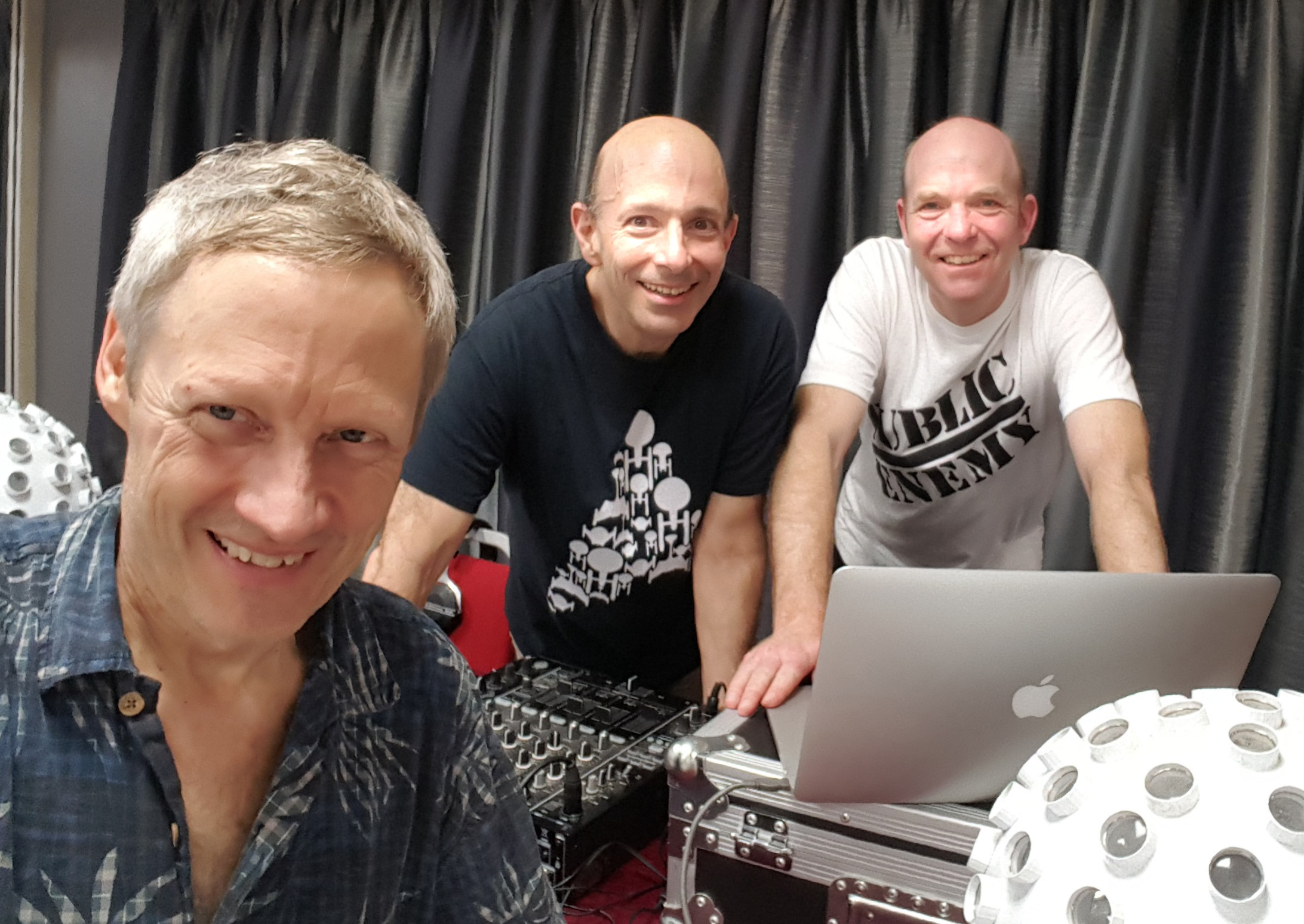
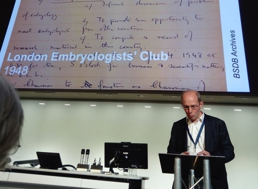
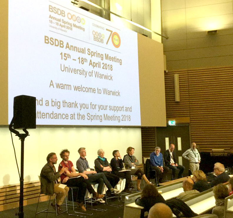
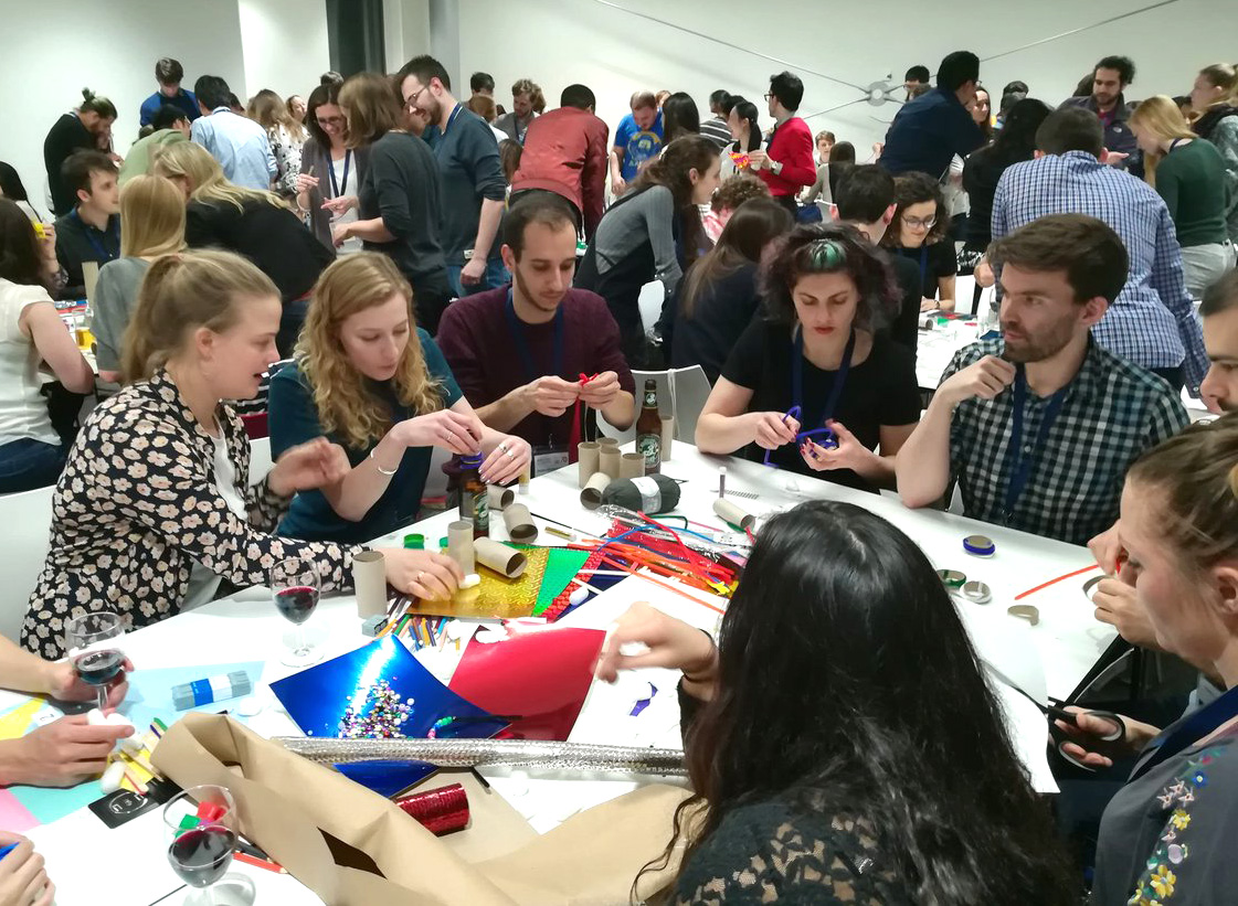
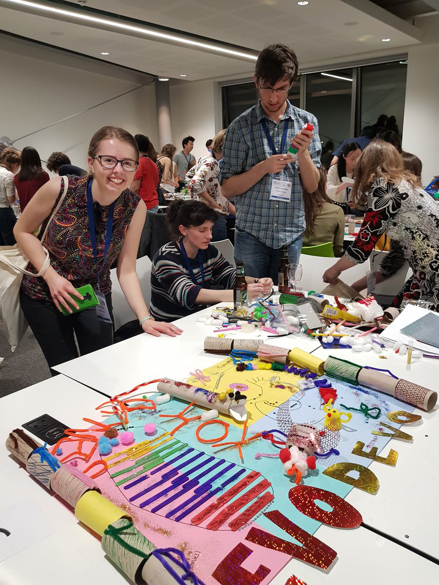
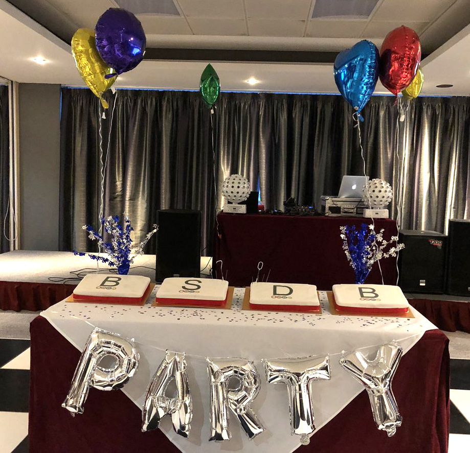
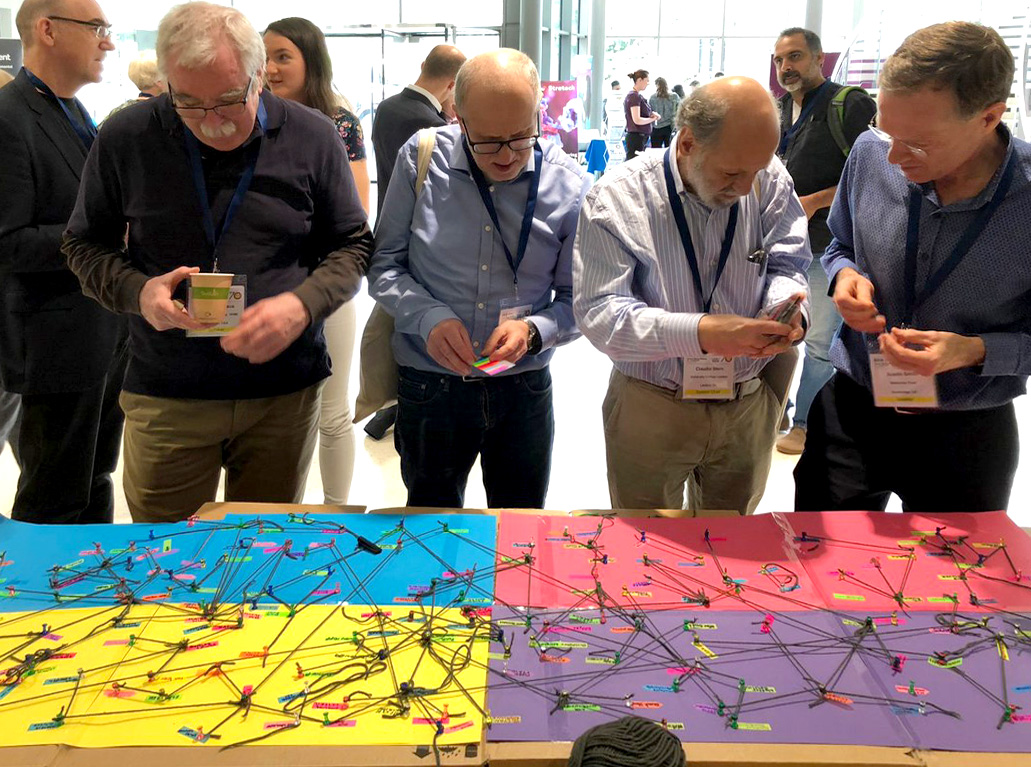
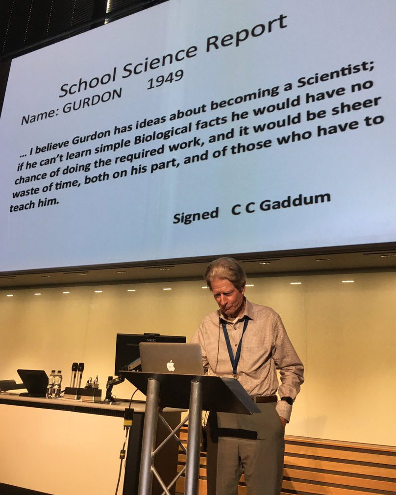
This year, is the BSDB’s 70th anniversary, and this was clearly reflected at our Spring Meeting, 15-18 April 2018 in Warwick! Apart from an outstanding speaker list, and the award of most BSDB medals & prizes of 2018, we saw a history session which looked back at the past, to then project into the future of Developmental Biology. Other specials were a very engaging PhD and postdoc social, the weaving of pedigree networks (with little labels, pins and threads of wool), and the live presentations of DB raps composed and sung by Jerry aka Gerald H Thomsen PhD and produced and mixed by Philip Larsen. See below a few image impressions of the meeting (taken from your many tweets!) and/or listen to the two raps! A detailed meeting report will follow.
Rap 1: BSDB History (part I & II)
Rap 2: Morphogen Mix
Gerald Thomsen, Josh Brickman and Philip Larsen
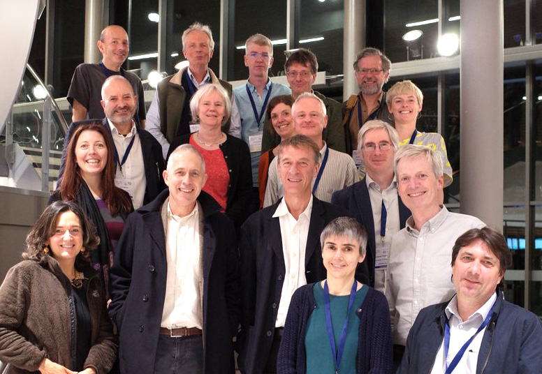
Only a tiny selection of participants
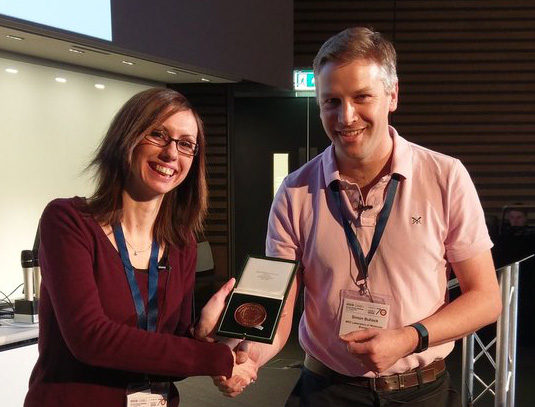
Emilia receives the Beddington medal from Simon Bullock
Nick Hopwood
The panel discussion
The PhD and postdoc social
PhD and postdoc social
Gearing up for the conference dinner
Weaving pedigree networks: Eric Wieschaus, David Ish-Horowicz, Claudio Stern, Austin Smith
A Wellcome Trust funded Postdoctoral Research Associate position is available in the lab of Dr. Matt Towers at the University of Sheffield, UK, to work on vertebrate limb development. How embryonic development is timed and scaled remains enigmatic. Understanding the underlying mechanisms remains a central challenge in biology, since these mechanisms coordinate the growth and patterning of all tissues and organs, and when disrupted are likely to form the basis of many congenital disorders. Our recent work on the embryonic chick wing demonstrates a pivotal role for intrinsic timers in limb development (Chinnaiya et al, Nat Comms 2014, Saiz-Lopez et al, Nat Comms 2015; Saiz-Lopez et al, Development 2017, Pickering et al, Biorxiv 2018). The aim of this project is to use a combination of classical grafting techniques and modern genomic analyses in chick embryos to understand the molecular mechanisms of how cells measure time in vivo.
A position (#123768) is available immediately for a Postdoctoral Scholar to contribute to our multidisciplinary studies aimed at elucidating the molecular basis of chick neural crest and placode cell development. The postdoc will conduct independent research and assist in the training of students in the laboratory of Dr. Lisa Taneyhill at the University of Maryland.
Laboratory skills: Molecular biology and biochemical assays (e.g., recombinant DNA/cloning; DNA, RNA, and/or protein blotting); immunohistochemistry; and/or in situ hybridization. Experience with microscopy and spectroscopy, chick embryology (including microdissections and electroporation), and tissue culture is desirable. For more information on the lab, please see http://www.ansc.umd.edu/people/lisa-taneyhill.
Qualifications: An advanced degree (Ph.D.) in Developmental, Molecular and/or Cell Biology is required. Fluency in spoken and written English is required. Compensation: Salaries are highly competitive, negotiable and commensurate with qualifications. Fringe benefits offered. Applicants must apply through eTerp at https://ejobs.umd.edu. Applications will be accepted until a suitable candidate is identified.
 The focus of training will be on construction and automation of BioImage Analysis workflows, using as examples more than one toolbox and different exercises. The schools will be held in Edinburgh 16-19th of October 2018, hosted by the MRC Center for Regenerative Medicine/Imaging facility and co-organized by the University of Edinburgh and University of Dundee.
 (No Ratings Yet)
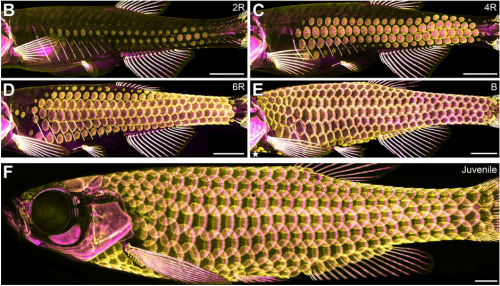
 (18 votes)
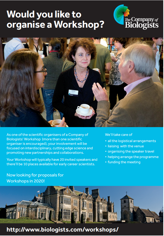
 (1 votes)