Interested in exploring cell adhesion, cytoskeletal regulation, or Wnt signaling & their roles in development and oncogenesis, as a graduate student or postdoc? The Peifer lab at UNC-Chapel Hill is a great place to explore these. You can learn more about our work at: http://peiferlab.web.unc.edu and a graphic summary is at: http://peiferlab.web.unc.edu/files/2017/11/PeiferLabPoster2017sm.pdf
I’ll be at the ASCB/EMBO Meeting in December-drop me a line to meet or email at peifer@unc.edu
On August 2013, I took my first one-way trip departing from Puerto Rico. Although I have always been passionate about travelling to as many places (the cheapest way) possible, embracing the PhD-journey in a new city I would have to call “Home” was very intimidating. Close to the start of my journey at the Georgia Institute of Technology I joined the Garcia Lab due to my interest in the engineering of materials to study tissue development and regeneration. A couple more months later and I have already found myself very engaged in a new project in collaboration with the Nusrat Lab. The project involved the engineering of a synthetic hydrogel to direct mouse intestinal stem cell differentiation in vitro and serve as an in vivo cell-delivery vehicle that promotes intestinal wound repair, overcoming the translational limitations associated with natural matrices. This project aimed to provide potential therapeutic options by facilitating intestinal mucosal wound repair which is central in many pathologic states, such as inflammatory bowel disease.
At the time I was very much enjoying the work and, considering my heavy fundamental engineering background, it was all very novel to me as I faced a steep learning curve of very exciting biological concepts. In addition, having such amazing collaboration with cell biologists and clinicians placed me in the forefront of multiple fields that collide for a single goal.
It was all fun and games until I found myself two years later stuck in the same first-stage of my project because the mouse intestinal tissue did not survive within (or basically, hated) my hydrogels. As I continued to assimilate that we weren’t going to reach our desired experimental results (or in colloquial words, that “the project wasn’t working”) we met Jason Spence. He introduced us to the novel technology of in vitro generation of human intestinal organoids (HIOs) from human pluripotent stem cells (hPSCs). We grew to what is now a three-lab interdisciplinary collaboration involving bioengineers, clinicians, cell biologists and developmental biologists. And that’s where this journey took a different turn.
With experiments happening mostly at the University of Michigan I now had the opportunity to merge into another new environment with new collaborators and, just as significant, a different weather. Must say that for a Caribbean islander (despite having such amazing hosts) spending winter in Ann Arbor made the experience quite more challenging. Furthermore, due to the complexity of the hPSC culture I used to travel to Michigan in very short (sometimes even with one-week) notice. Thus, having to figure out the logistics for the trip (from lodging to shipping reagents; from all I was leaving behind in Atlanta to the experimental planning in Michigan) was very challenging. Once I was there, there was no time to waste. The clock was ticking, Miguel and I had an experimental plan to accomplish despite any (or the many) complications we faced and, to me, returning to Atlanta saying, “I didn’t have enough time to do that” was not an option. Nevertheless, after several trips to the beautiful (and cold) Ann Arbor, lots of planning and experimenting, research started to move forward, my project kept evolving and, most importantly, “it was working”…
Ricardo Cruz-Acuña examines differentiating human intestinal organoids (HIOs) under a microscope. The research may lead to a new technique for treating injuries caused by gastrointestinal diseases. (Credit: Rob Felt, Georgia Tech)
The Science
In this project we studied the in vitro generation of human intestinal organoids (HIOs) from human pluripotent stem cells (hPSCs). This technology offers strategies for generating multi-cellular 3D structures that recapitulate important features of the human intestine. For example, HIOs can be used for the establishment of chronic intestinal disease models, such as inflammatory bowel disease (IBD), and provides a platform for functional modeling and repair of genetic defects in intestinal development. As part of the method to generate HIOs, hPSCs must be cultured and differentiated in a MatrigelTM-coated substrate, giving rise to 3D intestinal spheroids which are collected and encapsulated within MatrigelTM for expansion into HIOs. MatrigelTM suffers from lot-to-lot variability and an undefined tumor-derived nature which limits its clinical translational potential. Therefore, we implemented a synthetic hydrogel based on a four-armed, maleimide-terminated poly(ethylene glycol) macromer (PEG-4MAL), developed in the Garcia Lab, as a substitute to MatrigelTM for encapsulation of intestinal spheroids and further generation of HIOs. Our synthetic material offers significant advantages over natural materials due to its well-defined structure, cytocompatibility and minimal toxicity in vivo, overcoming the limitations of MatrigelTM.
In order to generate the HIOs using our synthetic hydrogel, floating intestinal spheroids were collected from the culture plate and encapsulated within PEG-4MAL macromers that were functionalized with adhesive and crosslinking peptide motifs for expansion into HIOs. The controlled stoichiometric incorporation of peptides to the synthetic material allowed me to independently modify the biophysical and biochemical properties of the hydrogel to ultimately identify an engineered material that supports in vitro generation of HIOs from hPSC-derived spheroids without the need of MatrigelTM. Both biophysical and biochemical properties of the synthetic matrix were important to intestinal organoid formation, and allowed me to engineer an optimal formulation that supports intestinal spheroid survival, expansion and epithelial differentiation into HIOs and differentiation into mature intestinal tissue in vivo to similar levels as MatrigelTM (Figure 2).
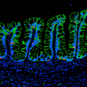
Figure 2: Human intestinal organoids (HIOs) generated in the hydrogel matrix differentiate into mature intestinal tissue and present main human intestinal cell types, such as enteroendocrine cells (CHGA; red), after transplantation into an animal. (Credit: Miguel Quirós, University of Michigan)
In addition (and probably my favorite part of the project), we established the use of the engineered hydrogel as a delivery vehicle for HIOs to murine intestinal mucosal wounds using a murine colonoscope. The tunable reaction time scales of this material allowed injection of hydrogel liquid precursors and HIOs to mucosal wounds resulting in an in situ polymerized hydrogel that supported localized organoid engraftment and enhanced wound repair. These findings form a basis for the development of HIO-based therapies to treat gastrointestinal diseases in humans involving intestinal epithelial wounds (e.g., IBD).
The (More) Questions
As we have established a synthetic hydrogel to generate hPSC-derived HIOs and treat intestinal injuries, many new questions arise: Can we use this material to study matrix contributions to gastrointestinal disease and repair? Can we use this material as an HIO-delivery vehicle to treat chronic gastrointestinal diseases? Can we establish this material as a platform to generate different types of human organoids? And the journey continues…
The Thanks
Now back in (the warmer weather of) Atlanta, I could go on for about three paragraphs naming everybody and everything that somehow contributed to what is, so far, my major achievement research-wise. Nevertheless, I feel is more than fair to primarily thank my homeland, Puerto Rico, and the University of Puerto Rico at Mayagüez, as they are the foundation of where I am today and to where I am going. I will forever be in debt to my island (that little piece of heaven) and will always hold it in my heart, proudly, wherever I am.
Join invited speakers and showcase your work to celebrate the 30th HGM. We particularly welcome presentations from budding scientists to foster networking and collaboration opportunities.
This 2-day meeting will end with the Peter Thorogood Memorial Lecture by Prof. William Harris.
29-30 January, 2018 – UCL Great Ormond Street Institute of Child Health, London, UK
Abstract submission for oral or poster presentation: 15th December, 2017
Conference Registration: 8th January, 2018
BackgroundtotheMeeting
Following the 1987 BSDB Meeting on “Craniofacial Development” organized by Peter Thorogood and Cheryll Tickle, the organizers and other colleagues (Mark Ferguson, Andrew Lumsden, Gudrun Moore, Alisdair Ivens, Julian Lewis and Gillian Morriss-Kay) decided to start holding annual informal meetings to increase communication between laboratories working on various aspects of the development of the head, to offer the opportunity to young scientists to present their work in a friendly and stimulating environment and promote collaborative research. The group has met annually since 1989, and the initial number of participants, about 20 people, rose very rapidly through word of mouth with more groups asking to be included in the mailing list. Over the last few years there have always been over one hundred people attending the meeting.
A postdoctoral research position is available starting in 2018 with Asst. Prof. Timothy Saunders’ group at the Mechanobiology Institute, Singapore. The Saunders lab has been active since 2013 and studies the fundamental processes shaping organs and tissues during development.
One major area of study in the lab is spatial position – how do developing embryos know where to position boundaries? One mechanism is the morphogen gradient, but, despite significant work over the past 20 years, there is still little reliable data about the dynamics of morphogen gradient formation. We are part of a major five-year grant focused on understanding the dynamics of morphogen gradient formation in Zebrafish embryogenesis. The project is a close collaboration between experimentalists, biophysicists and computational biologists. We are looking for a talented post-doctoral researcher interested in working at the interface of developmental biology and biophysics. In particular, the position will involve extensive imaging and image analysis of the early Zebrafish embryo, including light-sheet microscopy. This offers an exciting opportunity for a dedicated researcher to be part of a genuinely interdisciplinary project that will advance our fundamental understanding of development.
Candidates should have strong experience in at least one of the following, and display a willingness to learn the other: (1) advanced microscopy techniques and imaging developing organisms; and/or (2) experience with image analysis and handling large datasets.
The Saunders’ lab is a young group at the Mechanobiology Institute. This provides an exciting prospect for a motivated post-doc to be involved in developing research directions. The post-doc will also be expected to guide and help the graduate students in the laboratory. More information can be found at: http://labs.mbi.nus.edu.sg/mod/.
Qualifications:
1) A Ph.D in Biophysics, Developmental Biology, Computational Biology, or related subject.
2) At least one first-author paper in English submitted to an international peer-reviewed journal.
3) Experience in: (i) Advanced microscopy; (ii) Image analysis and handling large datasets; and (iii) Imaging developing organisms.
Salary and benefits are commensurable to educational qualifications and working experience of the candidates. Benefits include annual leave, medical and flexi-benefits, etc.
Please submit your curriculum vitae and a statement about your research interests to
Lee lab has been studying on cell specification process using human pluripotent stem cells, particularly nervous system and skeletal muscle cells to study human diseases and to develop new drugs.
We are looking for highly motivated postdoc(s) who have experience on one of the following fields, including Alzheimer’s disease and tau pathology.
Previous experience on human pluripotent stem cells is not required (but basic cell culture experience will be great), and we value more on the ‘non-stem cell’ expertise. Please send your application (CV and three reference contact info) to Gabsang Lee (leelabjob@gmail.com).
Neurodevelopmental disorders are a group of different conditions in which the development of the central nervous system is disturbed. This includes developmental brain dysfunction, which can manifest as neuropsychiatric problems (Autism Spectrum Disorders, schizophrenia, fragile-X syndrome, down syndrome), or impaired motor function, learning, language or non-verbal communication. The generation of the appropriate diversity of neural cell types, their migration to correct sites in the brain and the establishment of precise connectivity with target cells are key developmental processes that may go awry, leading to brain dysfunction and neurological diseases.
During this advanced 3-week course, we will provide participants with a comprehensive theoretical and experimental/practical training on the most advanced methodologies and models in developmental neurobiology, in health and disease. World experts (both seniors and juniors) in the field of neural stem cells, brain patterning, neuronal migration, axon guidance or the genetics of human neurodevelopmental disorders will give stimulating lectures and design mini-projects to be carried out by the students.
This will give students a unique opportunity to become familiar with cutting-edge in vivo gene manipulation methods (electroporation, Crispr/Cas9, genetics…), 3D imaging technologies (SPIM and light sheet microscopy, tissue clearing, two-photon imaging…), time-lapse microscopy and cell tracking, advanced pluripotent stem cell culture (iPSCs, cortical progenitors and neurons derived thereof, organoids, organotypics…).
Deadline: 18 December 2017 (Midnight Brussels time)
The Johnson Lab in the Department of Developmental Biology at Washington University School of Medicine (http://devbio.wustl.edu) is seeking applications for NIH funded Research Scientist positions. Our lab uses multiple model systems to uncover molecular mechanisms of muscle development and disease. We are looking for highly motivated individuals to begin new projects using mouse models and human ES cells to probe mechanisms of muscle disease.
DUTIES:
Assists in the training and development of technicians, acting as a group leader.
Independently generates mouse strains and collects and analyses histology data.
Develops human ES cell differentiation methods using published protocols.
Performs complex analysis/projects according to research protocols, explaining methods and procedures to other technicians.
QUALIFICATIONS:
Master’s degree with 2 years of experience in a laboratory setting (or equivalent combination of education and experience equaling 7 years)
Experience with mouse husbandry and mammalian cell culture
Please send a cover letter, CV, and list of 3 references to anjohnson@wustl.edu
Glaucoma is a devastating retinal degenerative disease without effective treatments, and its causes remain elusive. Our laboratory uses mice as a model to study glaucoma pathogenesis at the molecular level. A NIH-funded postdoctoral position is available immediately to investigate glaucoma pathogenesis in mice. In addition, our laboratory also studies the molecular mechanisms underlying adult stem cell self-renewal and differentiation. The candidate also has a unique opportunity to investigate the potential of using stem cells to treat retinal degenerative diseases. Our research has been published in high-profile journals, including Science, Nature, Cell Stem Cell and PNAS. Preferably, the candidate has a Ph.D. degree in molecular biology, developmental biology and/or genetics (research experience with mouse research and vision research is a plus). We offer highly competitive salary plus medical benefits. Our lab is accessible to state-of-art expert-run facilities, including high throughput sequencing, microarrays, proteomics, histology, FACS and imaging. For more information about the research in Dr. Xie’s laboratory, please visit the website: http://www.stowers.org/faculty/xie-lab. If interested and any questions, please send an email to Dr. Ting Xie attgx@stowers.org.
Zika infection in humans is associated with birth defects including microcephaly. Zika has two major lineages – the Asian lineage, which has been associated with birth defects, and the African lineage, which has not – but the relative effects of each strain on brain development, and the effects of the related dengue virus that co-circulates with Zika, are not understood. This week we feature a paper published in the latest issue of Development that uses a mouse model to compare the individual effects of these viruses. Co-first authors, Qiang Shao and Stephanie Herrlinger, and PI Jian-Fu (Jeff) Chen told us more.
Qiang Shao, Stephanie Herrlinger and Jeff Chen
Jeff, can you give us your scientific biography and the main questions your lab is trying to answer?
JFC My lab is interested in mechanisms underlying microcephaly and neurodegeneration. We use mouse and human pluripotent stem cell-derived neural cells to model neurological disorders followed by mechanistic studies at molecular, cellular, and circuit levels.
Qiang and Stephanie, how did you come to join the Chen lab?
SH I have always been very passionate about studying the brain and I was looking for a mentor who was as enthusiastic as I was. Jeff’s previous work studying brain development and neural tube defects gave me the opportunity to explore brain development and embryology, and his enthusiasm for understanding biology is infectious. After the first week of my rotation with his lab, it was obvious that I was going to do my PhD with Jeff.
QS I did my PhD in China, focusing on the discovery of antiviral genes and drugs. I am always passionate about brain development and neuro-infectious disease. Before my graduation, one of my committee members, Dr. He, recommended me to Jeff for a postdoc position, knowing that he is an expert in neurodevelopmental disorders. After a few rounds of interviews, Jeff offered me this position. And here I am studying Zika virus and microcephaly.
What was known about the differences between the two lineages, both in the lab and in the human population, before your work?
JFC We knew that Asia Zika virus (ZIKV) is sufficient to cause microcephaly and additional brain abnormalities from both animal models and the human population. On the other hand, Africa ZIKV had not yet been associated with any birth or brain defects. Before our work, we also knew there was about a 110 amino acid difference between the Africa ZIKV isolate and Asia isolate in our lab, but we had no idea about their comparative toxicity and their impact to the developing brain.
Can you give is the key results of the paper in a paragraph?
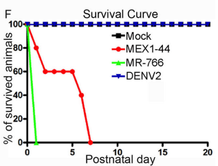
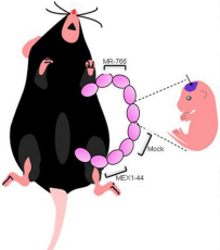
JFC The key findings are that Africa Zika virus (ZIKV) grows faster, causes more cell death in neural progenitor cells (NPCs) and neurons, and leads to more severe brain damage and postnatal death than Asia ZIKV. Both viruses similarly infect NPCs and trigger microglial activation and astrogliosis. Meanwhile, Dengue virus also infects NPCs and grows robustly in the developing brain, but fails to cause brain damage and postnatal death. Therefore, Africa ZIKV and Asia ZIKV have some intrinsic differences that account for their differential impacts on the developing brain.
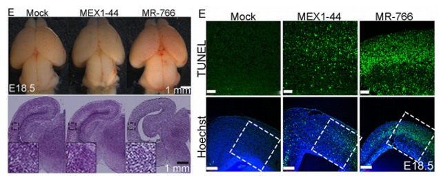
Africa Zika causes a smaller cortex and increased progenitor and neuronal death than Asian Zika. From Figures 4 and 7, Shao et al, 2017.
Why did you test this hypothesis in a mouse model as opposed to using human cerebral organoids?
JFCWe previously have established the first postnatal microcephaly mouse model associated with ZIKV infection (Shao et al., Development, 2016). It is handy for us to use this system. It is absolutely important to further test this hypothesis using human cerebral organoids, which have the advantage of modelling early human brain development.
SH Cerebral organoids are a great system to study NPCs and NPC behaviours at early stages; however, the developing brain includes a diverse set of neural progenitor, immune, vascular, and glial cell types. With our mouse model, we are able to specifically ask how the heterogeneous cell populations of the brain responds and contributes to ZIKV infection pathology and disease progression.
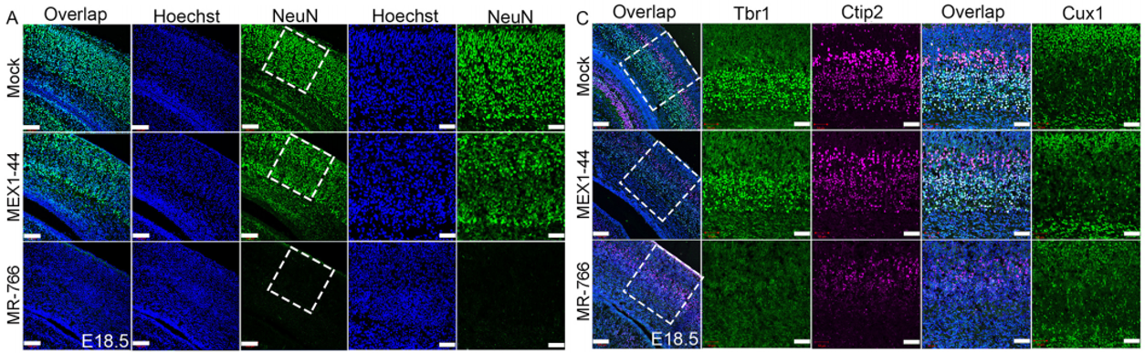
Africa Zika causes more neuronal loss than Asia Zika. From Figure 5, Shao et al, 2017.
You mention cases of co-infection between Zika and dengue, do you have plans to model this in mice and what effect do you hypothesise it will have on neural development?
JFC We would like to model it and we expect that co-infection between Zika and dengue will yield an intermediate brain defect.
What do you hypothesise the genetic/molecular basis might be for the different in virulence between the strains?
JFC We expect that certain mutations in one or more structural/non-structural viral proteins accounts for the difference. We are actively pursuing this direction now.
When doing the research, did you have any particular result or eureka moment that has stuck with you?
SH When we first saw the much more dramatic pathology of the African ZIKV strain on the developing brain, we were very surprised! The fact that this virus was not previously associated with birth defects was a big part of why we hypothesized that the Asian-ZIKV would be more deleterious than the African-ZIKV strain.
QS It was how the ZIKV African strain caused embryonic lethality that struck me the most. Before this study, there was no evidence suggesting an association between ZIKV African strain and microcephaly. I was expecting to see a mild phenotype in ZIKV African strain infected mouse embryos compared to Asian strain. It turned out that none of the ZIKV African strain infected embryo survived to postnatal stage. I was shocked by the phenotype of embryonic lethality. After this project, I realised unexpected results often lead to interesting and important discovery.
Postnatal survival of mice infected with the different viruses. From Figure 4, Shao et al, 2017.
And what about the flipside: any moments of frustration or despair?
SH It was frustrating at first to find that the African-ZIKV infected embryos did not survive gestation as the Asian-ZIKV infected embryos did. We were initially concerned that it was due to some complication of the surgeries as they can be arduous and the embryos are sensitive.
QS ZIKV African and Asian strain side by side comparison surgery is the most difficult part of this study. Statistical analysis requires three biological replicates. So I had to inject three embryos with mock, ZIKV-African, and ZIKV-Asian, respectively, which means nine embryos in one litter. This presents with many technical and experimental challenges including the potential of early embryo loss. It took me a whole month to optimize surgical conditions such as polishing glass needles, reducing viral titer and adjusting injection sites. Fortunately, through a long process of trial and error, I was able to obtain enough samples to work with.
Infection of embryonic mice. From Figure 4, Shao et al, 2017.
What are your career plans following this work?
SH I successfully received a predoctoral to postdoctoral transition award from the NIH (D-SPAN F99/K00) to study the impact that ZIKV has on post-transcriptional regulation in infected neural progenitor cells. In the coming year I plan to graduate and move on to my postdoctoral career.
QS I am currently writing a research proposal to further study the mechanism underlying ZIKV caused neurovascular defect. Specifically, I am interested in understanding the role of individual ZIKV protein in neurovascular development disruption. This work will provide important molecular mechanism of ZIKV pathogenesis.
And what next for the Chen lab?
JFC We are trying to figure out the molecular mechanisms underlying Zika virus-induced microcephaly and associated neurological disorders.
Finally, what do you three like to do when you are not in the lab?
JFC I always like to spend more time with my family.
SHI like to spend time going to see live music, my family, hiking, and travelling.
QS I enjoy spending time with my partner and friends. I love cooking at home. It’s kind of like doing experiments in the kitchen with different ingredients. I find those moments very relaxing and full of delight.
The Mokalled lab in the Department of Developmental Biology at Washington University School of Medicine is hiring at all levels (http://www.mokalledlab.com/). Our lab uses zebrafish and mouse model systems to study neural regeneration after spinal cord injury or disease. Candidates with enthusiasm for neuroscience, regenerative biology, and zebrafish research are encouraged to forward a cover letter, CV, and list of 3 or more references to mmokalled@wustl.edu.
 (1 votes)
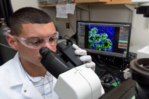
 (No Ratings Yet)