The community site for and by
developmental and stem cell biologists

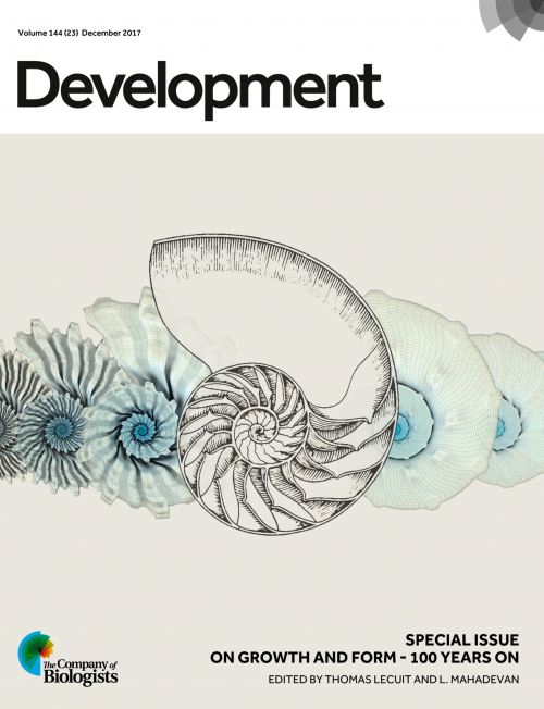
On Growth and Form at 100: Perspectives from the field

Posted by , on 28 November 2017

What explains the lasting legacy of D’Arcy Thompson’s ‘On Growth and Form’? A century on from the publication of the first issue, we reached out to authors from Development’s special issue celebrating the centenary and asked what the book meant to them.


A rare first edition of On Growth and Form held in the D’Arcy Thompson Zoology Museum in the University of Dundee. Find out more about the museum in our interview with its curator Matthew Jarron here.


Arkhat Abzhanov

Natural History Museum (London) & Imperial College London

“There are ideas which make their impact and give way to new and better ideas. They have their day and disappear into history.  However, there also ideas that have a much more profound and enduring effect and continue to influence the way we think for many yeas, sometimes for generations. “On Growth and Form” by D’Arcy Thompson is a book full of such brilliant everlasting ideas (and so eloquently written).  I remember reading it for the first time as a half-fledged undergraduate awed by the notion that living organisms and their parts do not have “random” shapes but could be related to each other in precise mathematical terms. What could be a more powerful and visual evidence for evolution than a gradual transformation of a skull of a primitive horse into a modern one via a series of shapes of the intermediate species?  This book asked many important questions but gave few concrete answers, instead outlining ways of addressing problems.  My own current research is very much driven by the quest to explain Thompson’s “laws of growth” and their role in evolution while using his “theory of transformations” in the form of modern geometric morphometrics.  For example, the origin and subsequent diversification of the uniquely-shaped skull of modern birds can be better understood by comparing avian skull shapes with each other and with their reptilian ancestors – all within the same “morphospace”, the concept invented by D’Arcy Thompson.  Thus, his ideas in many ways are still surprisingly relevant and stimulating today as ever before. I hope that the 100th anniversary will bring this book more attention from the new generation of scientists curious about biological shapes.”


Arkhat’s review – The old and new faces of morphology: the legacy of D’Arcy Thompson’s ‘theory of transformations’ and ‘laws of growth’


Douglas DeSimone

University of Virginia 

“I spent several summers in the early 1980’s undertaking my doctoral research on early sea urchin development at the Marine Biological Laboratory (MBL) in Woods Hole. I remember fondly the diverse array of sea life that John Valois and his crew in the supply department would collect for seasonal investigators over the course of each summer. Their labors afforded opportunities to observe and handle not only adult marine animals – many of which I had never seen alive before – but also, on occasion, their eggs and embryos. It was against this backdrop that I developed a career-long fascination with the problem of morphogenesis, and first became acquainted with D’Arcy Thompson’s “On Growth and Form”. Back then, there was a small bookstore on Water Street that was unusual for carrying not only the light summer reading demanded by island-bound tourists, but also a selection of titles reflective of the greater MBL and Woods Hole Oceanographic Institute scientific communities. One August, as the exodus of summer investigators, students and tourists began to transform Woods Hole back to its less frenetic state, I purchased a paperback edition of Thompson’s book, which I began reading on the beach and would continue back at my home institution that Fall. Thompson’s perspective inspired me to appreciate the problem of development as more than an unfolding of genetically-encoded programs but one that is also subject to physical rules and open to mathematical description. In many ways, I feel that my research and that of others interested in morphogenesis has moved inexorably toward a view of embryos as biomechanical “machines”. Thompson likely could not have imagined the modern tools and approaches now being developed to measure, analyze and model forces generated by cells and tissues but it is safe to say he would have approved of where the field has headed since the publication of his influential work.”


Paper from the DeSimone lab – ‘Mechanical and signaling roles for keratin intermediate filaments in the assembly and morphogenesis of Xenopus mesendoderm tissue at gastrulation


François Graner & Daniel Riveline

Université Denis Diderot, Paris and IGBMC, Strasbourg

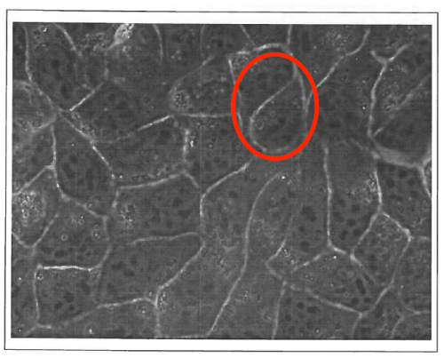
“DR is an experimentalist biophysicist, working on acto-myosin as well as cell adhesion and motility in vitro; FG is at the interface between tissues and foams, and between theory and experiments. We have been knowing each other 20 years ago, while we were in the same lab in Grenoble, and our first discussions in 1999 made already clear that MDCK cells monolayers cellular outlines were not following rules of shapes for bubbles (see our original MDCK image below).


Tissues as foams ? The original 1999 image with MDCK epithelial cells observed by phase contrast microscopy which triggered the first discussions between the authors. In the highlighted zone, these cellular outlines would not be possible in foams.


We both then moved to different scientific sites, and we started to interact again in 2010 around a new Master program launched by DR in Strasbourg University, where we gave interdisciplinary lectures to the same students. DR suggested to share classics pages with students on considering living matter as a matter, such as Thompson book proposes, with modern echoes in developmental biology. FG was reluctant in considering these pages as actually seminal: he rather considered that they had been causing a waste of time over the century, by triggering inadequate searches for simplicity and perfection. These distinct appreciations of ‘On Growth and Form’ nurtured between us an animated debate. We decided to focus on two chapters which were central for us, i.e. on shape and packing of cells in tissues. Our disagreement was so strong that it was difficult to imagine that we would agree on a common flow; initially, we had no agenda whatsoever to wrap up this discussion in an article one day. We turned this debate into a scientific approach, by carefully listing specific arguments and counter-arguments.

And then, we both moved from our original respective postures. DR recognized that Thompson had been merging phenomena under the same umbrella, with often analogy as a guideline and limited care about physical mechanisms. FG acknowledged the influence of Thompson in general and even on his own work for several aspects, and particularly the illuminating ability of Thompson to act as honest experimentalist representing tissues through their essential read-outs – cell contours, representing them as they actually are. We had just decided to write an article when, by coincidence, Development launched a call for its special issue.

A long way has been done since our very first discussions, and we view a posteriori our debates as one of the most productive discussions we had in recent years, with no other ambition than understanding the text and its scope a century after its first publication. And we can only encourage others to perform the same patient task for other chapters of this book or other classical texts in Biology.”


François and Daniel’s review – ‘The Forms of Tissues, or Cell-aggregates’: D’Arcy Thompson’s influence and its limits


Olivier Hamant

University of Lyon

“The field of development has reinvented itself several times over the past decades, building from grafting/morphogen-centered experiments to molecular genetics, cell biology and now including more quantitative and predictive aspects, through biophysics and computational modeling. With the rise of quantitative approaches, the current era is very much in line with D’Arcy Thompson persistent words:


“Dreams apart, numerical precision is the very soul of science, and its attainment affords the best, perhaps, the only criterion of the truth of theories and the correctness of experiments”


D’Arcy Thompson certainly was a (maybe too) strong advocate of the role of physical forces in development, but as we are collecting more and more evidence of a widespread role of mechanical signals in morphogenesis and cell behavior, his take on development also offers a source of inspiration for the young generation of interdisciplinarians in development.

One key quote that was influential to me is a simple one:


“The form of an object is a “diagram of forces””


The simplicity of these words has many implications. It underlines the fact that changing shape is not only a geometrical problem: it also involves a structural change, and thus, that changing patterns of forces must be managed by cells. Not as immediately obvious, this statement also implies that a living object will usually try to resist such forces. There are now many examples of cells displaying such resistance strategies, from changes in cell growth rate, to cell polarity or extracellular matrix stiffening. This also is true at tissue and organ scales, the most obvious example being Wolff’s law, which is mentioned in D’Arcy’s book, where bones build their intricate trabeculae network along maximal mechanical stress lines. We, and others, found similar responses in plants, with cells and tissues reinforcing their structure in the direction of maximal stress too, making this statement from physics, a true biological theorem. D’Arcy Thompson was a supporter of the organismal theory of development, and this quote is also consistent with that option, diagrams of forces spreading, and arguably, synchronizing, populations of cells. While this leaves out many subtleties, it remains a very powerful reminder of the fact that cells manage communication, nutrition, reproduction, etc. and while doing all this, also need to maintain their own structural integrity, which calls for synergies between biochemical and mechanical signalling.

One last take home message from D’Arcy Thompson’s book is his didactic choice: the use of many analogies makes his book very accessible to a wide audience, truly bridging biology, mathematics and physics. This is a remarkable piece of work, and despite several questionable interpretations 100 years down the road, “On growth and form” remains a landmark in developmental biology, and very much worth a read today, for biologists, mathematicians and physicists alike.”


Paper from the Hamant lab – ‘Phyllotactic regularity requires the Paf1 complex in Arabidopsis


Akatsuki Kimura & Kazunori Yamamoto

SOKENDAI & National Institute of Genetics, Mishima, Japan

AK “When I was a post-doc, I first read “On Growth and Form” in the Japanese version translated by Dr. Tomomichi Yanagida et al. and published in 1973. In the translators’ preface, Dr. Yanagida wrote the following: “Initially, we were not sure whether the book is worth translating, as the book is about 70-years old and the biology had been progressed dramatically since then.” For me, having read many textbooks on molecular biology, reading this book was an eye-opening experience. Explaining features in sizes and shapes of various organisms at the cellular level (or beyond), using mathematics and physics was an area I wished to pursue as a scientist. Many topics of study in this 100-year-old book are currently pursued by many quantitative biologists. In fact, our manuscript in this special issue of Development was inspired from the chapter “The Forms of Tissues, or Cell-aggregates.” I hope that our manuscript has provided a novel insight into the topic covered in this chapter. Dr. Yanagida concluded the preface as follows: “Nowadays, there is a flood of biology textbooks, but this book is one of a few books that is truly inspirational”; I completely agree with this statement.”


KY “I learned about this book for the first time when I was a graduate student. At that time, I was skeptical of pursuing research in molecular biology because I found it difficult to believe that the essence of the organisms I wished to work on could be revealed by simply focusing on the function of a single gene. The book looked at the question from a different point of view. Therefore, I found the philosophy of the book attractive. Indeed, for many people who like to watch organisms including myself, one of the interesting aspects of organisms is the various beautiful structures they create. The premise of the book that the shape of different structures, such as a cluster of cells, the skeleton of planktons and sponges in the ocean, and the body shape of jellyfish, might be explained by surface properties of liquids, is indeed enthralling. The association of surface tension with the broad range of biological shapes, from cell shapes (micron-size) to animal body shapes (meter-size), intrigues me. Now, I deeply respect D’Arcy Thompson’s abundant knowledge and speculations regarding organismal morphology based on physical and mathematical viewpoints, and I shall remember him every time I observe dynamic behaviors of organisms under a microscope.”


Kazunori and Akatsuki’s paper – ‘An asymmetric attraction model for the diversity and robustness of cell arrangement in nematodes


Rusty Lansford

Keck School of Medicine, University of Southern California & Children’s Hospital Los Angeles

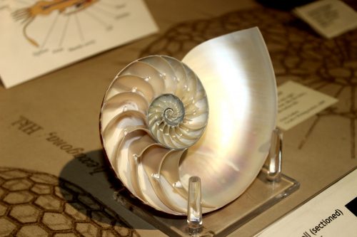
“I first ‘read’ Thompson’s ‘On Growth and Form’ as an undergrad. I found it rather daunting in a kind of War and Peace thick science text way. I tepidly thumbed through the pages, mostly stopping at interesting figures, especially the ones about spirals in nature and the accompanying mathematics. Soon I was up the N. California coast abalone diving with friends. We were there to collect and enjoy the yummy gastropods with local spirits. We always enjoyed the iridescent nacre that lines the inner shell. However, on this trip, my mind drifted back to Thompson’s figures of Haliotis, and its precise patterns of growth. My perspectives had forever changed, even on road trips with friends, to see patterns and morphometrics everywhere.”


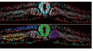
Paper from Rusty Lansford and colleagues – ‘Multi-scale quantification of tissue behavior during amniote embryo axis elongation


Julien Vermot

IGBMC, Strasbourg

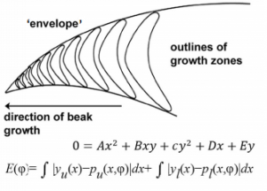
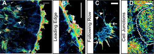
“I was phD student the first time I heard from on Growth and Form. Thomas Lecuit gave a very nice seminar in our institute. I remember him showing the famous drawings describing how environmental forces may affect the overall shape of an adult fish (it should correspond to the Figure 517 to 524). As a PhD in developmental biology, it had a strong impact on me. At the time, we were struggling with in situ hybridization and our understanding of how shape is emerging out of these expression profiles was clearly not satisfactory. I cannot say if it was the drawings themselves or if the field had already shifted, but the idea of forces affecting shape led to lots of discussion in the institute. It definitely influenced my post doctoral work and current research.”


Paper from the Vermot lab – “Anisotropic shear stress patterns predict the orientation of convergent tissue movements in the embryonic heart


Thanh Thi Kim Vuong-Brender

Labouesse Lab, IBPS, Paris

“Throughout the book, which I read in a French translation of the abridged John Tyler Bonner version, D’Arcy Thompson tried to use physics and mathematics to explain the shape of life forms. This was an audacious project, since the living world remained a big mystery at that time. D’Arcy Thompson foresaw the great utility of physics and mathematics in studying growth and form, while admitted that they could not explain all the living phenomena. He stressed the importance of material and mechanical nature of life, considering that a living form was nothing else than “a force diagram”. D’Arcy Thompson illustrated the beauty of a wide variety of species through mathematical description. He pointed out the similarities between seemingly unrelated things, linking the natural phenomena with physical laws. Modern research on morphogenesis has proved that many of these intuitions were correct.

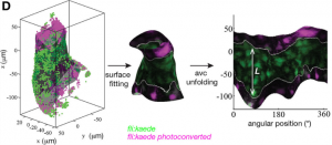
As a researcher working on a particular morphogenetic problem (elongation of C. elegans embryo), I also look at the biological phenomenon from a physical point of view, in terms of “materials”, “forces” and “force distribution and evolution”. Like D’Arcy Thompson, I also try constantly to relate the morphogenetic mechanisms with physical laws and physical phenomena that we see in daily life. Some paragraphs of the book are directly related to my work, for example the one stating that the formation of shapes different from a perfect sphere may results from either a difference in internal pressure (inside the object) or the anisotropy of the surface envelope. The relevance to my particular case on the anisotropic deformation C. elegans embryos shows that D’Arcy Thompson’s observations can still inspire future generations of scientists. Although many of the theories in “On Growth and Form” were simplistic and unproven, the observations and reasoning are a good incentive to further investigate the nature of the phenomenon. The book deserves to be read broadly, not only for its pioneer ideas on morphogenesis but also for its enjoyable elegant writing style.”


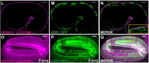
Thanh’s paper –  ‘The apical ECM preserves embryonic integrity and distributes mechanical stress during morphogenesis


What does On Growth and Form mean to you? We’d love to hear your thoughts in the comments section below 


On Growth and Form at 100 on the Node:

The story behind the cover

An interview with Matthew Jarron

Morphogenesis one century after On Growth and Form


Thumbs up (3 votes)
Loading...

Tags: ,
Categories: Discussion

Navigate the archive

Use our Advanced Search tool to search and filter posts by date, category, tags and authors.

On Growth and Form at 100: An interview with Matthew Jarron

Posted by , on 28 November 2017

In late September I boarded a tiny propeller plane to Dundee to meet Matthew Jarron, who curates the University of Dundee’s D’Arcy Thompson Zoology Museum. We had seen Matthew speak about On Growth and Form at a Royal Society event over the summer, and realised he would be a perfect person to interview to give the work some context in our special issue celebrating its centenary (published today!). On the taxi from the airport, through the fog I could just about make  the Tay Rail Bridge, which was rebuilt in 1887 after collapsing in a storm in 1879. D’Arcy Thompson’s book contains a section comparing dinosaur skeletons to railway bridges – how does the form of an object reflect the mechanical strains placed on it? – so I could almost imagine a sleeping dinosaur stretching south towards St. Andrews. Matthew was a great source of insight into the book and the man behind it; I’d encourage anyone who visits Dundee to go see the museum and its wonderful collection of items, many of which inspired Thompson’s ideas.


Matthew Jarron (left) and a portrait of D’Arcy Thompson by David Shanks Ewart (right; courtesy of the University of Dundee Museum Services).


D’Arcy Thompson was born in 1860, trained in Edinburgh and Cambridge, and held positions in Dundee and St Andrews, where he worked until his death in 1948. On Growth and Form, his classic work on the mathematical patterns and physical rules underlying biological forms, was first published in 1917. To learn more about the book’s context, we met Matthew Jarron, Curator of Museum Services at the University of Dundee, in the University’s D’Arcy Thompson Zoology Museum. Surrounded by specimens, many of which were collected by Thompson himself, we discussed the legacy of On Growth and Form and the life of the man behind it.


Matthew, can you tell me how you first came to encounter D’Arcy Thompson’s work?

Before I came to Dundee, I was a curator of a local history museum in St Andrews, which had been gifted a medicine chest from his days as a student in Edinburgh by the last surviving of his three daughters. This wonderful chest was my first encounter with D’Arcy, and as I gradually read more stories about him, I realised what a fascinating character he was. When I came to Dundee, I discovered we had what was left of his zoology collection. This was once one of the largest collections of zoological specimens in the country but had become dispersed in various displays and was not really accessible to the public. It wasn’t until the department of Life Sciences moved building in 2007 that we were able to create a proper version of the museum, have it open to the public and start telling people more about D’Arcy’s work. The more I read about him, the more I realised how incredible his influence has been in so many different fields in biology and beyond. In the museum, we’ve been very keen to use his collection in as many different ways as we can, to get people interested in his life and work. The centenary of his book has provided a great opportunity to do that, and you can find out about the various celebratory events here in Dundee as well as globally on the anniversary website (www.ongrowthandform.org/).


How did the idea for On Growth and Form come about?

From very early childhood, D’Arcy was fascinated by the natural world, partly because a number of his family were vets, but also because his father was a great classical scholar and had introduced him to classical biologists like Aristotle. Originally, he went to Edinburgh University to study medicine, but quite quickly realised that he wanted to specialise in biology, and transferred to Cambridge, which was really the only place where you could focus on the natural sciences at the time. He came to the University of Dundee (known then as University College Dundee, which had opened in 1883) at a very early age – he was only 24 when he was appointed to the first chair of biology in 1884. It was quite a small university, but they assembled a really extraordinary and dynamic group who were able to think in interdisciplinary ways. So, from very early on I think he had that broader interest, paired with his classical training and the associated idea that naturalists could look at broader themes.

As for On Growth and Form, we know that in 1889 he wrote a letter to one of his students saying that he had taken to mathematics. He had been looking at foraminifera, single-celled organisms that grow these extraordinary ‘tests’ (external shells) that often form geometric shapes, and he realised that there were very specific patterns that kept recurring. That was probably when he started to think that not everything in biology could be explained by Darwin’s ideas of evolution, which by that point had become quite widely accepted. These foraminifera, which presumably had similar evolutionary pressures, adopted quite diverse geometric forms; for D’Arcy this suggested that there was clearly something more going on here. He began to think about the physical forces acting on these organisms and realised that they formed specific mathematical patterns during their development.


Thompson started to think that not everything in biology could be explained by Darwin’s ideas of evolution


At the time he realised that these ideas would be quite controversial, and also that no one would really have much interest in applying them. He wasn’t completely keeping it to himself, but it wasn’t until 1908 that he published anything at all – a paper in Nature on the shape of eggs. Then, in 1915, he published a fairly substantial paper in the Royal Society of Edinburgh’s Transactions called ‘Mathematics and Morphology’, which is essentially what forms the final and most celebrated chapter of On Growth and Form. In 1911, he had been asked by Cambridge University Press to write a small book on the biology of growth and form. They had a series of popular science works that cost something like a shilling and were perhaps a hundred pages long, but as he started writing, it just got bigger and bigger, taking many years to write, and when he finally sent the text back to Cambridge they must have been somewhat horrified to find how enormous it had become! They did, however, agree to publish it separately.


The shell of a nautilus, one of the species that D’Arcy Thompson included in his section on spirals in nature, from the D’Arcy Thompson Zoology Museum (University of Dundee, UK).


And what does the book actually contain?

D’Arcy introduces the book with his case for looking at biology from a mathematical standpoint, to understand form from the point of view of physics and of mathematical laws, even though this was something that naturalists didn’t particularly want to do. The next chapter is concerned with size, and he makes the point that gravitational forces are much more important at larger scales, while surface forces, and in particular surface tension, play a much more important role at smaller scales. He starts with the smallest organisms, individual cells and their internal structures, and gradually works his way up, for instance exploring how the formation of clusters of cells is analogous to how soap bubbles come together. Throughout the book, and particularly here, he takes an organic form and compares it with an inorganic one; where he sees the same patterns, he reasons that the same physical forces are causing these shapes to appear. He goes on to look at individual parts of organisms – for instance the shells of foraminifera and nautilus, or antlers, horns and tusks – and explores how growth rates might cause their different curves and spirals. Then he looks at larger organisms, and famously compares the structure of quadruped skeletons with the structure of bridges. There is a chapter (which is missed out of the abridged edition) on plants and their phyllotaxis in relation to the Fibonacci sequence, and then in the final chapter he presents his theory of transformations. When dealing with large entire organisms, he admits he clearly can’t explain all their differences through mathematics, but he can take two related organisms and see how one could mathematically have changed into the other. He looks at the organism as a system, and explores how that whole system will transform; the famous diagrams are his attempt to explain that. He ends the book by saying that what he has described in the book is ‘a field which few have entered and no man has explored’.


A rare first edition of On Growth and Form, held in the D’Arcy Thompson Zoology Museum (University of Dundee, UK).


How was the book received?

When the book came out it was very widely praised. Everyone was hugely impressed by the vastness of his learning; one of the most extraordinary things about the work is how he brings together examples from recent research with examples from ancient history, that he views the biology of Aristotle as equally relevant to the biology of the time. There were reviews of the book not only in biology but also in engineering journals, as well as Country Life of all places, and I think this reflects a recognition that the ideas in the book could have a much wider application. But it’s notable that many of the reviewers were basically saying how well written it was, rather than how it was going to transform biology. While an enormous amount of learning had gone into the work, very few people could actually grasp how to take it forward. A key issue was that many biologists didn’t really know much mathematics; similarly, he struggled to get many mathematicians interested in the work. But there were some key people who picked up on the book and took forward the idea of mathematical biology, and with whom D’Arcy was in regular correspondence. Interestingly, these people were often working in different fields and using completely different kinds of mathematics from D’Arcy’s; for instance, early ecologists interested in using mathematical models to look at populations.


The D’Arcy Thompson Zoology Museum (University of Dundee, UK).


More and more people appear to be turning back to his work and finding useful things in it today

The first edition had sold out in 1923 – as far as we know it was limited to 500 copies, one of which is displayed here in the museum – but it was 20 years before he got round to writing his second edition. It’s no coincidence that both editions were published during wartime – it was only then that he had time to write, as so many of his students were away at the front and his administrative role lightened. The second edition includes more illustrations and examples – for instance, a section on animal coat markings that was a key influence on Alan Turing – but it does not actually contain that many new areas of research. In particular, he was criticised for ignoring all the developments in genetics since the first edition was published. It wasn’t that he didn’t know anything about genetics – he was completely up to speed with everything that was going on – rather, it was that he couldn’t see how he could incorporate the work into his thesis, and so he just ignored it. While he could get away with this in the first edition, by the time of the second edition it was less easy, and that rather doomed his work for a lot of biologists. Conversely, while the second edition was problematic for some biologists, it got very quickly picked up by artists, engineers, architects, geographers and anthropologists. So from the 1950s there was a renewed interest that slowly picked up, until recently when it seems to have taken off exponentially. It’s quite amazing that more and more people appear to be turning back to his work and finding useful things in it today.


The book argues that one can look at biology through a mathematical lens – what type of mathematics did Thompson employ?

The mathematics he knew was pretty much all classical: geometry and algebra. He certainly didn’t have any kind of detailed knowledge of contemporary mathematics, and indeed he acknowledges that himself. I think it was a real problem for him, but he just didn’t have the time to get into detailed mathematics of the kind that later biomathematicians did. He probably did not have the inclination either: I don’t think he ever felt that mathematical biology was the topic he’d spend the rest of his life on, as his main interest was in fisheries. Indeed most of his work was taken up with international diplomacy about fishing quotas and that sort of thing; he was well known as a great diplomatist in helping to resolve disputes while countries were dividing up the sea for different fishing rights. With On Growth and Form, he felt that he’d opened a gate into this new field, and was happy for other people to go in and explore it.

Richard Dawkins once said it was a great shame that the computer wasn’t invented in D’Arcy’s lifetime because his work cries out for it, and perhaps this is one of the reasons why so few people at the time actually picked up on what he was doing. It took more sophisticated mathematicians like Alan Turing and the development of computers to allow you to test these theories and build models.


What do you think are the book’s key contributions?

Largely, I think it’s the general idea that you can apply mathematics to biology. This has been so influential in biology as a whole, and particularly now in developmental biology, where its specific influence might not have been that great at the time. His holistic approach was influential for the whole organicist tradition of biology, and particularly for people like Conrad Hal Waddington, who was enormously influenced by the book and whose ideas about epigenetics have become so crucial to development. The idea of taking this vast and complex world of nature and reducing it down to fundamental rules is also obviously a key part of systems theory, and was hugely influential in its development, not to mention cybernetics and the start of computing, artificial intelligence and so on. These are all linked to D’Arcy’s work, and his ideas are still influential in art, architecture, geography and anthropology. Actually there are whole new areas of science that trace an influence back to him – things like nanochemistry; one of the field’s pioneers, Geoffrey Ozin, credits D’Arcy as an inspiration. It’s clearly something that people in a wide range of fields are continually coming back to, even if they are just taking general ideas rather than specifics.


Model of a radiolarian skeleton (Actinomma inerme) from the D’Arcy Thompson Zoology Museum (University of Dundee, UK); D’Arcy ordered a set of these from the Czech model-maker and natural history dealer Vaclav Fric.


Considering Stephen J. Gould’s opinion that biologists regarded On Growth and Form as ‘an unusable masterpiece doomed by excessive length and difficulty of application’, why should the modern reader pick it up?

It certainly is the case that very few people have read the book from cover to cover, in order, but in a sense that doesn’t really matter. For me there are two reasons to pick it up: one is that even if you’re never going to read through the mathematical bits, or the stretches of Latin, Greek and German that he doesn’t bother to translate for you, there are lots of very beautiful, poetic passages that still read very well, and which are just inspiring; the other is that it has so many amazing illustrations that are also just as inspirational. Frankly, if all you do is pick it up, flick through it and look at the pictures, that’s great – you’ll still be inspired by the mathematical beauty of nature, as D’Arcy wants you to be. There’s still lots there for the modern reader.


And what about D’Arcy the man – you’ve read much of his correspondence and what people wrote about him. Do you have any feel for his personality?

He had a great personality, and was a larger than life character in every sense. For one thing he was a large, tall man, standing six foot three, described as a veritable lion due to his beard. His students loved him as an eccentric teacher who would use bizarre props to help illustrate his points. There are many anecdotes about how learned he was, how in tutorials he would translate from Medieval Italian, that kind of thing. I think he had a great sense of humour, which certainly comes across in letters to his friends. He corresponded with so many different people, and though he was this great diplomatist, always very polite to people, when he was speaking to his closest friends he could be incredibly and hilariously rude about others! He was also a great populariser of science – he loved giving public lectures, and talking to children and showing them round his museum. Famously, in his old age he had a pet parrot that perched on his shoulder as he wandered the streets of St Andrews. So by all accounts he was a great character.

In a sense he was the last of an era. In the 19th century, it was quite common to have great erudite polymaths, but in the 20th century less so. I think he very much felt of himself as a man out of time, and this was perhaps one of the reasons he loved classics so much – he looked back on great figures like Aristotle who could take a broad look at life. The breadth of his knowledge was extraordinary, and he was constantly championing ideas of interdisciplinarity and emphasising how important it is to look beyond your own field. Holistic overviews like the one provided by D’Arcy are, I think, still important, and it would be a shame if we totally lost them.


Do you think there are any misconceptions about D’Arcy Thompson and his work?

Well, one is that he is somehow anti-evolution. It’s not that he thinks that evolution is wrong or doesn’t exist, it’s just that he’s saying that Darwinian evolution can’t explain everything. The transformation diagrams were, I guess, the part that most obviously appeared to contradict Darwinian evolution – whereas Darwin emphasised slow, gradual change, with each particular part responding to natural selection, D’Arcy says that that can’t always be the case, and there must have been times when there was more of a sudden transformation from one kind to another. But even if you argue with this point, his work emphasises that, however something evolves, it will evolve according to certain mathematical patterns.

There’s also a general misconception – and of course I’d say this being here in Dundee – that he was some kind of maverick loner in a remote backwater writing this book without any support. This simply isn’t true – he drew hugely on his fellow professors here in Dundee; for instance, the engineering professor Thomas Claxton Fidler helped him with his ideas of dinosaurs and bridges, and the physics professor William Peddie was enormously valuable in helping to shape the entire book.


If Thompson were alive today, what would you ask him?

From a purely personal point of view, working here in the museum, I would ask him where he got each one of these specimens – he never catalogued his museum, it was just all in his head! Occasionally, we’ll find a letter where he’ll describe some gorilla he’d just acquired, but for a vast amount of stuff we just have no written record.

The other thing I find mysterious about him was his position on the great debates in biology at the time around ideas of vitalism, mechanism and organicism. It is never entirely clear where D’Arcy sits with regard to them – on the one hand, On Growth and Form seems to be quite a mechanistic book, reducing nature down to rules, but at the same time D’Arcy is very keen to point out that mechanism doesn’t have all the answers, that there are things that you can’t explain in nature. In that sense, he takes elements of vitalism, while a lot of the organicists saw him as a key influence. It probably comes back to his diplomacy – he very deliberately doesn’t come down on one side or the other. So I’d like to sit him down in a room and find out what he really thought about these issues.


On Growth and Form at 100 on the Node:

The story behind the cover

Perspectives from the field

Morphogenesis one century after On Growth and Form


Thumbs up (1 votes)
Loading...

Tags: ,
Categories: Interview

On Growth and Form at 100: Morphogenesis one century after On Growth and Form

Posted by , on 28 November 2017

This editorial by Thomas Lecuit and  L. Mahadevan originally appeared in Development’s Special Issue: On Growth and Form – 100 Years On


Morphogenesis, the study of how forms arise in biology, has attracted scientists for aeons. A century ago, D’Arcy Wentworth Thompson crystallized this question in his opus On Growth and Formusing a series of biological examples and geometric and physical analogies to ask how biological forms arise during development and across evolution. In light of the advances in molecular and cellular biology since then, a succinct modern view of the question states: how do genes encode geometry?

Understanding this fascinating problem requires insight into how shape emerges when molecular information and physical forces are regulated over many different scales in space and time. To address this requires an appreciation of the enormous ‘morphospace’ of potential shapes and sizes that living forms can take up. In parallel, we need to consider the large diversity in the genetic space of potential regulatory interactions that influence form. While the conceptual framework of developmental patterning explains how cells acquire information and how this defines their behaviours, Thompson’s agenda of describing biological processes in mathematical terms is based on understanding how instabilities and patterns in physical systems might be harnessed by evolution. Consequently, the subjects of morphological (phenotypic) and regulatory (genotypic) diversity that are separated by many orders in length scales, have not been sufficiently coupled intellectually.


100 years after the publication of On Growth and Form, we are in a position to better encapsulate phenotypes and genotypes under a unified conceptual and mechanistic framework


Now, 100 years after the publication of On Growth and Form, we are in a position to better encapsulate phenotypes and genotypes under a unified conceptual and mechanistic framework. This entails a search for a potentially low-dimensional phase space for the description and control of shape over developmental and evolutionary time scales. Any parametrisation of the processes at play must have both physical as well as regulatory bases in terms of biomolecular processes that respond to and control these physical parameters. A fundamental challenge therefore is to connect these different scales while deducing the dimensionality of these ‘morpho-genetic’ spaces underlying the development and evolution of shape.

The past two decades have seen an increasing influx of physicists, mathematicians, engineers and computer scientists into the field of developmental biology, who are all attempting to determine the correspondence between the parameters that describe shape and those that define its generation and transformation. There are three major areas in which they have made contributions. First, they have developed mathematical and algorithmic tools for the quantitative description of shape, i.e. morphometrics, directly inspired by the last chapter in Thompson’s book, titled ‘The Theory of Transformations’. This has led to the modern field of pattern theory and statistical shape analysis. Second, they have contributed theoretical and experimental tools to describe and measure the collective biophysical properties, instabilities and patterns of active living matter. Finally, they have pushed forward the efforts to describe morphogenesis using a limited set of relevant physical or mechanical parameters and relate them to biological regulatory processes, initiating a transition towards a predictive developmental biology.

This Special Issue celebrates this synergy by providing insights into the genetic underpinning of embryo and tissue patterning, the biological basis of cell and tissue dynamics, and a physical framework to capture these processes operating across scales. The issue begins with an interview with Matthew Jarron, curator of the D’Arcy Thompson museum in Dundee (Maartens, 2017). This Spotlight article sets the scene – introducing the reader to Thompson’s life and ideas, as well as his legacy.

The Review and Research papers that follow cover a wide spectrum of topics across developmental biology. Coen and colleagues (Coen et al., 2017) directly address the question laid out at the beginning of this Editorial – how genes regulate geometry – with a particular focus on plant tissues. Continuing the theme of plant morphogenesis, three research papers apply mathematical approaches to phyllotactic patterning (Fal et al., 2017), sepal growth (Tsugawa et al., 2017) and cell packing and topology in the leaf (Carter et al., 2017). Graner and Riveline review chapters VII and VIII of Thompson book, ‘The Forms of Tissues, or Cell-aggregates’, providing an overview of the mathematical and physical principles underlying epithelial cell shape and packing in both historical and modern contexts (Graner and Riveline, 2017).

Also focussing on epithelial tissue, a research paper from Dye and colleagues (Dye et al., 2017) provides a quantitative analysis of growth and patterning in Drosophila wing, which is one of the best-studied epithelial tissues, and Irvine and Shraiman (2017) review our understanding of tissue growth from a mathematical and mechanical perspective, using planar shapes such as wings and leaves as examples.

In their Review, Heer and Martin (2017) discuss how tension and contractility influence morphogenesis. Complementing this, but on a very different scale, Felsenthal and Zelzer (2017) review how the developing musculoskeletal system influences its final form and function. Revisiting the last chapter of Thompson’s book, ‘Theory of Transformations’, Abzhanov (2017) argues that to understand both ontogeny (development) and phylogeny (evolution), an appreciation of the intrinsic ‘laws of growth’ is essential to frame our models of adaptation and speciation across evolutionary time. Complementing this, Sharpe (2017) provides an overview of the kinds of computational tools and problems that Thompson might have used and addressed had he been alive today.

The mechanics of developmental processes involves multiple scales, and a number of papers discuss examples of this: Boselli and colleagues consider the role of fluid flows and shear stress in orienting tissue movements (Boselli et al., 2017), Nelson and colleagues look at the effects of pressure on early branching processes (Nelson et al., 2017), Ruiz-Herrero and colleagues provide a general framework for size control of growing tissue cysts (Ruiz-Herrero et al., 2017), Lefevre and colleagues analyse multi-scale branching in the mammalian kidney (Lefevre et al., 2017), and axis elongation in the avian embryo is the focus of the work of Bénazéraf and colleagues (Bénazéraf et al., 2017).

In their Review, Engler and colleagues (Kumar et al., 2017) provide an overview of the latest advances of our understanding of how such forces can regulate stem cell fate. Several papers directly address the role of mechanics in growth, form and fate, focusing on the role of the extracellular matrix (Chlasta et al., 2017Vuong-Brender et al., 2017), cytoskeletal dynamics and cell-cell contacts (Sonavane et al., 2017), and the mechanical phenotype of cells during reprogramming and differentiation (Urbanska et al., 2017). In addition to some of those studies already mentioned above, problems ranging from gradient establishment in the Drosophilaembryo (Carrell et al., 2017), eggshell shape in Caenorhabditis elegans (Yamamoto and Kimura, 2017), and patterning of stem cell colonies in culture (Tewary et al., 2017) showcase the variety of physical and mathematical approaches that modern development is beginning to embrace.


Problems yield only when appropriate tools can be developed to solve them


Problems yield only when appropriate tools can be developed to solve them. Thompson’s pioneering vision was exemplified in his statement that ‘[The] problems of form are in the first instance mathematical problems, and [the] problems of growth are essentially physical problems; and the morphologist is, ipso facto, a student of physical science’. Today’s developmental biologists have much more sophisticated tools at their disposal, relative to those a century ago, when we had almost no way to measure gene expression patterns, to image at the subcellular level, to understand physical instabilities and patterns in nonlinear systems, or to use computers to help simulate them. Indeed, looking at the papers in this issue, we can see the influence of quantitative experiments coupled with mathematical modelling and simulations everywhere. For example, the mathematical and computational tools deployed range from topology (Carter et al., 2017Graner et al., 2017), complex analysis (Irvine et al., 2017), reaction-diffusion theory (Carrell et al., 2017), agent-based models (Yamamoto et al., 2017) and graph theory and lineage analysis (Lefevre et al., 2017) among others.

On Growth and Form raised the question of the origin of biological shape in a physical framework. Since then, advances in our understanding of the biochemical basis of the laws of heredity have provided the modern conceptual understanding for how shapes develop anew at each generation, from a single cell – thus surviving the death of an individual through its offspring. As this Special Issue illustrates, we are now beginning to understand how genes encode geometry. As morphology both enables and constrains function, a natural next question is how biology creates functional (and plastic) shape that begins to link morphology to physiology and behaviour. As you mull this question, we would like to thank all the authors and referees of the articles in this Special Issue for their contributions, and we hope you enjoy reading it!


On Growth and Form at 100 on the Node:

The story behind the cover

Perspectives from the field

An interview with Matthew Jarron


Thumbs up (No Ratings Yet)
Loading...

Tags: ,
Categories: Discussion, News

Embryonic development inspires regenerative medicine

Posted by , on 27 November 2017

  • The European Molecular Biology Lab (EMBL) in Barcelona and IRB Barcelona bring together leading scientists in the conference “Morphogenetic Engineering”, an event supported by the BBVA Foundation.
  • The merge of various disciplines of developmental biology is key to enhancing our knowledge of tissue development and repair, processes that find their most direct applications in regenerative medicine.
  • “One day we will be able to re-build damaged organs and keep the whole body fit and healthy. But right now we must strengthen our knowledge of how tissues are built and how they are maintained,” say James Sharpe, Head of EMBL Barcelona, and Marco Milán, coordinator of the Cell and Developmental Biology programme.

Developmental biologists seek to unravel how animals generate and repair their organs and issues. Tissue engineers, on the other hand, endeavour to understand how damaged tissue in the adult organism can be built and repaired. One approach that may help us to understand how to build new tissues is to “learn from the embryo. This view is shared by James Sharpe, director of the European Molecular Biology Laboratory (EMBL) in Barcelona, and Marco Milán, ICREA researcher at the Institute for Research in Biomedicine (IRB Barcelona) and it is the idea underlying the organisation of the Barcelona Biomed Conference entitled “Morphogenetic Engineering”.

From 27 to 29 November, the Institut d’Estudis Catalans de Barcelona will bring together a group of international experts on embryos, tissue morphogenesis, gene regulation and developmental mechanics, organoids, regeneration and engineering. The “Morphogenetic Engineering” meeting is part of the Barcelona Biomed Conference Series, which is organised by IRB Barcelona and supported by BBVA Foundation since 2006, and is the 31st gathering of leaders in biomedicine in this series.

Copying development with 3D mini organs

Organoids—3D mini organs built in the lab—are developmental models that provide a “very good” way to bring these fields together. “Organoids perform simple morphogenetic processes—not in a very natural manner but in a controlled lab environment amenable probing, perturbing, tinkering and engineering,” says Sharpe.

The main question to be tackled by the experts is how tissues and organs can be built. But this question encompasses a series of more specific scientific questions that have not been fully answered to date: How do cells know which decisions to make? How do they use molecular and mechanical cues to know where they are, or which direction they should move? How do large numbers of cells, each with only limited local information, collaborate to create something much larger and more complex than themselves?

The experts explain that many of the state-of-the-art technologies are required to tease out relevant information from model systems, whether these be organoids or “classical animal models such as the fruit fly, zebra fish or mouse,” adds Milán. They give special emphasis to 3D and 3D imaging, multi-cellular transcriptomics, and computer modelling, among others.

Biomedical promises and health

“One day we will be able to re-build damaged organs and keep the whole body fit and healthy,” says Sharpe. He admits that this will take many years of basic biology research along with more translational studies into tissue engineering. “In the shorter term, within the next decade, we should be able to boost the self-healing properties of our tissues, and start to replace and repair small regions of damaged tissue,” he says. Marco Milán adds that, adds that there is a link not only between embryonic development and regeneration but also between the latter and tumour development.

Both researchers agree that the Barcelona Biomed Conference “Morphogenetic Engineering” is an excellent way to reflect on the growing and exciting fusion of ideas and scientific communities.


About the organizers:

Marco Milán, ICREA professor at IRB Barcelona. Head of the “Growth Control and Development Lab” and coordinator of the Cellular and Developmental Cell Biology programme. Using the wing development of the fly Drosophila melanogaster as a model, Professor Milán studies the signalling pathways and genetic circuits required for tissue development, and new pathways involved in regeneration and cancer.

James Sharpe is Head of EMBL Barcelona, which aims to study the dynamic interactions of multicellular systems that underlie how tissues build, maintain and fix themselves, how this can go wrong in disease, and how we can learn to build tissues through engineering. His own lab focuses on understanding vertebrate limb development, through imaging, experimental perturbations and computer modelling.

Thumbs up (No Ratings Yet)
Loading...

Tags: , , , , , ,
Categories: Events, News, Uncategorized

PhD position at the Palacios’s lab, SBCS, QMUL, London

Posted by , on 24 November 2017

Closing Date: 15 March 2021

My ultimate scientific aim is to contribute to the understanding of diseases, and I believe that in order to achieve this goal we need to understand fundamental cell biophysical mechanisms underpinning health. My lab applies a biophysical approach to studying how cytoskeletons (actin, microtubules and spectrin) collaborate in the establishment of cell polarity and tissue architecture at the mesoscale level. We use Drosophila as a model system to study the role that cytoskeletal forces, and their functional crosstalk, play in the development of the female germline, although we are also exploring the impact of our findings in other tissues (e.g., neurons) and organisms (e.g., mouse oocytes), in a collaborative manner.


Research in cell biology has become increasingly quantitative, and some areas, such as studying the highly dynamic cytoskeletons at the tissue level, require interdisciplinary collaborations. We are successfully collaborating with experimental and theoretical physicists, which allowed us to tackle the role of cytoskeleton dynamics on cellular self-organisation and tissue morphogenesis from a multidisciplinary point of view.


More specifically, there are two available PhD projects. The first one aims to understand the biophysical properties of the interplay between actin and microtubules in cells that are immobile and not dividing. We are studying the biophysical features of this interplay in the Drosophila oocyte, where the crosstalk between the two cytoskeletons impacts on the mechanical properties and self-organization of the female germline, and ultimately on its polarization and function. To further understand the coupling between motor-induced forces, fluid dynamics and cytoskeletal organisation, we are also extending our analysis to super-resolution microscopy and advanced motion and image analysis, as well as to the mouse oocyte. This interdisciplinary approach will allow the student to cover various aspects of quantitative biology, physical modelling and experimental design required to study the relation between the fluid mechanical properties of the cytoplasm and oocyte polarity.


The second project focuses on studying the role of the Spectrin cytoskeleton in epithelium architecture. The spectrin membrane skeleton is a mechanically deformable network, that crosslinks actin to the membrane, and although it has been greatly studied in erythrocytes, little is known about the function of this cytoskeleton in epithelia. We are studying the role of the spectrin cytoskeleton during epithelia morphogenesis using the Drosophila follicular epithelium as a model system. This germline-surrounding epithelium, which is essential for oocyte polarity, has emerged as a powerful model to study epithelial morphogenesis. Spectrins are conserved in all eukaryotes, with a greater conservation between Drosophila and mammalian non-erythroid spectrins than between erythroid and non-erythroid forms. We identified a primary role for the spectrin skeleton in controlling cell shape, specifically cell elongation. Furthermore, the spectrin cytoskeleton is key to maintaining a mono-layered epithelium, as spectrin mutant cells form a “tumour-like” multi-layered mass. We have found that increasing and reducing the activity of the actomyosin cytoskeleton enhances and decreases spectrin multi-layering phenotypes, respectively. Our hypothesis suggests that the spectrin cytoskeleton is essential to balance adequate forces, probably by modulating the actomyosin cytoskeleton, in order to maintain cell shape and epithelium architecture. We are currently studying the distribution of forces in the follicular epithelium, and how this distribution is related to the function of spectrins in regulating the actomyosin cytoskeleton.


http://www.sbcs.qmul.ac.uk/postgraduate/research/projects/display-title-455850-en.html


Thumbs up (No Ratings Yet)
Loading...

Categories: Jobs

Seeking postdoc in developmental biology

Posted by , on 23 November 2017

Closing Date: 15 March 2021

Postdoc position in Developmental Biology in Mukhopadhyay lab, UT Southwestern, Dallas.

The focus of the current research in the Mukhopadhyay laboratory is to understand mechanisms of cellular signaling at the level of the primary cilia, and its relevance to human health and disease. The first cellular organelle to be described in biology, the primary cilium was long mistaken as a vestigial appendage. The primary cilia are now considered as vital sensory organelles for detection and transmission of a broad range of chemical and mechanical signals in most cells. Signaling mediated by the primary cilia plays fundamental roles in cellular differentiation, polarity and cell cycle control. We utilize a variety of biochemical, cell biological and reverse genetic approaches to understanding signaling mediated by cilia, and dissecting their role during normal development and carcinogenesis. A detailed description of current lab projects can be found at: http://www.utsouthwestern.edu/labs/mukhopadhyay/ and a recent invited review in MBoC.

One postdoctoral position is available in our laboratory to study the role of cilia and cilia-generated signaling in multiple developmental processes. We are a closely-knit group of scientists with diverse sets of expertise and passionate about solving the particular biological problem, often embarking on newer methods and paradigms as necessary. We are located in the Department of Cell Biology in UT Southwestern Medical Center, Dallas. UT Southwestern, one of the premier academic medical centers in US, integrates pioneering biomedical research with exceptional clinical care and education. The institution’s faculty includes many distinguished members, including six who have been awarded Nobel Prizes since 1985.

Developmental biology questions include but are not restricted to the role of cilia in limb bud formation, skeletal morphogenesis including intramembranous bone formation, neural tube patterning, cerebellum and brain development.

Our recently accepted paper (in press in Development) on orphan cilia-localized GPCR, Gpr161 and limb development is available to interested candidates upon request. Here we demonstrate that Gpr161 promotes forelimb formation, regulates limb patterning, prevents periarticular chondrocyte proliferation, and drives osteoblastogenesis in intramembranous bones in a cilium-dependent manner.

Another paper on Gpr161 and cerebellum development and tumorigenesis (currently in review) is also available upon request. Here we demonstrate that Gpr161 restricts cerebellar granule progenitor production by preventing premature and sonic hedgehog-dependent pathway activity, highlighting importance of basal pathway suppression in medulloblastoma pathogenesis.

Candidates must have a recent Ph.D. or M.D./Ph.D., with less than three years of prior postdoctoral experience, and a demonstrated research record with at least one first author publication. Preference will be given to applicants with a strong background in cell and molecular biology or mouse genetics.

Interested individuals should email saikat.mukhopadhyay@utsouthwestern.edu a copy of your current curriculum vitae, contact information for references, and a cover letter highlighting your prospective research plan.

UT Southwestern Medical Center is an Affirmative Action/Equal Opportunity Employer. Women, minorities, veterans and individuals with disabilities are encouraged to apply.

Thumbs up (No Ratings Yet)
Loading...

Categories: Jobs

New resources section on the Node

Posted by , on 23 November 2017

Since its launch in 2010, the Node has functioned as a community resource for developmental biologists. When I started my 3 month internship in September (see my introductory post here), we decided to utilise my experience as an active researcher to redesign and update the Node’s resources page. This included transferring the resources from the British Society for Developmental Biology’s homepage, which had previously been curated over several years by Andreas Prokop and which gave me wonderful insight into the wide range of resources out there.

The new resources page is designed to provide many different links to explore. You can learn more about becoming involved in advocacy and outreach, or find links on new teaching resources for schools and databases for a wide range of species.

This new list is by no means comprehensive and we need your thoughts on how it can be improved, whether by content or user experience!

I still have two weeks left of this internship before returning to my PhD in Tom Pratt’s lab, and during this time I aim to continue working on the resources section. However this is a community resource and to improve it further I need the input of the community. Please get in touch to let us know what is useful to you, what needs to be added/updated and any way that the resources section can be improved. I hope you enjoy using it and it is useful to the developmental biology community in your future endeavours.

Thumbs up (4 votes)
Loading...

Tags: , , , ,
Categories: News, Resources

Postdoctoral position at the IIMCB Laboratory of Protein Metabolism in Development and Aging

Posted by , on 22 November 2017

Closing Date: 15 March 2021

International Institute of Molecular and Cell Biology in Warsaw

Laboratory of Protein Metabolism in Development and Aging

is seeking a talented Postdoctoral fellow


Location: Warsaw, a vibrant city with an international academic research environment. International Institute of Molecular and Cell Biology (www.iimcb.gov.pl) – one of the most dynamic and top ranked Polish research institutes.

Job descriptionLaboratory of Protein Metabolism in Development and Aging, which will be opening on August 2017, is seeking highly motivated and talented Postdoc to join young team investigating the protein homeostasis in development and aging. We use both genetic, molecular and biochemical approaches, primarily in the C. elegans, to study proteolytic networks. Postdoc fellowship is funded in frame of National Science Centre OPUS grant.

Summary: Organismal development or environmental stimuli challenge the homeostatic protein balance (proteostasis) of individual cells, tissues or the entire organism. The ubiquitin proteasome system (UPS) is a key determinant of proteostasis as it regulates the turnover of damaged proteins supporting cellular protein homeostasis and thereby maintains the proteome during stress and aging. Our long-term objective is to understand the mechanistic and developmental aspects of protein degradation pathways defined by combinations of particular ubiquitin ligases (E3). The identification of stress and aging-induced signals that coordinate the interplay between specific E3s will offer intriguingly new mechanistic insights how proteolytic networks are fine-tuned to maintain the cellular proteome and support development and longevity.

Qualifications:
• PhD (or be close to completion) in Molecular Biology, Cell Biology, Protein Chemistry, Genetics or a related discipline;
• experience in C. elegans or cell culture is an advantage;
• experience in Next Generation Sequencing techniques (RNA-Seq, ChIP-Seq) and genomic engineering is an advantage;
• good writing and oral communication skills in English, and competence in scientific writing.

How to apply: 
Please send your application including CV, motivation letter and the list of publications to the e-mail address: wpokrzywa@iimcb.gov.pl, until 10th December 2017. Thanking all applicants for their interest, we will contact only selected candidates for an interview.
Please include in your application the following statement: “In accordance with the personal data protection act from the 29th of August 1997, I hereby agree to process and to store my personal data by the Institution for recruitment purposes”.
The recruitment procedure fulfills the National Science Centre’s regulations on granting the scholarships to young scientists.

Selected publications:

Riga T*, Pokrzywa W*, Kevei E, Akyuz M, Vishnu Balaji, Svenja Adrian, Hoehfeld J, Hoppe T. (2017). The ubiquitin ligase CHIP integrates proteostasis and aging by regulation of insulin receptor turnover. Cell. 169: 470-482.

Ackermann L., Schell M., Pokrzywa W., Gartner A., Schumacher B., Hoppe T. (2016). E4 ubiquitin ligase specific degradation hubs coordinate DNA double strand break repair and apoptosis. Nat Struct Mol Biol. 23: 995-1002.

Kaushik S, and Cuervo AM (2015). Proteostasis and aging.  Nat Med. 21, 1406-15

Frumkin A, Dror S, Pokrzywa W, Bar-Lavan Y, Karady I, Hoppe T, Ben-Zvi A. (2014). Challenging muscle homeostasis uncovers novel chaperone interactions in Caenorhabditis elegans. Front Mol Biosci., doi: 10.3389

van Oosten-Hawle P, and Morimoto RI (2014). Organismal proteostasis: role of cell-nonautonomous regulation and transcellular chaperone signaling. Genes & Dev. 28: 1533-43.

Segref A, Kevei E, Pokrzywa W, Mansfeld J, Schmeisser K, Livnat-Levanon N, Ensenauer R,  Glickman M.H, Ristow M, Hoppe T. (2014). Pathogenesis of human mitochondrial diseases is modulated by reduced activity of the ubiquitin/proteasome-system. Cell Metab. 4:642-652.

Pokrzywa W. and Hoppe T. (2013). Chaperoning myosin assembly in muscle formation and aging. Worm. 2:e25644.

Gazda L*, Pokrzywa W*, Hellerschmied D, Loewe T, Forné I, Mueller-Planitz F, Hoppe T, Clausen T. (2013). The myosin chaperone UNC-45 is organized in tandem modules to support myofilaments formation in C. elegans. Cell. 1, 183-195.

Kuhlbrodt K, Janiesch PC, Kevei E, Segref A, Barikbin R, and Hoppe T (2011). The Machado-Joseph disease deubiquitylase ATX-3 couples longevity and proteostasis. Nat Cell Biol. 13, 273-81.


Thumbs up (No Ratings Yet)
Loading...

Tags: , , , ,
Categories: Jobs

Senior Laboratory Research Scientist at the Crick Institute

Posted by , on 22 November 2017

Closing Date: 15 March 2021

We are looking for a highly motivated Senior Laboratory Scientist to join the quantitative cell biology laboratory headed by Dr Silvia Santos. The lab is a new addition to The Francis Crick Institute in London and focuses on understanding control principles in cell decision-making. Current areas of research include understanding control of cell division and differentiation, using human embryonic stem cells as a model system. There is a strong focus on single cell analysis and live cell imaging approaches. The team is currently composed of three PhD students and two post-doctoral fellows.

For more information please see

https://www.crick.ac.uk/research/a-z-researchers/researchers-p-s/silvia-santos/

and quantcellbio.wordpress.com


THE CANDIDATE

The successful post holder is expected to drive his/her own research, help with lab management and training and support on-going research projects. The ideal candidate is likely to be an energetic, focused and productive individual with a desire to work on interesting biological problems in a collegial and collaborative work environment. Excellent time management and organisation skills are essential.


PROJECT SCOPE
The decision to divide is a fundamental cellular decision and the conserved networks that trigger cell division adapt and remodel in a variety of biological contexts including developmental transitions and malignancy. We have been exploring spatio-temporal control of cell division in mammalian cells and remodelling of cell cycle networks during developmental transitions, using embryonic stem cells as a model system.
Embryonic stem cells have the propensity to differentiate into the three germ layers. The switch between pluripotency and differentiation in these cells has been our paradigm of choice to understand how protein and gene networks decode cellular signals and thereby encode irreversible commitment to different cell fates.
The molecular basis of these decisions is of fundamental biological importance and have significant clinical applications.


Informal enquires can be sent to Silvia Santos at: silvia.santos@crick.ac.uk


If interested please apply here with a cover letter stating your background and motivation, CV and names of two referees.

https://jobs.crick.ac.uk/pls/corehrrecruit//erq_jobspec_version_4.display_form?p_company=1&p_internal_external=E&p_display_in_irish=N&p_applicant_no=&p_recruitment_id=006424&p_process_type=&p_form_profile_detail=&p_display_apply_ind=Y&p_refresh_search=Y

Thumbs up (No Ratings Yet)
Loading...

Categories: Careers, Jobs, Uncategorized

Looking back on the adventure: exploring cell fate conversion with single cell RNA-seq

Posted by , on 21 November 2017

The story behind our recent paper: Liu Z*, Wang L*, Welch JD*, Ma H, Zhou Y, Vaseghi HR, Yu S, Wall JB, Alimohamadi S, Zheng M, Yin CY, Shen WN, Prins JF, Liu JD, Qian L (2017). Single cell transcriptomics reconstructs lineage conversion from fibroblast to cardiomyocyte. Nature, 551, 100-104 *: co-first author. https://www.nature.com/articles/nature24454


The Journey Started

In February 2014, I graduated from Indiana University with a PhD in Microbiology and Immunology. Being fascinated by regenerative medicine and how cell fate can be changed/ reversed during reprogramming, I joined Dr. Li Qian’s lab at University of North Carolina (UNC) in March 2014. Until today I still feel things just happened so fast and I am such a lucky person to be a part of this group. So I started my new journey at the beautiful small town of Chapel Hill to become a “reprogrammer.” Previously, Dr. Qian discovered that introduction of three cardiac transcription factors Mef2c, Gata4, and Tbx5 into mouse heart after myocardial infarction could significantly improve the heart’s function by converting the local fibroblasts in the heart to beating cardiomyocytes – which we termed as the process of “direct cardiac reprogramming” or “induced cardiomyocytes (iCM) reprogramming.” In addition to iCM, other researchers have reported the conversion of fibroblasts into induced neurons, hepatocytes, β cells, and so on. Direct lineage conversion not only offers a new strategy for tissue regeneration and disease modeling, but also provides a unique platform for understanding cell fate.


The Cell, the Single Cell

The cell is the most basic functional unit of an organism. The human body contains more than 40 trillion cells that are from different origins, form different organs, and carry out different functions. Each cell has its unique transcriptome, proteome, and functional role even though all cells originate from the same zygote and in theory share the same genome. Due to technical limitations, researchers were only able to check either the expression of limited number of genes at the single cell level (like immunofluorescence staining) or the transcriptome but at the price of averaging signals from a population of cells (like bulk RNA-sequencing). As the dawn of the era of single cell OMICs, we realized that we could do more now to understand the mechanisms of cell fate determination.


Direct Cardiac Reprogramming

Direct cardiac reprogramming shows promise as an approach to replenish lost cardiomyocytes in diseased hearts and its utilization of local scar-forming fibroblasts adds to its chance of potential clinical application. Considerable efforts have been made to improve the efficiency and unravel the underlying mechanism. However, it still remains unknown how the conversion of fibroblast into cardiomyocyte is achieved without following the conventional cardiomyocyte specification and differentiation. The reprogramming process is inherently heterogeneous in that the starting cells (primary fibroblasts) exhibit uncharacterized molecular heterogeneity and they don’t reprogram at the same pace, rendering it difficult to study using conventional bulk RNA-seq. Therefore, we decided that we were going to leverage the power of single-cell transcriptomics to really dissect the cellular and molecular mechanisms of iCM reprogramming.


Figure 1. In vitro conversion of neonatal mouse cardiac fibroblasts into iCMs. Fluorescent images of the cardiac reporter αMHC-GFP and two cardiac structural proteins cTnT and α-actinin were shown. Nuclei were stained with DAPI. Scale bar = 200 µm. The middle panels are enlarged from left panels showing striated sarcomere structure. Figure adapted from paper: Wang L*, Liu Z*, Yin C, Asfour H, Chen O, Li Y, Bursac N, Liu J, Qian L (2014). Stoichiometry of Gata4, Mef2c, and Tbx5 influences the efficiency and quality of induced cardiac myocyte reprogramming. Circulation Research, 116 (2), 237-244. *: co-first author. http://circres.ahajournals.org/content/116/2/237.full


Collaborator Hunting

Me and Li Wang at old well, UNC

Everything was perfect and exciting except that we had never done single cell RNA-seq at that time. Not many people around have either. So now, it’s time for the wonderful collaborators to come to the stage! First is the co-first author of the paper, my dear colleague and friend Dr. Li Wang, also from the Qian lab. This wonderful lady joined the lab earlier than me; she set up the experimental platform for everyone who joined after her and literally taught everyone hand-by-hand all the technical details to become a “reprogrammer”. This was our second project to co-first author. We designed and performed all the single cell experiments together. We discussed the results along the way and encouraged each other when things were not working. With the help of the UNC Advanced Analytics Core, we managed to get the experiments done with high quality. Nevertheless, throughout the time span of this project including the revision, we still went through difficulties such as changing the type and amount of control RNA spike-in due to evolvement of the technology and standard in the field, changing the version of the microfluidic chip because of a new release from Fluidigm, and revising our experimental design to take into account the difference in starting material (mRNA abundance) between different treatments.


Now we have the data! But how should we analyze it? In order to do data analysis, I took a great two-week workshop organized by UNC High Throughput Sequencing Facility that jumpstarted my bioinformatics and enabled some basic analyses by myself. But as we all understand, a two-week workshop and some self-learning are not sufficient for a high-quality paper and we needed a real expert at that time, desperately. So here is the magic how we found our wonderful collaborator, the computer modeling expert and the other co-first author of the paper, Dr. Josh D. Welch . To tell the story, I had to mention Haley Ruth Vaseghi first. Haley is a graduate student in the Qian lab. She knew that we were looking for help with single cell RNA-seq data analysis and her best girlfriend’s husband happened to be an computer modeling expert with a strong interest in analyzing single cell omics data. So she basically connected us by email and said:” Hey, maybe you will want to talk to each other~” Then we found our amazing collaborator Josh, who was still a graduate student in Dr. Jan Prins’s lab in the Computer Science Department at UNC at that time, now a postdoc at the Broad Institute and Assistant Professor-to be at University of Michigan. I knew immediately after our first meeting that Josh was not those graduate students that you say “em…” or ”fine.” He was one of those few graduate students that you say “wow!” His passion, expertise, and sparkling ideas turned out to tremendously help the project. From experimental design to data processing and normalization, then to data modeling and analysis and manuscript writing, his input to this project ensured the quality of data analysis in the paper.


Our wonderful collaborator Josh! & graduate student in the lab, the “matchmaker” Haley!


Back to the Science

With everything ready, we explored the mechanisms of iCM reprogramming using single cell RNA-seq. We analyzed the transcriptome of 513 single mouse neonatal cardiac fibroblasts that undergo reprogramming for 3 days. Combined with computational modeling and experimental validation, here are what we found:


  1. Using unsupervised dimensionality reduction and clustering algorithms, we identified molecularly distinct subpopulations of cells during reprogramming, including an novel intermediate cell population pre-iCM that express both fibroblast (the start cell) and cardiomyocyte (the target cell) genes. Our findings here suggest that iCM reprogramming is different from the induced pluripotent stem cell reprogramming, which requires early down-regulation of fibroblast markers for reprogramming to proceed.
  2. We constructed the route of iCM formation using SLICER, an algorithm for inferring nonlinear cellular trajectories, and calculated pseudotime of reprogramming progress for each cell based on the trajectory. The trajectory revealed a bifurcation of reprogramming and proliferation states of cells.


Figure 2. Single-cell RNA-seq reconstructs iCM reprogramming and identifies intermediate cell populations. Three-dimensional trajectory constructed by SLICER showing hierarchical clustering/PCA cell groups (d) or pseudotime (e). Fib, fibroblast; iFib, intermediate fibroblast; CCI, cell-cycle inactive; CCA, cell-cycle active; LLE, local linear embedding; NP, non-proliferating; Pro, proliferating. Figure adapted from paper: https://www.nature.com/articles/nature24454


  1. We followed up on the trajectory and delineated the relationship between cell proliferation and iCM induction. Our data suggest that decreased proliferation or cell cycle synchronization promote iCM reprogramming and that increased proliferation inhibits reprogramming. This finding could be important for potential clinical application of reprogramming. Because after myocardial infarction, cardiac fibroblasts become activated and proliferative but their proliferation gradually decrease along with time post injury.


Figure 3. Impact of immortalization of cardiac fibroblasts and hence accelerated proliferation on iCM induction. Schematic for the experiment (m) and fluorescent images showing expression of two cardiac structural proteins cTnT and α-actinin (n). Nuclei were stained with Hoechst. Scale bar = 100 µm. CF, cardiac fibroblast; ICC, immunocytochemistry; puro, puromycin; zeo, zeocin. Figure adapted from paper: https://www.nature.com/articles/nature24454


  1. We performed nonparametric regression and k-medoid clustering to identify clusters of genes that are significantly related to and show similar trends over the reprogramming process. We found immediate and continuous downregulation of genes involved in the basic cellular machineries of protein translation/ biosynthesis, modification and transportation. This is consistent with another observation during the data analysis that total mRNA abundance decreased by 40% upon reprogramming. These changes are probably to balance for increased energy requirements during the cell-fate switch and/or to transit from a protein production and ‘secretion factory’ (a fibroblast) to an energy-consuming ‘power station’ (a cardiomyocyte).
  2. Further analysis of global gene expression changes during reprogramming revealed unexpected downregulation of factors involved in mRNA processing and splicing. We therefore performed a loss-of-function screen against a library of major splicing factors and identified Ptbp1 as the top candidate. Detailed functional analysis revealed that Ptbp1 is a critical barrier for the acquisition of cardiomyocyte-specific splicing patterns in fibroblasts. Concomitantly, Ptbp1 depletion promoted cardiac transcriptome acquisition and increased iCM reprogramming efficiency.


Figure 4. Identification of Ptbp1 as a barrier to iCM splicing repatterning. Six gene clusters were identified to change during reprogramming (a) with gene ontology analysis of cluster 1 shown (b). False discovery rate (FDR) < 0.05. The number of genes in each cluster/ GO term is shown in parentheses. (h) Fluorescent  images of cTnT and α-MHC–GFP of MGT-infected cardiac fibroblasts treated with shRNA against Ptbp1 (shPtbp1) or shRNA non-targeting control (shNT). Nuclei were stained with Hoechst. Scale bar = 200 µm. Figure adapted from paper: https://www.nature.com/articles/nature24454


  1. Additional quantitative analysis of our dataset revealed a strong correlation between the expression of each reprogramming factor and the progress of individual cells through the reprogramming process, suggesting that reprogramming is a process highly associated with/determined by the expression levels of reprogramming factors.
  2. Correlation analysis led to the discovery of new surface markers for the enrichment of iCMs such as the top negative selection marker candidate Cd200. Combinatorial use of these negative selection markers with positive selection cardiac reporters might enable convenient enrichment of iCMs using e.g., FACS sorting.


Figure 5. Negative selection marker of iCM identified from single cell RNA-seq. Fluorescent images (e) and quantification (f) of Cd200 and α-MHC–GFP during reprogramming. n = 20 images. Scale bar = 100 μm. Figure adapted from paper: https://www.nature.com/articles/nature24454


In summary, we used single-cell transcriptomics analysis to gain insights into the heterogeneity of cells within an unsynchronized cardiac reprogramming system. The findings show promise for improving the efficiency and detection of iCM formation. We also anticipate that the experimental and analytical methods presented here, when applied in additional cell programming or reprogramming contexts, will yield crucial insights into cell fate determination and the nature of cell type identity.


Everyone’s Efforts Make Things Happen— the Collaborative Qian Lab

Throughout this project especially during the revision process, everyone in the Qian lab and our collaborators all help a lot. In addition to the other two first authors mentioned above, Dr. Yang Zhou, another postdoc in the lab helped with a lot of the proliferation assays; Dr. Hong Ma helped with most of the Ptbp1-related experiments; Haley, as mentioned above, helped with the shRNA screen; our lab manager Dr. Chaoying Yin helped with cloning and a bunch of other things throughout the project; Dr. Weining Shen in University of California-Irvine, expert in stats and my college classmate helped with the statistical analysis; several other students and volunteers Blake, Sahar, Michael, and Shuo helped with different experiments. The two most important people behind this project are surely Dr. Jiandong Liu and Dr. Li Qian. They are not only a couple of passionate and talented scientists, but also caring mentors always supporting and guiding us on our way of scientific adventure. The most amazing part of my experience in the lab besides the quality and productivity of research is actually the unbelievable collaborative, encouraging, and happy environment Li and Jiandong have created. I think that is the exact reason why things can happen this way and everyone is enjoying both life and research in this lab. I feel blessed for being a part of the lab.


The Qian and Liu lab at the award ceremony of 2017 UNC Postdoc Mentoring Award! All five postdoc in the two labs nominated Dr. Qian together and she became the youngest winner of the award, showing her effort and excellence in mentoring trainees in the lab! Upper row left to right: Sahar, Michael, Blake, Chaoying, and Hong. Lower row left to right: Yang, Li Wang, Ziqing, Li Qian, Jiandong, Haley, Tiffany and Peisen. Here is the lab website: http://uncliqian.web.unc.edu/


Our other wonderful collaborators Dr. Jan Prins at UNC  and Dr. Weining Shen at UCI 


What Is Next?

Decades of accumulation of bioinformatics and computational modeling algorithms and improvement on sensitivity of each step in the sequencing pipeline finally led to the quantum leap that resulted in the advent of single cell OMICs. It is like the invention of smart phone – a completely new era of biological discovery has dawned. Single cell OMICs provide access to new biological questions or revisits of old biological questions but at an unprecedented resolution. Examples of questions to re-ask include “what defines a cell?”, “ultimately how is the fate of a cell determined?” and “what are molecular cascades to establish the cell fate once determined?”

In the world of big data, changes in technology and computer modeling algorithms are rapidly re-shaping the world around us and the way we do research. Interdisciplinary projects adopting combinatorial approaches of both biological experiments and computational analyses will open new windows of biological discoveries and inspire new ideas on both sides. In the future, we expect to continue this strategy of interdisciplinary collaboration and further explore cell fate control in development, regeneration, and disease.


Thumbs up (5 votes)
Loading...

Tags: , , , ,
Categories: Highlights, Lab Life, Research