In plants, the vascular cambium, a bifacial stem cell niche, drives wood formation by generating the xylem on one side and the phloem on the other. In this post, Ari Pekka Mähönen, Peter Etchells and Kirsten ten Tusscher tell the story behind their paper “Identification of cambium stem cell factors and their positioning mechanism”.
Ari Pekka Mähönen:
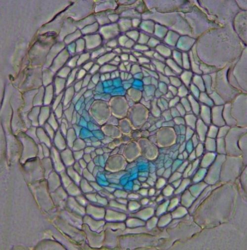
In the autumn of 2009, I returned from my post-doctoral period in Ben Scheres’ lab, located then in Utrecht. During my postdoc I was working on the roles of PLETHORA/AINTEGUMENTA-LIKE (PLT/AIL) transcription factors in stem cell regulation in the root meristem. Moving back to Finland my idea was to study whether any of these factors are expressed in Arabidopsis root cambium, the meristem I intended to study in my newly established research group. It was exciting to see that PLT5 showed very specific expression in the dividing cambial cells (Figure), however, further studies had to wait for several years due to my focus on finalizing ongoing projects. After obtaining funding for my research group, a PhD student, Gugan Eswaran, started to work on the project, and he discovered that ANT, PLT3 and PLT7 are also expressed in the cambium, suggesting genetic redundancy in cambium development. Unfortunately, ant single mutants and plt triple mutants showed only slightly reduced secondary growth. I was expecting a stronger phenotype from putative stem cell factors of cambium. In subsequent attempts, generation of the quadruple mutant failed due to unexplained lethality, and an artificial microRNA approach did not provide stronger phenotypes either. This was disappointing and thus Gugan started to focus more on his side projects. Then, rescue for the project came a few years later from a technical innovation. Xin Wang, a PhD student in my lab, invented an inducible genome editing system (IGE) for plants (Wang et al Nature Plants 2020). This IGE system was used to generate a conditional quadruple ant/plt mutant. Finally, this mutant showed severely reduced radial growth, something one would expect from loss of stem cell factors. This was great, but of course now we had a new question to answer – what regulates CAMBIUM-EXPRESSED AINTEGUMENTA-LIKE (CAIL) (the name we gave to ANT, PLT3, PLT5 and PLT7) expression in such a narrow, specific domain? I think, this was the point where you Peter contacted me, or was it even earlier than when we got the conditional quadruple mutant results?
pPLT5-GUS shows expression in dividing cambial cells.
Peter Etchells:
I got in touch while Gugan was making the IGE construct. I had been working on putting the pieces of PXY signalling together since joining Simon Turner’s lab in Manchester in 2007, and this continued when I moved to Siobhán Brady’s lab at UC Davis in 2013. Over that whole period, through my work and that of others in Hiroo Fukuda’s lab, a series of PXY-downstream targets, WOX4, WOX14, BES1, LBD4 and TMO6 were identified (Hirakawa et al Plant Cell 2012; Etchells et al Development 2013; Kondo et al Nature Comms 2015; Smit et al Plant Cell 2020). However, I was never satisfied that all the transcriptional targets of TDIF-PXY had been uncovered. PXY is homologous to CLAVATA1, which famously regulates the shoot apical meristem via regulation of the homeodomain transcription factor WUSCHEL (WUS). WOX4 and WOX14 are homologous to WUS, so they were a natural focus for investigation, but wox4 wox14 mutants only have a mild cambium phenotype. BES1, LBD4 and TMO6 are also only responsible for regulation of a subset of the pxy phenotypes. It seemed like we were missing something. The key was a transcriptomic experiment which demonstrated that CAIL genes were differentially expressed in both pxy and TDIF over-expression lines, performed just as I was transitioning out of Siobhán’s group to start my own lab in 2015. To test for a genetic interaction between the CAILs and TDIF-PXY, we crossed the TDIF over-expression line, which is characterised by ectopic cambium, to plt357 mutants. Although the plt357 line alonedid not have a cambium phenotype, it did suppress phenotypes associated with TDIF over-expression, which, combined with the CAIL expression patterns that Ari Pekka’s group had, demonstrated that the CAIL genes did have a cambium function and were likely controlled by TDIF-PXY. It was not long after that Gugan’s IGE line came through which sealed the deal. Still, the story was incomplete because the PXY expression domain is so broad relative to that of the CAILs.
Ari Pekka Mähönen:
So, now we knew that CAILs are regulated by the TDIF-PXY ligand-receptor pathway. However, we still did not know how come CAILs are only expressed in such a narrow region in stem cells, given that the PXY receptor expression domain spans from the stem cells into the xylem. A few researchers in my lab participated to hunt for the mechanism underlying this tight spatial control, and we indeed found a few regulatory feed-back mechanisms that could help excluding the CAILs from the xylem. On top of this, we wondered whether efficient sequestration of diffusing TDIF peptide by the PXY receptor could play a role in focusing CAIL signalling. With all the feedback regulation and a possible sequestration of TDIF, we were quite unsure which one of these mechanisms (or whether any of these mechanisms) could contribute to narrow CAIL expression in planta. Therefore, I contacted Kirsten ten Tusscher, a computational biologist, with whom I had collaborated before on addressing the role of PLT genes in root zonation (Mähönen, ten Tusscher et al. Nature 2014). I suggested that we could address these different scenarios in TDIF-PXY-CAIL signalling in a computational model.
Kirsten ten Tusscher:
As I had greatly enjoyed our previous collaboration, and questions on patterning are the bread and butter of computational biology, this was of course an offer I could not refuse. Luckily, a talented PhD student in my group, Jaap Rutten, was quite far already with the results for the main project of his PhD thesis and waiting for experimental data. Thus, it was not hard at all to convince him to broaden his horizon beyond the control of root meristem size that we were working on together with Sabrina Sabatini to the control of cambium patterning and positioning together with Ari Pekka and Peter. To investigate the importance of different feedback mechanisms as well as the potential of ligand sequestration in defining the narrow domain of CAIL cambium expression, we started building a model incorporating all the important molecular players and the regulatory interactions between them, using both new and previously published data. As a start we developed a model for a single cell and tested whether it could model xylem, phloem and cambium cells depending on the incoming signals. However, for non-modelers it often seems that if models are complex enough and you tweak parameters you can make them do anything you want. Therefore, it was important to show that the models’ capacity to simulate phloem, xylem or cambium forming cells was a very generic property of the modelled network architecture, not of precise parameter values or details. To achieve this Jaap performed a whopping 1,768,593,750 simulations to extensively test different model settings, occupying some of our computers for weeks, and show that overall model behaviour remained the same. In the process we could already confirm that some of the feedback uncovered by Ari Pekka’s team indeed limited cambium formation and promoted xylem formation. As a next step we could now move to a one-dimensional model of a strip of cells spanning from xylem to phloem and start testing the ligand-sequestration hypothesis. Key to this was to include an auxin-dependent PXY gradient starting from the xylem end of the tissue, and a TDIF gradient arising from the diffusion of TDIF from TDIF producing phloem cells into our model. With this in place, Jaap demonstrated that if binding of TDIF ligand to PXY receptors is sufficiently strong, at the tissue position where TDIF meets the first low levels of PXY receptors, TDIF is bound and effective diffusion is halted, preventing TDIF-PXY interaction further towards the xylem. Interestingly, this also explains why cambium stem cell patterning is robust under various cambium sizes: when the xylem and phloem are further apart, the TDIF will diffuse further before it reaches the first PXY receptors and until that time it diffuses freely ensuring it will still meet PXY receptors! However, an important issue remained: the regulatory feedback mechanism uncovered earlier could to some extent limit CAIL expression. So, to test which of these potential mechanisms occurs and/or is most important in planta, we tested in silico what would happen if we decreased PXY expression or elevated TDIF levels. Next these experiments were also performed in the lab, with lab outcomes matching the predictions made by the sequestration-based model. This finally enabled us to cement the importance of sequestration for defining the CAIL expression domain.
Ari Pekka Mähönen, Peter Etchells, Kirsten ten Tusscher:
So, now we could confidently say that sequestration of TDIF is the key to focusing the TDIF-PXY signalling and thus CAIL expression in a narrow domain to define the stem cells. The manuscript was submitted, and we got constructive comments from the reviewers, especially on providing more evidence for the sequestration mechanism. Xixi Zhang, a post doc in the Mähönen lab, had already earlier started to work on the generation of PXY reporter lines. She noticed, among other findings, that the translational reporter pPXY:PXY-YFP has a significantly sharper gradient within the cambium than the transcriptional reporter line pPXY:erYFP, indicating that the PXY-YFP fusion protein is more unstable in phloem-side cambium cells than in the cells on the xylem-side of the cambium. Since TDIF ligand originates from phloem, this suggest that the TDIF binding to PXY could make PXY-YFP unstable. Thus, regulation of TDIF-PXY stability could be the key mechanism for the sequestration, and this is what Xixi is studying now, as a follow up of the published work.
In the end, seeing this paper published was particularly satisfying, both because of the long journey it took to finalize it and because of the enjoyable collaboration we had while working together on this project.
Two years ago, we organized the inaugural symposium dedicated to Women in Tunicate Biology. It was a joyful event, celebrating women scientists from the 19th century to the present. A special issue of the journal genesis was published in November 2023, collecting the biographies and research talks from the symposium (https://onlinelibrary.wiley.com/toc/1526968x/2023/61/6).
We would like to announce that the second edition of this symposium will take place on Tuesday March 25th and Wednesday 26th, 2025 by Zoom. In this upcoming event, we will include talks by graduate students and postdocs working in the field of tunicate biology, as well as PIs.
If you are interested in participating, whether as a speaker or attendee, please let us know. All researchers are welcome to attend!
Thanks for your attention and best wishes, Anna Di Gregorio and Marie Nydam adg13@nyu.edu mnydam@soka.edu (1 votes) Loading...
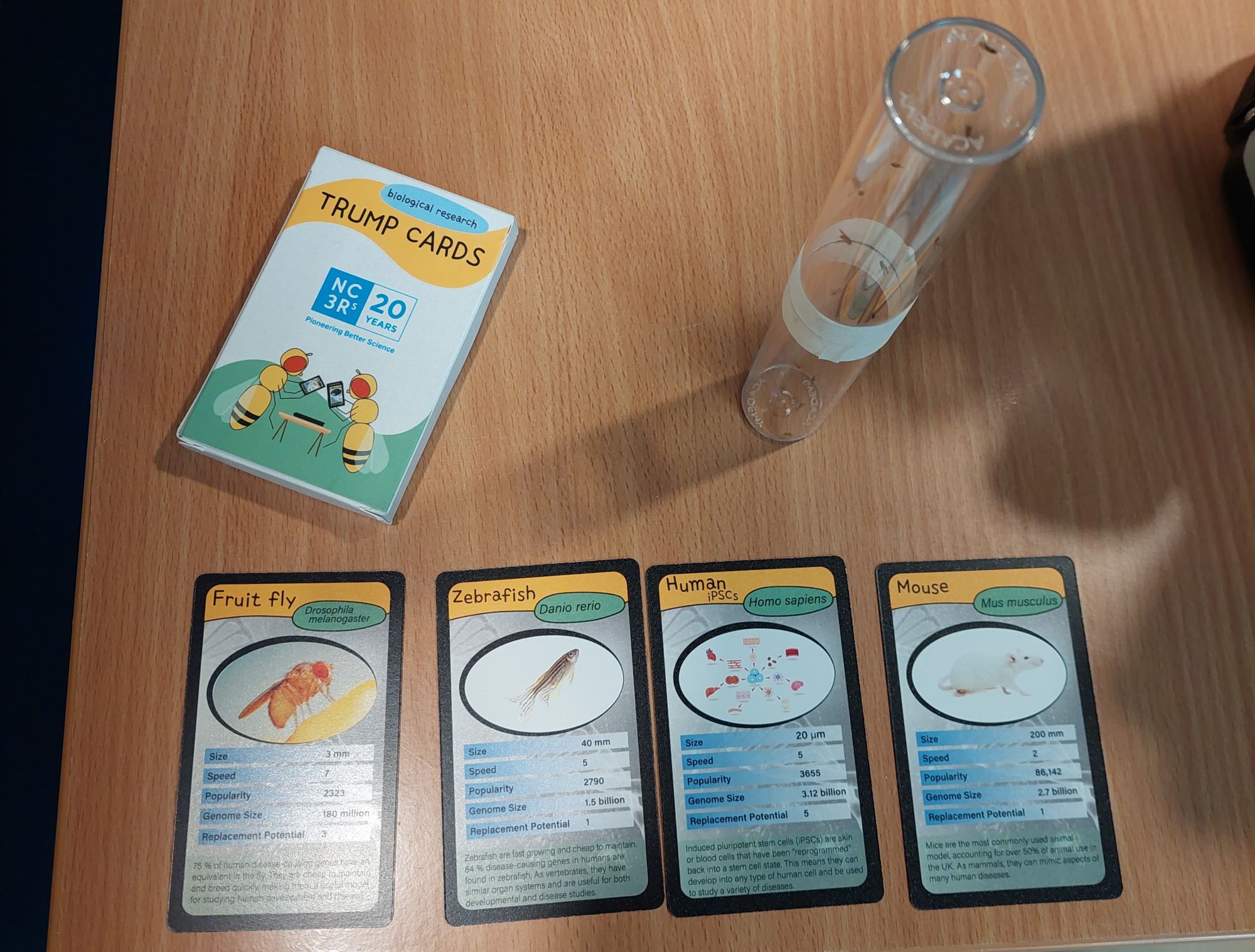
In 2023 I was awarded a NC3Rs 20th Anniversary Public Engagement Award to develop and print a “Biological Research Trump Cards” game. I have now designed and printed 100 packs of the game, and my aim is to share them with the scientific community and educators. I hope they will prove a fun and engaging public engagement tool in a variety of different settings.
Pleaseget in touchif you are:
A researcher in an academic setting who would like to use the cards in your outreach programme
A teacher at primary or secondary school and would be interested in having some packs / hosting a workshop (see below for an example)
Interested in using them but unsure if they are suitable for your audience
So what are “Trump Cards”?
There are few people who have gone through life without encountering Top Trumps in some form. First published in 1978, there are now hundreds of different varieties, from football teams to dinosaurs. The gameplay is simple, with users comparing numerical data to try and trump and win an opponent’s card. It is this simplicity, along with the easily adaptable format, that makes it ideal as a customisable public engagement tool.
Why this format?
I have always wanted to create a card game on a scientific theme that is both fun and educational, and Trump cards are an obvious choice – no complicated rules, compact, and the numerical categories offer the opportunity to convey a lot of information on a single card. The idea for the specific theme of these cards came to me after attending an NC3Rs early career researcher event, where I learnt about all the different research models and systems scientists were using for their research.
How was the game developed?
After extensive research into different research models, I came up with a full set of Top Trumps, including systems ranging from mathematical models to sea urchins. I decided to focus the game on the3Rs message, specifically the replacement of animals in research. Each card has 5 categories, including “replacement potential”, which is based on whether the system is an animal, partial replacement, or full replacement. Together with the NC3Rs team, I fine tuned the cards, making sure it conveyed the 3Rs message in a clear and accessible way. The other categories are “genome size”, “speed”, “size”, and “popularity”, with “speed” referring to how quickly experiments can be carried out, and “popularity” based on the number of articles published in 2019. I designed the cards myself using Adobe Illustrator, and spent many evenings deciding the perfect colour scheme, fonts, and drawing cartoons of fruit flies playing cards.
What is in the pack?
Each pack contains a set of Trump cards, along with explanatory cards for each of the categories, what the NC3Rs is and their mission, and a “how to play” card.
Who are the intended audience?
Top trumps are a well-loved game by children and adults alike, and no prior knowledge of scientific research is necessary to engage with the activity. Most adults and secondary school pupils will have some understanding of how we use animals in research and may have opinions about this, but I anticipate that they will not have been introduced to the myriad other model systems that scientists use. The aim is to promote discussion around the use of animals and alternatives. Younger children will enjoy the pictures and facts about unusual animals, and hopefully it will pique their interest in scientific research.
How do you use the cards?
I have 100 packs for distribution to schools and other researchers for use in their public engagement activities and would be delighted to share them with you. Their use is not limited to playing the full game from start to finish- here are some other examples of ways they can be used:
Short format – one card is chosen at random by each player and one turn is played. This would be most appropriate for stands at science fairs, for example, where people are passing through quickly.
As illustrations – if you are focusing on one or a few different model systems, the cards can be laid out, or images of them displayed on screen, as a quick way to convey a lot of information about that system. In addition to the numerical categories, there is a description on the bottom of the card explaining what the organism or system is used for.
Workshops in schools – in addition to simply playing the game, they are a useful tool to get students thinking about why scientists might use different model systems. For example, I have designed a workshop where students are given three scenarios and they have to choose what they think the best three models are for each research aim. This gets them thinking and discussing the advantages and disadvantages of different models, with support from scientists leading the workshop.
Trump Cards in action at an Motor Neuron Disease Association Legacy event at the Sheffield Institute of Translational Neuroscience. Here we used the cards as illustrations for the different models we use to research MND, as part of a stand showcasing fruit flies and mouse models.
Example workshop
I have designed a workshop aimed at secondary school and sixth form students, which is available for you to download. It can easily be adapted to suit different abilities. The workshop begins with a short introduction to modelling and why we do it, followed by examples. It also touches on what to consider when choosing a model. The main activity involves the students choosing three models for each research aim. During this activity, I would allocate one volunteer per group if possible to sit with the students whilst they discuss. This is helpful because they may have technical questions about different systems that would influence their choices. Additionally, you can probe their reasoning and get them to think about less obvious choices. For example, they might not know that fruit flies can be used for exercise experiments, or consider that mathematical modelling could be used for looking at the relationship between diet and motor neuron disease.
Xiao-Feng Zhao, Rafi Kohen, Eljo Y. Van Battum, Ying Zeng, Xiaolu Zhang, Craig N. Johnson, Karen Wang, Brian C. Lim, Juan A. Oses-Prieto, Joshua M. Rasband, Alma L. Burlingame, R. Jeroen Pasterkamp, Matthew N. Rasband, Roman J. Giger
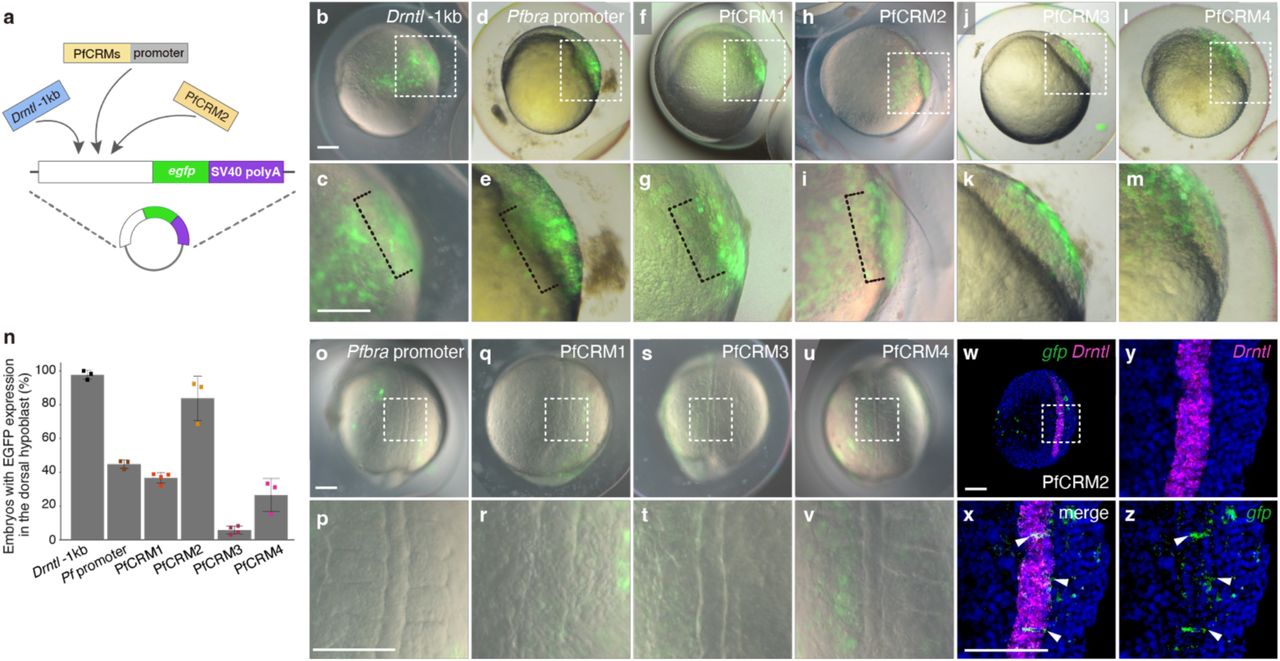
Marie Zilliox, Gaëlle Letort, David Sanchez, Christian Rouviere, Pascale Dufourcq, Frédérique Gaits-Iacovoni, Anne Pizzoccaro, Violaine Roussier-Michon, Patrick Blader, Julie Batut
Renata Coutinho-dos-Santos, Daniele G. Santos, Lupis Ribeiro, Jonathan J. Mucherino-Muñoz, Marcelle Uhl, Carlos Logullo, A Mendonça-Amarante, M Fantappie, Rodrigo Nunes-da-Fonseca
Luca Caputo, Cedomir Stamenkovic, Matthew T. Tierney, Maria Sofia Falzarano, Rhonda Bassel-Duby, Alessandra Ferlini, Eric N. Olson, Pier Lorenzo Puri, Alessandra Sacco
Rachel Forman-Rubinsky, Wei Feng, Brent T. Schlegel, Angela Paul, Daniel Zuppo, Katarzyna Kedziora, Donna Stoltz, Simon Watkins, Dhivyaa Rajasundaram, Guang Li, Michael Tsang
Archana Prabahar, Connie S. Chamberlain, Ray Vanderby, William L. Murphy, William Dangelo, Kulkarni Mangesh, Bryan Brown, Barsanjit Mazumder, Stephen Badylak, Peng Jiang
Byron W.H. Mui, Joseph Y. Wong, Toni Bray, Lauren Connolly, Jia Hua Wang, Alexander Winkel, Pamela G. Robey, Kristian Franze, Kevin J. Chalut, Mekayla A. Storer
Asya Bastrich, Daniil Antonov, Aleksandra Podzhilkova, Darya A. Petrova, Svetlana V. Pylina, Dmitriy N. Laptev, Elena A. Sechko, Sergey N. Kuznetsov, Ekaterina A. Vetchinkina, Natalia G. Mokrysheva
Ruiqi Hu, Linda L. Boshans, Bohan Zhu, Peiwen Cai, Yiran Tao, Mark Youssef, Gizem Inak Girrbach, Yingnan Song, Xuran Wang, Alexander Tsankov, Joseph D. Buxbaum, Sai Ma, Nan Yang
Georgios Tsissios, Marion Leleu, Kelly Hu, Alp Eren Demirtas, Hanrong Hu, Toru Kawanishi, Evangelia Skoufa, Alessandro Valente, Antonio Herrera, Adrien Mery, Lorenzo Noseda, Haruki Ochi, Selman Sakar, Mikiko Tanaka, Fides Zenk, Can Aztekin
Madhura P Nijsure, Brendan Tobin, Dakota L Jones, Annemarie Lang, Grey Hallström, Miriam Baitner, Gabrielle I Tanner, Yasaman Moharrer, Christopher J Panebianco, Elizabeth G Seidl, Nathaniel A Dyment, Gregory L Szeto, Levi Wood, Joel D Boerckel
Erin N. Sanders, Hsuan-Te Sun, Saman Tabatabaee, Charles F. Lang, Sebastian G. van Dijk, Yu-Han Su, Andrew LaboD, Javeria Idris, Marco Marchetti, Shicong Xie, Lucy Erin O’Brien
Harshita Mangal, Kyle Linders, Jonathan Turkus, Nikee Shrestha, Blake Long, Xianyan Kuang, Ernest Cebert, J. Vladimir Torres-Rodriguez, James C Schnable
Antoine Nicolas, Panagiotis Papadopoulos, Matteo Caroulle, Bernard Adroher, Magali Goussot, Anne-Sophie Sarthou, Nicolas Arnaud, Aude Maugarny, Patrick Laufs
Joel Rodríguez Herrera, Kenia Aislinn Galván Alcaraz, Ramsés Uriel Albarrán Hernández, Juan Pablo Villa Núñez, Gustavo Rodríguez Alonso, Svetlana Shishkova
Melissa Dipp-Alvarez, J. Luis Lorenzo-Manzanarez, Eduardo Flores-Sandoval, Domingo Méndez-Álvarez, Annie Espinal-Centeno, Jesús León-Ruiz, Fernando Olvera-Martínez, John L. Bowman, Mario A. Arteaga-Vázquez, Alfredo Cruz-Ramírez
Markéta Luklová, Marieke Dubois, Michaela Kameniarová, Klára Plačková, Jan Novák, Romana Kopecká, Michal Karady, Jaroslav Pavlů, Jan Skalák, Sunita Jindal, Ljiljana Tubić, Zainab Quddos, Ondřej Novák, Dirk Inzé, Martin Černý
Tomás Urzúa Lehuedé, Victoria Berdion Gabarain, Miguel Angel Ibeas, Hernan Salinas-Grenet, Romina Acha, Tomas Moyano, Lucia Ferrero, Gerardo Núñez-Lillo, Jorge Perez, Florencia Perotti, Virginia Natali Miguel, Fiorella Paola Spies, Miguel A. Rosas, Ayako Kawamura, Diana R. Rodríguez-García, Ah-Ram Kim, Trevor Nolan, Adrian A. Moreno, Keiko Sugimoto, Norbert Perrimon, Karen A. Sanguinet, Claudio Meneses, Raquel L. Chan, Federico Ariel, Jose M. Alvarez, José M. Estevez
Katie A. Long, Ashleigh Lister, Maximillian R. W. Jones, Nikolai M. Adamski, Rob E. Ellis, Carole Chedid, Sophie J. Carpenter, Xuemei Liu, Anna E. Backhaus, Andrew Goldson, Vanda Knitlhoffer, Yuanrong Pei, Martin Vickers, Burkhard Steuernagel, Gemy G. Kaithakottil, Jun Xiao, Wilfried Haerty, Iain C Macaulay, Cristobal Uauy
A.P Lipinska, G. Cossard, P. Epperlein, T. Woertwein, C. Molinier, O. Godfroy, S. Carli, L. Ayres-Ostrock, E Lavaut, F. Marchi, S. Mauger, C. Destombe, M.C. Oliveira, E.M. Plastino, S.A. Krueger-Hadfield, M.L. Guillemin, M. Valero, S.M. Coelho
Guy Teichman, Mor Sela, Chee Kiang Ewe, Itai Rieger, Sarit Anava, Yael Mor, Péter Szántó, David H. Meyer, Hila Doron, Or Shachar, Vladyslava Pechuk, Hila Gingold, Meital Oren-Suissa, Matthew McGee, Michael Shapira, Björn Schumacher, Oded Rechavi
Feline W. Lindhout, Hanna M. Szafranska, Ivan Imaz-Rosshandler, Luca Guglielmi, Maryam Moarefian, Kateryna Voitiuk, Natalia K. Zernicka-Glover, Daniel J. Lloyd-Davies Sánchez, John Minnick, Mircea Teodorescu, Madeline A. Lancaster
Aleksandra Babicheva, Ibrahim Elmadbouh, Shanshan Song, Michael Thompson, Ryan Powers, Pritesh P. Jain, Amin Izadi, Jiyuan Chen, Lauren Yung, Sophia Parmisano, Cole Paquin, Wei-Ting Wang, Yuqin Chen, Ting Wang, Mona Alotaibi, John Y.-J. Shyy, Patricia A. Thistlethwaite, Jian Wang, Ayako Makino, Y.S. Prakash, Christina M. Pabelick, Jason X.-J. Yuan
Oliver Arnolds, Eve M. Carter, Madison Edwards, Edvard Wigren, Evert Homan, Pauline Ribera, Kirsty Bentley, Martin Haraldsson, Nmesoma Theo-Emegano, Peter Loppnau, Magdalena M Szewczyk, Michelle A Cao, Dalia Barsyte-Lovejoy, Karen Vester, Anna Thrun, Alexandra Amaral, Ralf Lesche, Jens Münchow, W. Felix Zhu, Louisa Temme, Christoph Brenker, Timo Strünker, Michael Sundström, Matthew H. Todd, Aled M Edwards, Claudia Tredup, Opher Gileadi
Eva L Simpson, Ben Wetherall, Liam P Cheeseman, Aleksandra Byrska, Tania Mendonca, Xiaomeng Xing, Alison J Beckett, Helder Maiato, Alexandra Sarginson, Ian A Prior, Geraldine M Hartshorne, Andrew McAinsh, Suzanne Madgwick, Daniel G Booth
Yan Huang, Nina Bucevic, Carmen Coves, Natalia Felipe-Medina, Marina Marcet-Ortega, Nikoleta Nikou, Cristina Madrid-Sandín, Maria Lopez-Panades, Carolina Buza, Neus Ferrer Miralles, Antoni Iborra, Anna Pujol, Alberto M Pendás, Ignasi Roig
Paula Fernandez-Guerra, Pernille Kirkegaard Kjær, Simone Karlsson Terp, Jesper S. Thomsen, Blanca I. Aldana, Herma Renkema, Jan Smeitink, Per H. Andersen, Johan Palmfeldt, Kent Søe, Thomas L. Andersen, Moustapha Kassem, Morten Frost, Anja L. Frederiksen
Danielle Pi, Jonas Braun, Sayantan Dutta, Debabrata Patra, Pauline Bougaran, Ana Mompeón, Feiyang Ma, Stuart R Stock, Sharon Choi, Lourdes García-Ortega, Muhammad Yogi Pratama, Diomarys Pichardo, Bhama Ramkhelawon, Rui Benedito, Victoria L Bautch, David M Ornitz, Yogesh Goyal, M. Luisa Iruela-Arispe
Antonia Weberling, Natalia A. Shylo, Bonnie K. Kircher, Hannah Wilson, Melainia McClain, Marta Marchini, Katherine Starr, Thomas J. Sanger, Florian Hollfelder, Paul Trainor
Jessica C. Edge, Olga Amelkina, Haidee Tinning, Gianluca Giovanardi, Elena Mancinelli, Samantha Gardner, Elton JR Vasconcelos, Virginia Pensabene, Karen Forbes, Mary J O’Connell, Peter Ruane, Niamh Forde
Luke TG Harland, Tim Lohoff, Noushin Koulena, Nico Pierson, Constantin Pape, Farhan Ameen, Jonathan Griffiths, Bart Theeuwes, Nicola K Wilson, Anna Kreshuk, Wolf Reik, Jennifer Nichols, Long Cai, John C Marioni, Berthold Gottgens, Shila Ghazanfar
Vincent Boudreau, Ashley R. Albright, Therese M. Gerbich, Tanner Fadero, Victoria Yan, Ben T. Larson, Aviva Lucas-DeMott, Jay Yung, Solène L.Y. Moulin, Carlos Patiño Descovich, Mark M Slabodnick, Adrien Burlacot, Jeremy R. Wang, Krishna K Niyogi, Wallace F. Marshall
Alma Zuniga Munoz, Kartik Soni, Angela Li, Vedant Lakkundi, Arundati Iyer, Ari Adler, Kathryn Kirkendall, Frank Petrigliano, Bérénice A. Benayoun, Thomas P. Lozito, Albert E. Almada
Luiz Fernando Silva Oliveira, Radhika S. Khetani, Yu-Syuan Wu, Venkata Siva Dasuri, Amanda W. Harrington, Oluwabunmi Olaloye, Jeffrey Goldsmith, David T. Breault, Liza Konnikova, Shannan J. Ho Sui, Amy E. O’Connell
Christopher J. Panebianco, Maha Essaidi, Elijah Barnes, Ashley Williams, Karin Vancíková, Margot C. Labberté, Pieter Brama, Niamh C. Nowlan, Joel D. Boerckel
Dana E. Cobb-Lewis, Devin Synder, Sonya Dumanis, Robert Thibault, Barbara Marebwa, Elisia Clark, Lara St. Clair, Leslie Kirsch, Michelle Durborow, Ekemini Riley
Some of you may have been so fortunate as to receive gift cards for Amazon.com or local bookstores in your Christmas stockings. While I wouldn’t think of dissuading you from purchasing the latest Louise Perry mystery or the memoirs of pre-eminent singers and chefs, I would recommend that you consider a new intellectual thriller, Evolution Evolving.
Imagine if two outstanding evolutionary biologists realized that evolutionary theory cannot explain adaptation and biodiversity without incorporating developmental biology. Imagine them inviting three developmental biologists to work on a book with them to construct the foundations of a more complete evolutionary theory. This book will become Evolution Evolving: The Developmental Origins of Adaptation and Biodiversity, a volume co-authored by evolutionary biologists Kevin Lala and Marcus Feldman, together with evolutionary developmental biologists Tobias Uller, Nathalie Feiner, and me.
This is not a textbook. It is a symposium, a working out of ideas, such that the reader is in dialogue with the book. The book presents evidence for certain views — that plasticity is universal and fundamental for evolution; that organisms are multigenomic holobionts whose symbionts can create new phenotypes and reproductive isolation in the animals they co-create; that there are multiple pathways of inheritance, including symbionts, epialleles, culture, and parental effects, and that some of these modes of inheritance allow the transmission of environmentally induced traits. Most of us had trained to see evolution as changes in gene frequency and development as changes in gene expression. This book organizes evidence that these genocentric explanatory mechanisms are inadequate to explain adaptations or the diversity of life.
Reading the book should make one question and refine one’s own ideas, to question one’s assumptions. Yes, these developmental phenomena happen; but are these differences important enough to change the way you think about evolution, organisms, development, and science? This book presents evidence that these phenomena — developmental plasticity, developmental symbiosis, and epigenetic inheritance systems — are critically important and that evolutionary biology gains enormous explanatory power only if it fully incorporates them. Some people have agreed with us. Marc Kirschner has called the book “a tour de force,” and Jessica Riskin has nominated the volume as a Scholarly Book of the Year, calling it “exhilarating reading. It is not just a book but an intellectual revolution.” Some people have disagreed. Evolutionary biologist David Houle doesn’t think these phenomena are important enough to change the way we think about evolution; moreover, “they are difficult to study.”
Twenty-five years ago, I predicted that evo-devo would cease to exist because it would become part of normative evolutionary biology. This is now happening. Look at the recent articles in PNAS about the genes responsible for the cryptic and mimetic pigmentation of insect wings. They are not classified under “developmental biology,” or even as “evolutionary developmental biology.” Rather, they are listed as “evolution.” Similarly, an evo-devo paper on the rates of prehistoric human teeth and brain development is listed in the “evolution” category, not as “development.” It seems that evolutionary developmental biology is becoming part of evolutionary biology. This book shows the many ways in which these fields can be merged.
Evolution is undergoing a metamorphosis, retaining some features, while jettisoning and repurposing others. It is evolution, but not as we knew it. It is an evolution where proximate and ultimate causes co-mingle, and where developmental mechanisms can bias the directions of evolutionary change. It is an evolutionary biology where the environment not only selects the phenotype but helps construct it. Evolution Evolving is an evolutionary biology book where developmental mechanisms are major players in the evolutionary processes that create adaptations and biodiversity.
The application deadline for the workshop is the 24th of January.
This is a residential workshop at Chicheley Hall on 2nd- 4th April 2025.
An important part of science is getting your results and ideas across to others, through papers, presentations, theses, grant proposals, conversations and interviews. Your audience may include specialists in the field, those from other disciplines, industry, or the general public.
How can you best communicate your science?
This workshop brings together experts in different fields to help you explore and develop your communication skills.
Working together with others on the course you will learn how to structure stories, bridge disciplines, simplify concepts and communicate effectively with a range of audiences. You will also get in-depth tutoring and practice in storytelling and public talks, developing hands-on demonstrations and multimedia (podcasts/YouTube/TikTok).
The Genetics Society will cover travel (within the UK only), accommodation and meals for successful applicants.
Tutors will include: Helen Keen (Award winning comedy writer and performer; author of the Radio 4 series, “It Is Rocket Science!”) First Create the Media (Led by award-winning writer and broadcaster Kat Arney) Alison Woollard (Presenter of the 2013 Royal Institution Christmas Lectures and Lecturer at University of Oxford)
Organiser: Jonathan Pettitt (Professor in Genetics, University of Aberdeen; Winner of the 2020 Genetics Society JBS Haldane Lecture) Cristina Fonseca (Science Communicator)
Who can attend?
The course is open to PhD students and postdoctoral researchers working in genetics and related areas.
Carer’s Award. In recognition of carer’s responsibilities, an award of (up to) £60/day will be made available to enable participants with carer responsibilities to attend this workshop. Awardees can spend this money as they think will best support their attendance.
With almost 200 posts published on the Node in 2024, below are just a few of our highlights.
Thank you to everyone who has contributed to the Node in the past year.
Have you read a Node post that you really enjoyed this year? Let us know in the comment section!
Behind the paper stories
Every paper has a story behind it. In these posts, we discover the highs and lows, the unexpected turns, and the fascinating discoveries from the breadth of developmental and stem cell biology.
Using an image or a video as a hook, these short posts bring people’s attention to a paper, a technique or a location that is of interest to the developmental and stem cell biology community.
“No such thing as a standard career path” interview series
In this new series, we chatted to several developmental biologists who have had vastly different career trajectories. Check out all the interviews in this series so far.
The Node correspondents
Correspondents are researchers who are also interested in science communication. They work with the Node team to develop and create content on a broad range of topics. Here are a few highlights of posts produced by the correspondents:
Do you want to broaden your science communication experience alongside your research? We are looking for new correspondents for the Node. Find out more and apply by 20 January 2025!
Even though we have grouped posts into different series, we always welcome posts that don’t necessarily fit into any of our existing blog series.
Remember, the Node is your site: once you’ve registered, you can freely share your blog post, job advert or event notice with the community. If you have any questions, just get in touch.
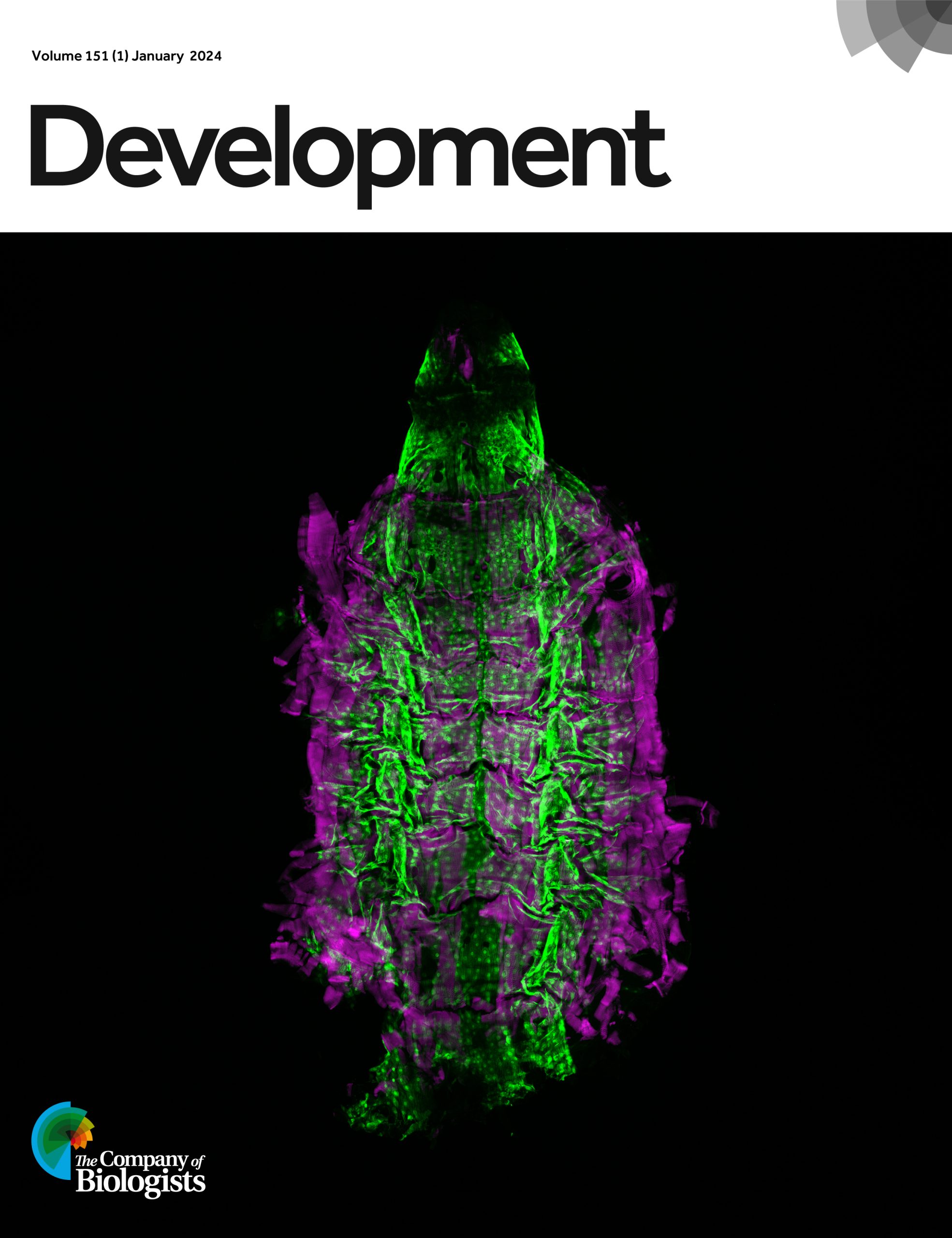
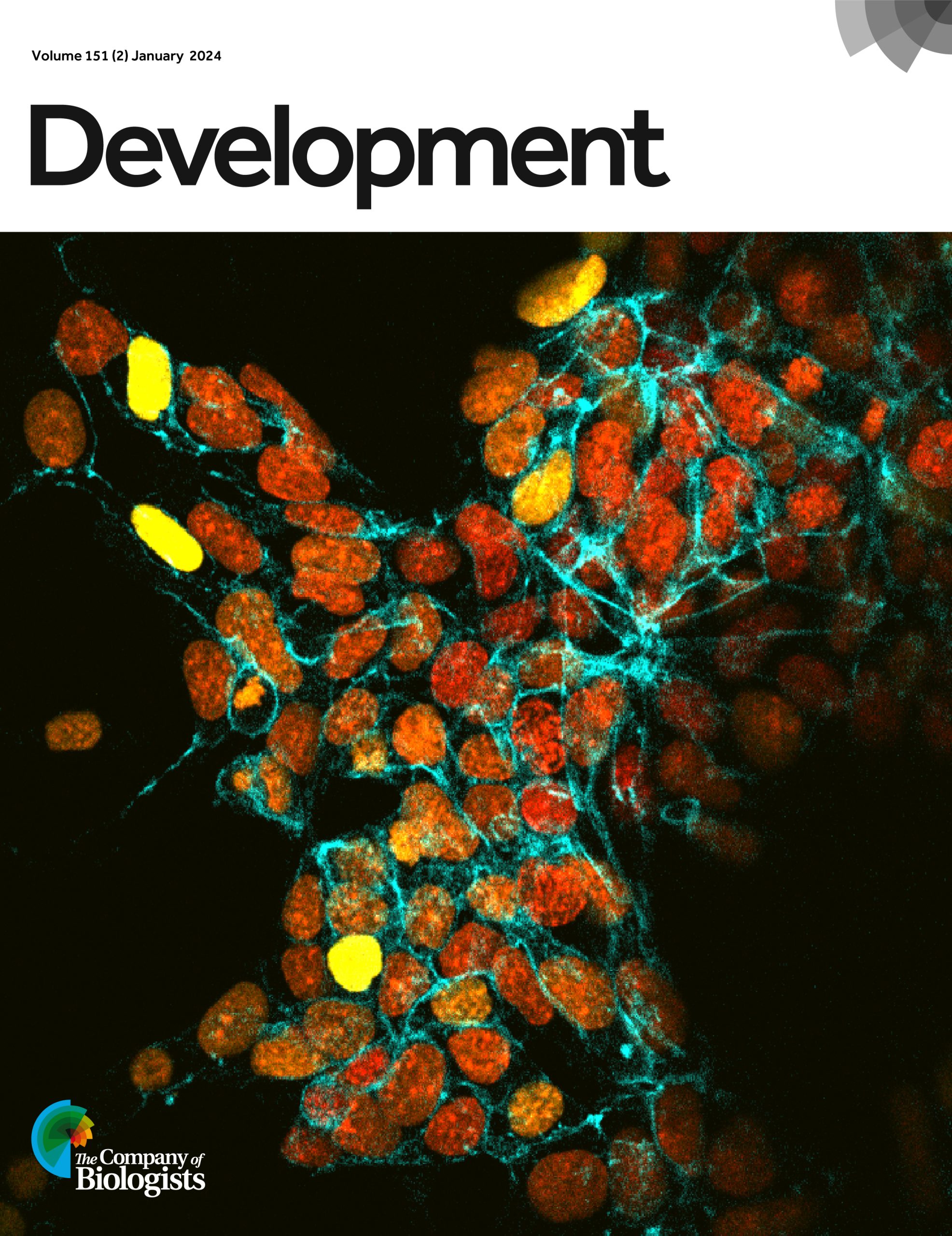
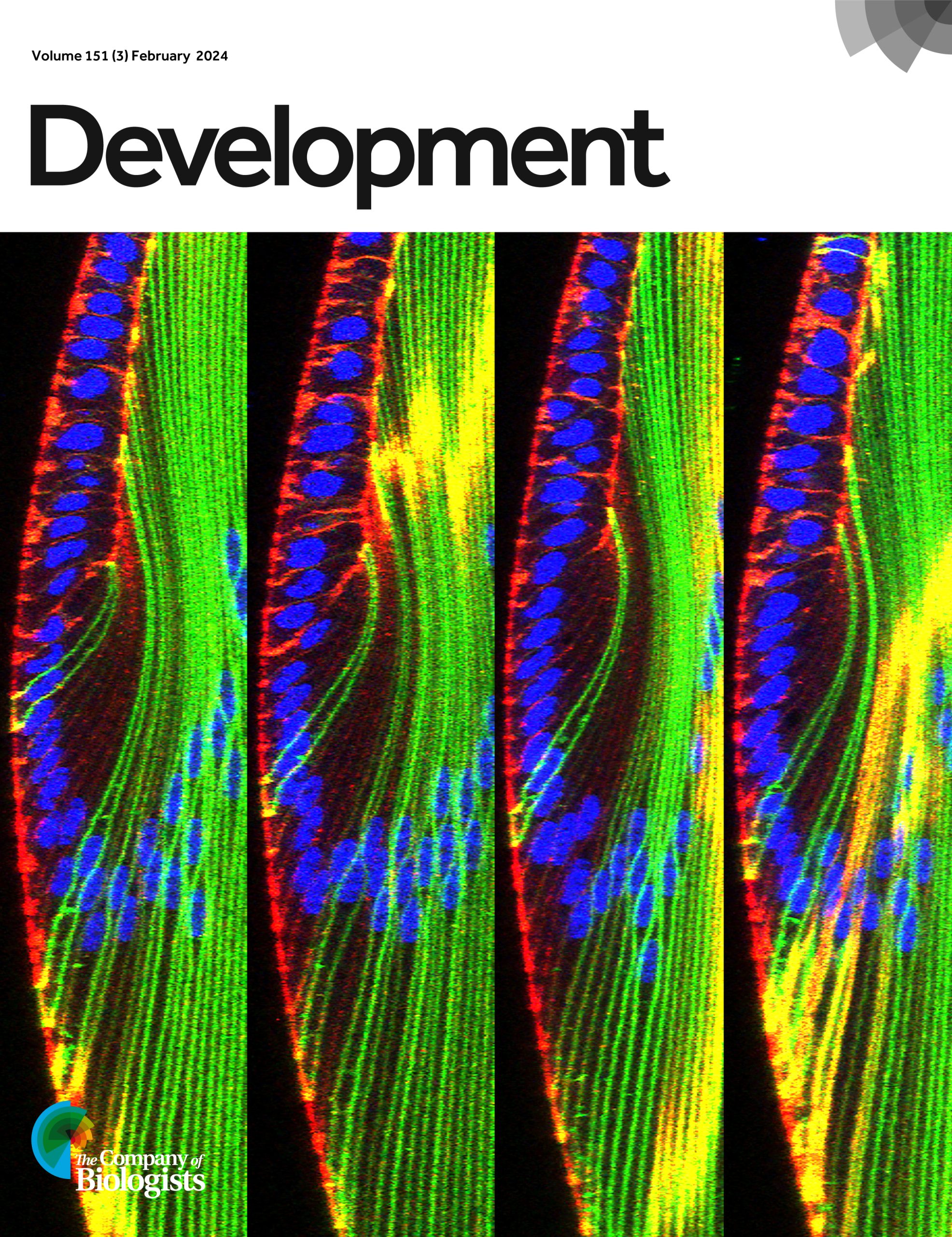
Over the past 12 months, Development has featured 24 cover images. Now, it’s your chance to pick your favourite!
To find out more about each cover image, you can visit Development’s 2024 issue archive. Thank you to everyone who’s contributed to this collection of wonderful images.
*The poll is now closed. Thank you to everyone who voted!*
Issue 1
Issue 2
Issue 3
Issue 4
Issue 5
Issue 6
Issue 7
Issue 8
Issue 9
Issue 10
Issue 11
Issue 12
Issue 13
Issue 14
Issue 15
Issue 16
Issue 17
Issue 18
Issue 19
Issue 20
Issue 21
Issue 22
Issue 23
Issue 24
Visit Development’s 2024 issue archive to find out more about each cover image.
Vote for the 2024 Development cover image of the year
*The poll is now closed. Thank you to everyone who voted!*
The final webinar of 2024 featured two early-career researchers working on gene regulation and will be chaired by Development’s Senior Editor, Alex Eve.
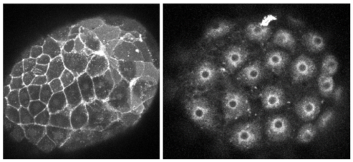
PRESS RELEASE: Millions of people around the world are affected by retinal degenerative diseases. In most cases, loss of vision is caused by damage to the macula, a region in the centre of the retina. The macula is rich in cone photoreceptors – cells important for perceiving colour and seeing finer details. Currently, there are no approved treatments to replace the damaged macula, despite its huge impact on the quality of life. Now, a team of researchers from the University of Montreal, led by Professor Gilbert Bernier, found that blind minipigs receiving retinal transplants made from stem cells showed signs of restored vision. They published their study in the journal Development on 5 December 2024.
In this study, Professor Bernier’s team developed a method to coax stem cells into forming sheets of cells that recapitulate the structure of the human retina. The type of stem cells they used are called human induced pluripotent stem cells – immature cells ‘reprogrammed’ from an adult (mature) cell that can differentiate into any type of cells in the body. Using the stem cells, the researchers made ‘retinal sheets’ that are enriched in immature versions of the cone photoreceptor cells, which could become mature cone cells when cultured in the lab.
After successfully creating the retinal sheets in a dish, the researchers tackled the next challenge: transplanting these sheets into minipigs with damaged macula. Professor Bernier explains, “To get as close as possible to human clinical application, we have chosen minipigs because the size of their eyes is near that of humans and the animals are about the same weight as humans. Hence, all surgeries in our study could be performed by a retinal surgeon.”
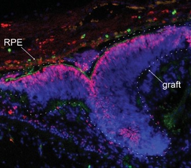
Upon transplantation, the researchers found that the retinal grafts were able to integrate into the minipig’s damaged retinal tissue. Encouragingly, the minipigs showed signs of restored vision: new neural connections were formed between the grafted photoreceptor cells and the minipigs’ neural cells, and the scientists could detect neural activity of the photoreceptors at the grafted area when the minipigs were placed in a well-lit room.
Given the pressing need to develop therapeutic interventions against vision loss, researchers around the world are testing different ways to repair damaged macula. “Some approaches use dissociated photoreceptor cells; others create micro-dissected retinal organoids, which are lab-grown ‘mini-organs’ in a dish,” says Professor Bernier. “In contrast, our method allows the spontaneous formation of a flat retinal tissue that is already polarised and organised, as in the human embryonic retina.” He adds that their method can generate large yields of retinal tissue for transplantation.
A limitation in this method lies in the difficulty of controlling the placement and orientation of the grafts during surgery. The macula is only 4mm in diameter – about the length of a grain of rice. “To properly orient, place and stabilise the graft in the retina remains a big surgical challenge,” says Professor Bernier. His team are now working to improve the transplantation success rate. They are validating an experimental retinal surgery device to ensure proper orientation and implantation of the graft at the correct retinal disease site. Although many challenges remain, this study demonstrates the potential of retinal sheet transplantation for treating retinal degenerative diseases.
Integration of a human retinal sheet (graft-dashed line) transplanted into a degenerated minipig retina, showing expression of the photoreceptor-specific markers CRX (in red) and PNA (in green) within the graft. Note the graft polarization and close association with the pig’s eye retinal pigment epithelium (RPE). Image Credit: Dr. Andrea Barabino
 (3 votes)
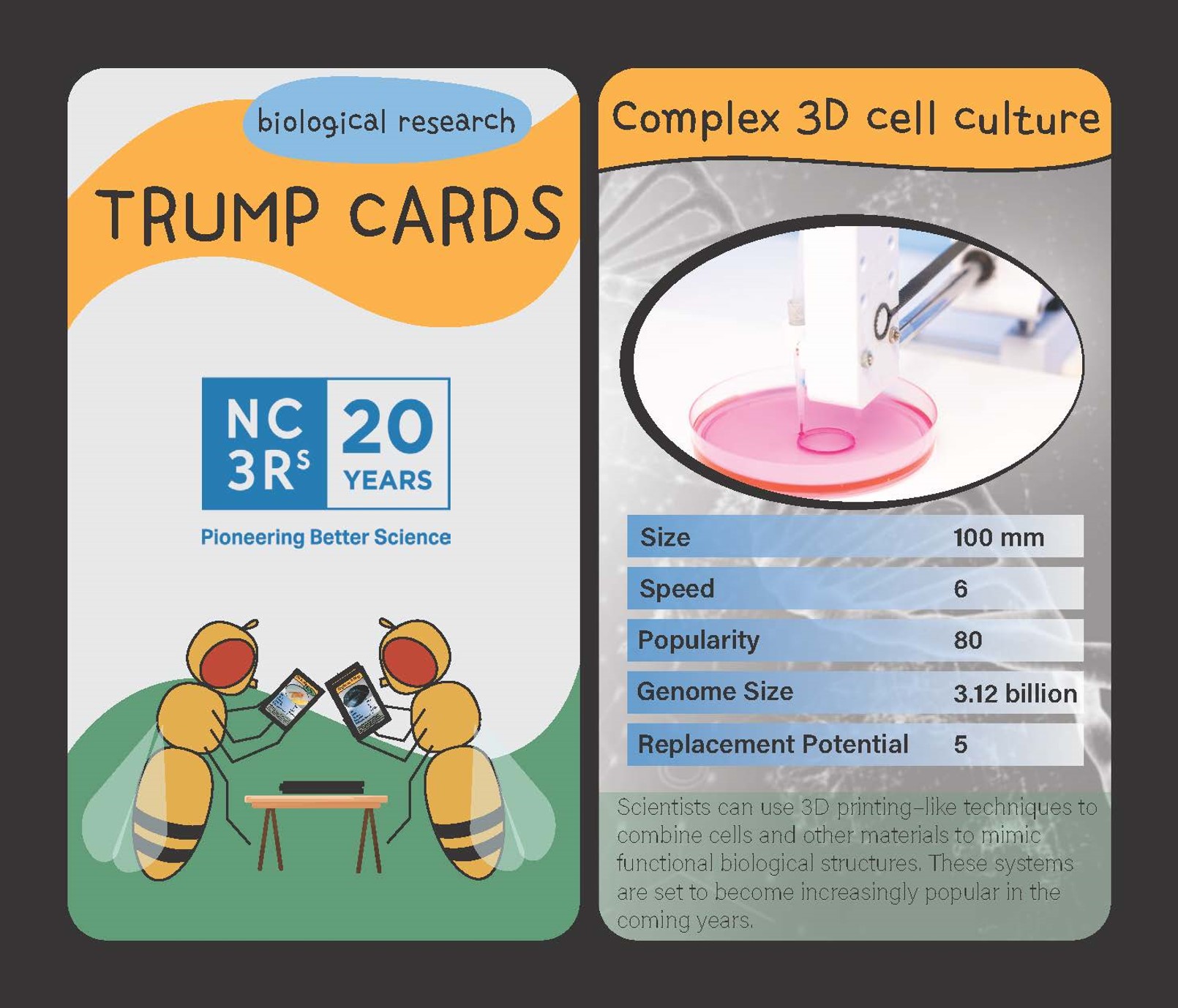

 (No Ratings Yet)