1-2 postdoc positions to study neuromuscular patterning during development & regeneration
Posted by cornelisond, on 10 March 2016
Closing Date: 15 March 2021
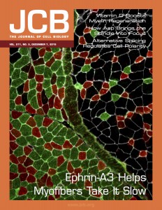 Each adult mammalian skeletal muscle has a unique complement of fast and slow myofibers, a consequence of cell-autonomous and non-cell-autonomous patterning decisions during development. Intriguingly, following either acute muscle regeneration or deinnervation/reinnervation, the proportionality and patterning of muscle fibers is largely preserved, suggesting a mechanism for ongoing maintenance of fiber type specification during homeostasis and regeneration/repair. We have recently reported that interactions between the repulsive guidance ligand ephrin-A3 [which is expressed on all and only slow (Type I) myofibers] and EphA8 [an ephrin receptor expressed by terminal Schwann cells at all and only neuromuscular junctions of fast (Type II) myofibers] promote and preserve slow myofiber identity by preventing stable innervation of slow muscle fibers by fast motor neurons [Stark et al, JCB 211:1077–91 (2015)]. Additional data from our lab suggest that variations on this mechanism may also direct slow fiber type-specific interactions with muscle satellite (stem) cells, and/or promote and preserve fast (Type IIb) myofiber identity during development and regeneration. The future potential of this work includes not only extending our basic understanding of muscle patterning during development, regeneration, neuromuscular disease, and aging but also has implications for targeted gene or cell transplantation therapies.
The Cornelison lab at the University of Missouri is recruiting 1-2 postdoctoral fellows to work on this NIH-funded project, addressing the significant unsolved question of how muscle fiber type is specified, maintained, and recapitulated in each muscle in that muscle’s unique pattern. The project will involve analysis of muscle fiber patterning in wild type, loss-of-function and gain-of-function mouse models during development and following muscle or nerve damage, using section immunohistochemistry, RNA in situ hybridization, timelapse videomicroscopy, flow cytometry, tissue culture and coculture, cell transplantation, and global gene expression and epigenetic profile analysis.
We are looking for highly motivated colleagues, ideally with a background in muscle and/or nerve development and regeneration, who are able to work both independently and collaboratively. A doctoral degree, strong background in cell and molecular biology, and proficiency in spoken and written English are required.
Columbia is a vibrant college town with a low cost of living that consistently appears on ‘Best Places to Live’ lists (Forbes, Livability, Outside). It is only two hours away from either St. Louis or Kansas City, and also has easy access to outdoor activities in-town (biking on the Katy Trail, rock climbing, camping) or nearby (climbing or canoeing in SW Missouri, boating on Lake of the Ozarks). The University of Missouri is a top-ranked public research university with the main undergraduate campus, the medical school, and the veterinary school all on the same campus, facilitating active collaborations in the life sciences. The Bond Life Sciences Center is a new multidisciplinary facility housing 38 labs from 14 different departments, as well as multiple University core facilities.
To apply, please send a CV, cover letter, and the names of three references to cornelisond@missouri.edu.
 (No Ratings Yet)
 (10 votes)
 I’ve just finished reading ‘