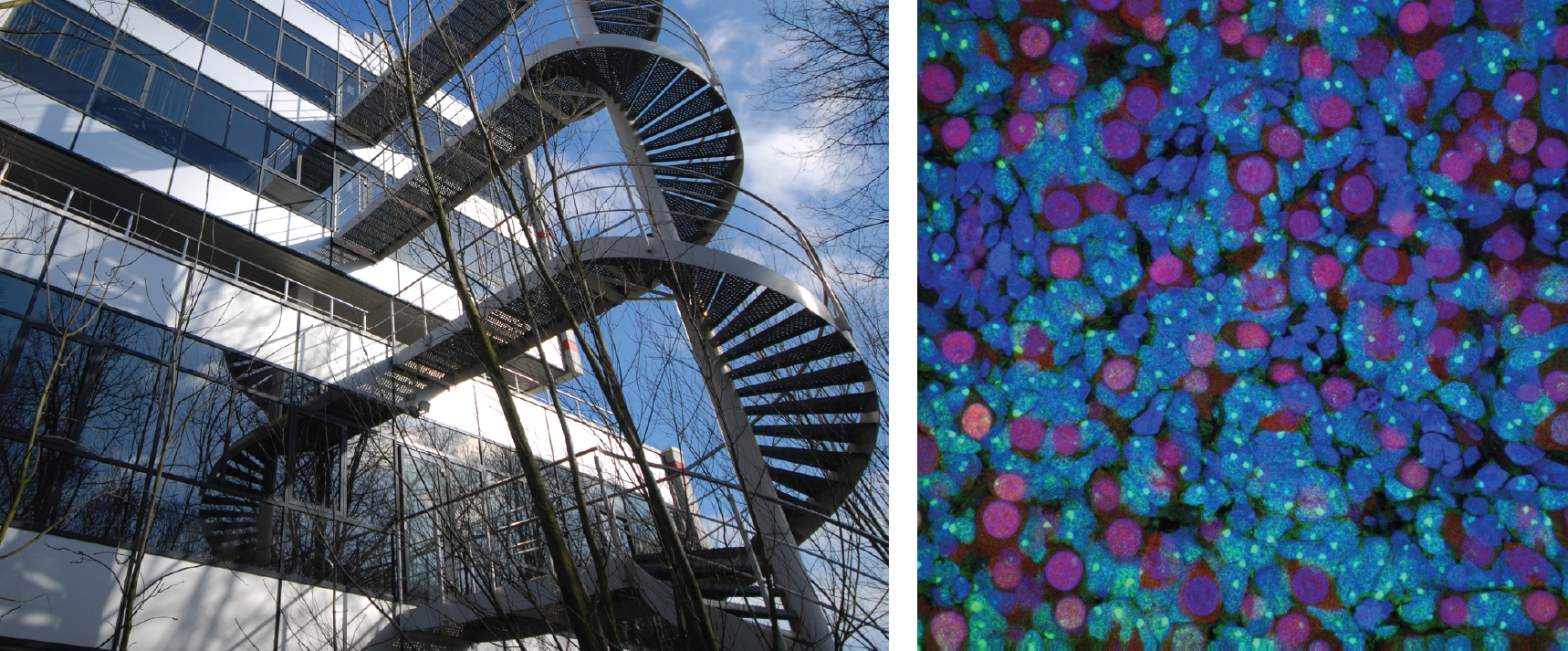
The “Nuit Blanche” in Paris. A city wide exposition of contemporary arts from dusk till dawn. Performances, light shows, dance, installations. Along the Canal Saint-Martin the visitors stroll from one exhibition to the next or sit down and take a break, chatting and drinking. A bit further up Boulevard Avenue Richerand the south-west entrance to the Hospital Saint Louis gives entry to a backyard. On the facade of the 17th century building, retinal pigmented epithelial cells perform their dance. Nothing but their cytoskeleton exposed in white before the dark building, magnified 10000-fold, accelerated 100-fold. One cell gets hold of a support beam, gradually spreading out over all its length. Loosing grip, rounding up and going into mitosis. Each daughter cell spreading out again. Other cells remain at one place, yet their cytoskeleton still moves, searching an exit from the confined space of a window.
When Manuel Thery came to me with the idea that we have to project cells onto a building – and cells which had been grown on micro-patterns in the shape of that same building – it was obvious that this idea simply had to be done. First, the general public has little access to all those beautiful images one takes when working with cells. Second, curiosity and “wanting to understand” needs to be promoted as a value by itself. Physics is not engineering, and biology is not R&D for pharmaceuticals. And the cytoskeleton with its appearing and disappearing regularities has the graphical power to draw you in and want you to grasp its function, without necessarily thinking about the benefit. Third, we try to find cues of how cells react to extracellular geometrical constraints in our day-to-day science, and we were curious ourselves, what we would see on these complex shapes. Fourth, as a structural scaffold for the cell (amongst other functions), the cytoskeleton shares some properties and constraints of engineering and architecture, and the metaphor has been made before. Fifth, simply, because we can.
The plan was simple:
1) take a photo of a public building
2) extract some interesting shapes from the building
3) produce micro-patterns of these shapes, the size of a single or several cells – micro-patterns are protein patterns for cell attachment on a substrate that otherwise repels cells
4) culture cells with fluorescently labeled cytoskeletal elements on these micro-patterns
5) do some live cell video microscopy
6) select the nicest videos and project the cells back on the original place of the pattern on the building, the size of the building, accelerated, for everyone to see.
Although that project may sound simple, it is by no means a one person, one weekend prep. At least, if you are a newbie to micro-patterning, live cell imaging, and your cell culture experience dates back more than a PhD’s time. Keep in mind that photo toxicity of the stain will kill the cells pretty quickly if you illuminate too strongly and frequently. On the other hand, if you want to play your time-lapse movies at a speed that people do not recognize it as a sequence of images, you need hundreds of frames for tens of seconds of footage, so you require days with an image every 5-10 min. And have cells survive under a microscope for 48h, on previously untested patterns of adhesive regions… is performing an experiment. And as such may just not work the first few times you try. Add the little pressure of a deadline (the day of the event that cannot be shifted).
But it gets even more involved with the work of the artists from the amazing Groupe Laps:
cut of the movie (the choreographies for the cells); giving the cells music to move to; correcting the image of the house for spherical aberration, so that the projection would align with the building; organizing the equipment for the projection; finding, in all those hundreds of movies, those that would be used; organizing the permissions to project onto the building; sticking non-reflecting foil over the windows on the day of the event; aligning the images with the features of the building; …
For someone having been involved in any sort of larger scale artistic event, all this may sound evident, but I was amazed by just how complex the basic idea would get.
Another change is the mindset between me and the artist. For them a cool sequence of cells would do. For me it also had to reflect a typical cell on these patterns – while still yielding footage with good enough contrasted structures to allow the projection.
Only on the evening itself did I see how well the composition by Groupe Laps had worked. It was very different from what I had imagined on the outset, and much richer for the fact that it did not look like all the images you would see in a publication – if you cared to project them on a house, that is.
Since we could not assume that the public would grasp that the images showed live cells, we also provided two information boards to describe the project in a short text as you might expect in an exhibition. However, we were surprised by the number of people who were at the event, and because a 10m high projection of something moving draws more attention than 1m high panel of written text, only a minority took advantage. Therefore, the level of understanding ranged from fellow scientists “yay, z-stack of microtubules”, over raising curiosity with many, to some complete ignorance that this had anything to do with biology. On the other hand, many spectators were clearly drawn in by the videos, watching the installation several times over. And I like to believe there was a lot wider spectrum of interpretation and thought, specifically because there was no classroom-ready explanation available.
In the case that you plan an outreach activity with collaborators foreign to your field or even science, my main advise would be to make sure you take the time to collaborate closely; to get to know each other and mutually showing additional possibilities and clarifying impossibilities on either side takes some iterations. This may apply less to smaller events, or repeating ones, where you can learn on the job and grow the event slowly. But for larger scale one-offs this is paramount. In fact, the sole regret I have about my experience with the nuit blanche project is that – because of distance and safety regulations – I could never show the involved artists our laboratories, so my work still remains abstract to them, and that we did not have regular “lab”-meetings, as is the case with my research.
If you ask yourself whether you should get involved in some outreach activity of the sort, involving people foreign to science, and specifically if you are not familiar with their line of work: Do it! It broadens the horizon and experiencing some recognition from someone outside of ones usual line of work has a special quality for both sides.
 This post is part of a series on science outreach. You can read the introduction to the series here and read other posts in this series here.
 (2 votes)
 Loading...
 (1 votes)
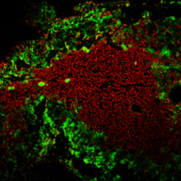

 Wnt, Fgf and retinoic acid signalling play a key role in patterning the posterior neural plate to form the midbrain, hindbrain and spinal cord. Despite intense study of Wnt signalling and neural patterning, only a few target transcription factors that mediate spinal cord development have been identified and the mechanism remains unclear. In this issue (p.
  The thymus is central to the adaptive immune system, but it is one of the first organs to undergo an age-related decline in function. Reduced expression of the thymic epithelial cell (TEC)-specific transcription factor FOXN1 has been associated with thymus degeneration, but whether restoration of FOXN1 expression can regenerate an aged thymus is unknown. Now, on p.
  Muscle stem cells, called satellite cells, are responsible for muscle growth and repair throughout life. Different subsets of satellite cells have varying degrees of self-renewal and differentiation potential, but how and when these different subsets arise has not been addressed in vivo. Now, on p.
  Unlike somatic cells, the nucleus of the oocyte and very early embryo contains a morphologically distinct nucleolus called the nucleolus precursor body (NPB). Although this enigmatic structure has been shown to be essential for normal mammalian development, its precise function remains unclear. In this issue, Helena Fulka and Alena Langerova now demonstrate (p.
  The WUSCHEL (WUS) family of transcription factors is well known for its role in stem cell maintenance in seed plants. There are two paralogues of the WUS-RELATED HOMEOBOX 13 (WOX13) gene in the moss Physcomitrella patens, but their function is unknown. Now, on p.
  Retinoic acid (RA) is essential for many developmental processes, but signalling levels must be tightly regulated since too much RA signalling can cause developmental defects. Cyp26 enzymes help to control this balance, metabolising RA and ensuring the correct specification of multiple different organs. Loss of Cyp26 activity can affect heart formation, and now (see p.
  Morphological asymmetry is a common feature of animal body plans, from shell coiling in snails to organ placement in humans. Many vertebrates use cilia for breaking symmetry during development: rotating cilia produce a leftward flow of extracellular fluids that induces asymmetric expression of the signaling protein Nodal. By contrast, Nodal asymmetry can be induced flow-independently in invertebrates. Here, Martin Blum et al ask when and why flow evolved, and propose that flow was present at the base of the deuterostomes and that it is required to maintain organ asymmetry in otherwise perfectly bilaterally symmetrical vertebrates. See the Hypothesis on p.
  Over the past 20 years, diverse roles for the Hippo pathway have emerged, the majority of which in vertebrates are determined by the transcriptional regulators TAZ and YAP. Accurate control of the levels and localization of these factors is thus essential for early developmental events, as well as for tissue homeostasis, repair and regeneration. Here, Bob Varelas provides an overview of the processes and pathways modulated by TAZ and YAP and outlines how TAZ and YAP contribute to organ homeostasis and regeneration. See the Review on p.