The Workshop will bring together researchers working on gene regulation, developmental biology, mathematical modelling and evolution. The invited researchers work on a variety of complex systems and are examining how these systems originated and have evolved over time. By comparing perspectives and experimental approaches to examine the evolution of their specific systems, we hope to draw common threads that may be applicable to most systems, and we aim to highlight these after the meeting in a Review article.
What defines the distinction between defect and difference? Between natural and unnatural? Between right and wrong?
When you are disabled, you are made to feel that you are wrong.
In my evo-devo PhD, my study species was a species of cichlid fish. When people unfamiliar with them ask me what on earth a cichlid is and why on earth I would study it, I launch into explaining what an adaptive radiation is with “you know Darwin’s finches? Like that but fish, and instead of islands, it’s lakes. They’re all sorts of shapes and colours to suit all sorts of ecological niches.” What’s so interesting about them is their diversity. In paper introductions we try to convey the extent of that diversity with adjectives like “remarkable”, “unparalleled”, and “astonishing”. They are words full of wonder. Even with our range of species in the lab, it is something I only really appreciated personally rather than academically when I finally went diving in Lake Malawi/Nyasa.
All of that diversity in form is the product of varying developmental processes, tweaked by genetic differences from an ancestral set of developmental trajectories. So another thing we sometimes say is that cichlids represent a “natural mutagenic screen”1. Nature has provided a set of morphological differences, so we can investigate the relationship between developmental processes and morphological outcome. Not only does this give us a better understanding of the role of development in evolutionary change, but also it may provide another window into the developmental processes that we still don’t fully understand. The implication is that by studying natural variation, you can better understand “normal” development. If you understand which genetic and developmental differences lead to natural biodiversity, you can gain insights into how genetic and developmental differences lead to pathological deviation from the norm2. Take cichlid craniofacial shape diversity, for example. The variation between cichlids reflects adaptations to different feeding strategies. One of the key findings of evo-devo research is that the same developmental pathways and programmes are used across even distantly related organisms. So studying craniofacial development in one organism, such as a fish, can shed light on craniofacial development in another vertebrate, such as humans. By extension, studying how craniofacial development differs between cichlids can shed light on parallel defects in human craniofacial development3.
Who decides what is normal development? Who decides what is natural biodiversity or pathological deviation? Who defines the distinction between defect and difference?
Developmental biology and disability have been two parallel paths in my adult life. Rather, they seemed to be parallel, until more and more ideas from each began to collide. I first stumbled across an article that made me realise I am neurodivergent one evening while undertaking an undergraduate summer research project in a developmental biology lab. Four years later I made it to the top of the waiting list at the NHS clinic and was diagnosed, during my PhD. In the intervening time, I was developing a better understanding of perspectives on evolution and development. In that same intervening time, I read up on neurodiversity theory and disability justice.
Disabled is one of those adjectives that is secretly a verb. Like exhausted, frightened, excited, there is an implicit action that caused this state. I am exhausted – I was exhausted by the swim. I am frightened – I was frightened by the news. I am disabled. The question is: disabled by what? The medical model of disability locates the disability in the person’s body. The social model, on the other hand, locates the disability in the world around me. The world disables me. It disables me by not meeting my access needs. It does not pretend my physiological condition does not exist – but it identifies that the problems related to that condition have external causes. The social model is freeing because it is actionable. If you are shortsighted, you probably don’t consider yourself disabled, and that’s likely because you have prescription glasses or contacts that mean you are not cut off from engaging with parts of the world. The social model is freeing because it means disabled is not a dirty word: you no longer consider it to be an inherent character flaw. Disability is not shameful. Inaccessibility is shameful.
You can repeat those sentences like a mantra as much as you want, but the internalised ableism is hard to shake. It is hard to shake the conviction that you are somehow innately wrong. That it is your fault.
Developmental biology is in the business of understanding how forms are built. With that remit comes the study of how form-building goes awry. Sometimes that’s a tool in the developmental researcher’s toolbox: “Break it, and we understand how a sequence is necessary”4. Sometimes understanding how it goes awry by itself is the motivation: so you can suggest how you could fix it. This is often given as justification for the blue-sky discovery science, and is sometimes the central motivation for the work.
About the same time as my diagnosis, I began to lose my hearing – a condition involving excessive bone remodelling that may or may not be inherited, may or may not be exacerbated by oestrogen, but definitely isn’t environmentally induced and that’s about as much as we know. After having covid, I began severely struggling to keep up with my work and life, and about a year later admitted defeat and took long-term sick leave to recover, thankfully returning to finish my PhD despite my fears at the point of intermission. I am accruing disabilities. It is as if I am collecting them like shiny cards. Shiny, shameful, cards.
One of the key perspectives on evolution and development that I like to make clear to people in my non-science life is that there is no such thing as best adaptation. It is a pervasive popular misconception that evolution is a series of advancements and that some species are more evolved than others. The misconception is understandable, given the original scientific thinking on evolution was imbued with the same ideas of advancement and progress. But there’s no such thing as adaptation in a vacuum. Fitness is a concept only in relation to the environment. In my first year undergraduate lectures, we were shown representations of fitness landscapes, and I had fun picturing the landscapes shifting when the environment changed. We stepped through the maths of sickle cell allele fitness in situations with malaria and without malaria. Everything shifts with context, and everything depends on that flimsy, ephemeral balance between organism and environment.
To be glib, a human is no more evolved than a fish, but is better adapted to running long distances to tire out prey, while a fish is better adapted to living underwater. Having lungs instead of gills is a problem if you want to be underwater. To see the cichlids in their natural environment, I donned heavy equipment to take the air down with me. That wouldn’t be something I’d need to consider if I had been looking for finches instead.
The social model of disability is analogous to this argument of adaptation: everything depends on the environment. I’m not arguing that disabilities are an adaptation to worlds that we haven’t yet built. I’m highlighting that the environment determines whether a difference is a disadvantage.
It doesn’t matter if you have insensitive hearing if you are communicating with hand signs. It is not a disadvantage to be sensitive to fluorescent lights if you don’t work in an office rammed full of them. Crucially for the social model of disability, we construct our environment. Humans generally can’t see in UV, so we don’t make road signs with UV markings. If starlings were in charge, things might be different.
Who defines the distinction between defect and difference?
There are many, many aspects of human variation. There are many ways to be disabled. The neurodiversity paradigm makes the argument the most strongly that just as there is bio-diversity between species, within humans there is neuro-diversity. This analogy was arrived at by autistic people on web forums in the 1990s5. The differences are physiologically neutral, but disabling because the world is set up to cater by default to the neurotypical neurotype.
Not every neurodivergent person feels the same about these things, of course. And people disabled in other ways feel differently too: people with chronic illnesses, particularly those with chronic pain and energy-limiting conditions, tend to find the social model doesn’t capture the problems they face. In these cases, there is often a dearth of research behind the disabling lack of treatment options. Disabled people are not listened to about which conditions need more research. And in particular, disabled people are not listened to about which aspects of their conditions they would find it useful to have research address. The fact that this research is not done (and sometimes instead unwanted, harmful research is done instead) is an extension of the social model: the lack of appropriate research is itself disabling.
Note my qualifiers: appropriate research, aspects of their conditions. I’ve watched developmental biology presentations where a gene has been identified as of interest through one method or another, and a condition or a whole collection of conditions are listed next to the gene name on the slide. Presumably these have shown up on a human GWAS somewhere. Sometimes it’s physical conditions like spina bifida, or polydactyly. Sometimes it’s neurological conditions like autism, schizophrenia, dementia. It is an off-hand list written to say: ‘look, there could be useful implications to this work! We could be useful and eliminate all these conditions!’ It flattens all these conditions into inherently bad, bad in the same way, with no nuance into what people might actually want to see for improvements to their lives. It is a gut-punch every time. I came to see a seminar about neurons, and I was casually told it would be better if I didn’t exist.
My brilliant friend wrote an article about the futility and danger of the simplifications of identifying genetic associations to complicated human traits in BlueSci, on the search for ‘gay genes’6. Not only it is unsurprising that complex, multi-dimensional traits are highly polygenic, but also this pathologising approach encourages the eugenicist perspective: find the cause of difference so you can eradicate it. (And consider: back when homosexuality was in the DSM, would that have shown up on the list of conditions next to the gene of interest in those presentations?) Often, disability-related research similarly enables eugenicist goals. The more I have come to understand this, the more wary I have become of efforts to introduce human embryo gene editing and unwittingly or wittingly make those eugenicist goals possible. With the last of my pre-intermission spare energy, I wrote an article for the Keppel Health Review on autism research, and how autism research funding is often spent on attempts to find the cause of autism, in a way that implies that autism can and should then be eliminated7. Rather than listen to criticisms or even make good use of the lived-experience insights from autistic people, high-profile autism research has continued the same research directions while simply pasting inclusive language on top. (Not exactly good practice Public and Patient Involvement!) I was excitedly gearing up to write about the use of language about disability in developmental biology and the intertwined history of eugenics when, ironically, I had to pause working due to my long-term illness.
I’m not arguing that disabled people don’t want research, or new treatments or interventions. I’m highlighting that disability can’t be flattened into something that is uniformly and inherently wrong, for the scientist to swoop in to save us all from.
Who defines the distinction between defect and difference?
Medical research can transform lives. We live in the age of miracles – I believe this genuinely and viscerally. Transformative medical advances can rest on developmental biology research. And more broadly, developmental and evolutionary biology concepts shape the way we see ourselves as human.
So, it is possible to do better. It is possible to engage with disabled people when you realise your development research is related to disability. Too often, attempts at engagement with marginalised groups relevant to research boils down to pasting inclusive language over the same narrow negative perspectives. Genuine engagement is as simple as listening. Listen to how disabled people feel about their conditions and their circumstances. Listen to what they would have change if they could. Listen to how they relate to their conditions and how they prefer to describe them. Listen to how they explain their experience of their conditions, because it might give you ideas and insights into what to research next. Listen to what kind of research they would find useful and what kind of research they would find harmful. Listen to people with the relevant conditions, because disability isn’t a singular experience. Listen to a range of people with those conditions because, again, disability isn’t a singular experience. And crucially, change your approach to be consistent with what you hear. Change your language, yes, but also interrogate what views underlie the language you were previously using and challenge those views. I think the field could be so much richer for it.
Who defines the distinction between defect and difference? Surely it should be those who live with those differences.
My favourite part of my PhD was any time I had the chance to look at my embryos under the microscope. They were strange, gorgeous things. Developmental biology is fundamentally beautiful. We are no less beautiful for our variation. Instead, perhaps we are more so. Perhaps we are remarkable. Perhaps we are full of wonder.
Kocher. (2004). Adaptive evolution and explosive speciation: the cichlid fish model. Nature Reviews Genetics 5, 288–298 https://doi.org/10.1038/nrg1316
Diogo, Guinard, Diaz Jr (2017). Dinosaurs, chameleons, humans, and evo-devo path: Linking Étienne Geoffroy’s teratology, Waddington’s homeorhesis, Alberch’s logic of “monsters,” and Goldschmidt hopeful “monsters”. J. Exp. Zool. (Mol. Dev. Evol.) 328B: 207–229 https://doi.org/10.1002/jez.b.22709
Powder, Albertson (2016). Cichlid fishes as a model to understand normal and clinical craniofacial variation. Developmental Biology 415, 338-346 https://doi.org/10.1016/j.ydbio.2015.12.018
Doctoral programs say they want the brightest, most creative, and motivated students. But once you enter, creativity often gets replaced by execution, independence by subordination, and discovery by survival. Why does this paradox exist, and what does it mean for the future of science?
The academic path looks simple on paper: PhD, to postdoc, to PI. In practice, most doctoral students never become principal investigators, and many who do spend more time chasing funding cycles than pursuing the questions that first drew them to science. Yet, in this high-pressure system, the bottleneck begins far earlier – during the PhD itself.
Doctoral training should be about developing the ability to ask new questions, test risky ideas, and learn through failure. Instead, many students are trained mainly as “hands”. They join ongoing projects, collect and analyze data, write papers, and keep the lab productive. That technical training is valuable, but it does not cultivate the creative independence required of a scientist.
This is a structural trap. A PhD student may be talented, motivated, and full of ideas, but the funding architecture rarely treats them as independent scientists. Instead, they are seen as extensions of their PI: useful hands within someone else’s grant, not originators of their own research. Their ability to explore depends entirely on whether a PI has the time, money, and open-mindedness to support side projects. Exploration in science rarely follows a straight line: when you begin working within a PI’s broader framework, small and unexpected findings often emerge. These fragments, seemingly minor at first, can combine later to sharpen or even overturn an initial hypothesis. But following up on them usually demands extra experiments, financial investment, and time – luxuries a student cannot access independently. Awarding small research grants directly to students could support such exploratory work, giving them the chance to refine an idea, craft a proposal, and navigate submission guidelines. This process itself is vital training in independence – not only in how to build a project, but also in how to cope with the inevitability of rejection and try again.
Funding systems reinforce the trap. In the US, there are prestigious opportunities such as the NIH F31 predoctoral fellowship or the NSF Graduate Research Fellowship Program; funding programs that allow students to pursue independently led scientific projects. These awards are fiercely competitive, but applying is itself a form of training: students must learn the system, engage with program officers, and craft proposals that stand a chance of success. Even without funding, the experience prepares them for future large-scale NIH or NSF applications. By contrast, in Europe such funding opportunities for student-led projects are scarce. Large initiatives like Marie Skłodowska-Curie Actions fund doctoral networks, but the money is formally awarded to the PI, not the student. Seed grants for doctoral candidates are rare, and existing options, such as EMBO or Company of Biologists travel grants, support mobility and training, but not the independent pursuit of a research project.
The result is a predictable cycle: new cohorts of doctoral students become experts in executing tools, presenting data, and meeting deadlines, but not necessarily in generating ideas that push science forward.
Lab culture compounds this problem. When the lab leader values only hierarchy, the PI’s ideas reign supreme. When junior researchers don’t feel safe or encouraged to voice critique, propose hypotheses, or share their own ideas – creativity is stifled. But in labs where every opinion is listened to, where mistakes are not punished but discussed, where funding applications from students are encouraged regardless of seniority – that is where scientific innovation grows.
This is not a problem of talent. PhD students are often able to push the frontiers of science, but only if given the resources and freedom to pursue new ideas. If doctoral training is to form scientists rather than technicians, then structures should be in place to make this possible: funding lines that support student-led discovery, PIs who act as co-mentors rather than gatekeepers, and programs that reward exploration as much as publication.
The paradox is clear. Doctoral programs attract creative minds, but the system too often suppresses the very qualities it claims to seek. And the consequence is equally clear: creative people leave academia for start-ups, biotech, and other environments where risky ideas are supported and failure is treated as progress. If this trend continues, academia risks not only losing its brightest people but also its role as the primary driver of scientific discovery.
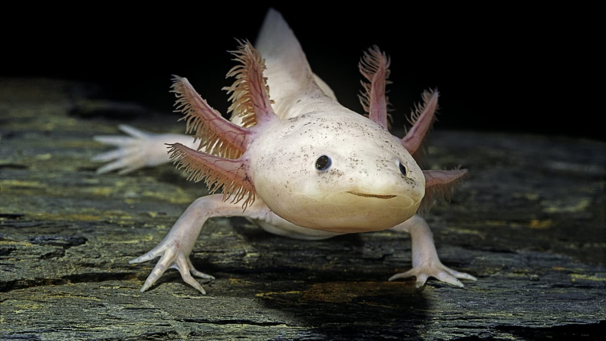
A recent paper by Otsuki and colleagues investigates the molecular mechanisms driving limb regeneration in axolotl
The question
You may know the axolotl (Ambystoma mexicanum), its funny face and gills floating around its head.
What you may not know is that it is also a model organism for organ regeneration thanks to its ability to regenerate many body parts, including its limbs. This is possible because cells know and remember where they are and can use this knowledge to inform the regeneration process. Cells found at the front of the limb possess so-called anterior identity, while those at the back hold posterior identity information. After amputation, cells from the anterior and posterior parts of the stub meet and trigger correct limb regeneration.
But how do cells know to produce a new limb after limb amputation, and not a tail or head instead?
The molecular bit
A recent study by Otsuki and colleagues1, highlighted in a News & Views article2, investigates the process of limb regeneration in axolotl through transgenic lines, transcriptomics and grafting experiments.
Otsuki and colleagues found that posterior identity in axolotl is established and maintained by a positive feedback loop that involves Hand2, a protein that controls the expression of other genes, and Shh (Sonic hedgehog), a signalling protein involved in limb growth. During development, Hand2 is expressed in posterior cells, and it is present at a steady state in adults. During regeneration, Hand2 is necessary and sufficient to induce the expression of Shh, which in turn activates Hand2 expression in nearby cells, sustaining the establishment of posterior identity in the new limb. After regeneration, Shh expression stops but residual Hand2 ensures lasting positional memory.
The unexpected discovery and why it matters
Interestingly, Otsuki and colleagues were able to rewire anterior-posterior memory, but only during regeneration and in one direction: anterior cells can stably acquire posterior identity when placed in posterior zones (or upon transient Shh signalling), but the opposite leads to defective limb regeneration.
The results presented by Otsuki and colleagues represent an important step forward in the understanding and manipulation of organ regeneration, and future studies into therapeutic applications in humans will benefit from this important work.
References
1. Otsuki, L., Plattner, S. A., Taniguchi-Sugiura, Y., Falcon, F. & Tanaka, E. M. Molecular basis of positional memory in limb regeneration. Nature 1–9 (2025) doi:10.1038/s41586-025-09036-5.
2. Wu, S. Y. C. & Whited, J. L. How axolotl cells ‘remember’ development to rebuild a lost limb. Nature d41586-025-01447–8 (2025) doi:10.1038/d41586-025-01447-8.
The Kahneman Chronicles #1: When a Nobel Laureate Fixed Our Lab’s Scheduling Disasters
Daniel Kahneman (1934-2024) was a legendary psychologist who revolutionized our understanding of human decision-making and became known as the “grandfather of behavioral economics.” Awarded the 2002 Nobel Prize in Economics, Kahneman’s groundbreaking research with Amos Tversky revealed systematic biases and mental shortcuts leading people to make irrational choices.
This article series imagines what would transpire when Daniel Kahneman took a sabbatical and worked in a fly lab. Part of “The Kahneman Chronicles: Lessons from a Fly Lab” – A report from our imaginary interdisciplinary fellowship program
On the day Nobel Laureate Prof. Daniel Kahneman arrived for his sabbatical, our Drosophila lab buzzed with nervous excitement. Here was the legend himself—extraordinary psychologist who’d won economics’ highest prize, revolutionizing our understanding of errors in decision-making.
The ghost of Thomas Morgan urged us to do our best. We’d prepared our most impressive experiments, polished our presentations, and practiced our pitch for explaining fly development.
What we hadn’t prepared for was Kahneman spending his entire first morning silently observing us work. Often he scribbled notes in a small black notebook with the focused intensity of Jane Goodall studying chimps.
Why do we spend so much time in the lab?
“I’ll just quickly mount these embryos—twenty minutes, tops,” announced postdoc Shweta. This became a two-hour odyssey involving broken coverslips and dried glue. Followed by an existential crisis, wondering if the fluorescent blob she saw was signal or autofluorescence from a properly developing embryo.
“Quick PCR setup, maybe thirty minutes,” declared grad student Fillip, before vanishing into an afternoon-long quest. Missing primers. Buffer math. Finding the thermal cycler waited on “infinite hold” since previous Tuesday. You know the drill.
“Fascinating,” Kahneman murmured after each wildly inaccurate prediction.
By day three, a pattern was undeniable. Every time estimate in our lab was spectacularly yet consistently wrong. “Simple” tasks morphed into epic quests.
The Intervention
Kahneman approached the whiteboard where we’d sketched our weekly schedule – optimistically planning seventeen different experiments into forty work hours.
“Let’s implement realistic time budgeting,” he announced with the calm authority of someone who’d spent decades studying how humans delude themselves. Our simple thirty-minute embryo injections were now allocated one-hour blocks.
The room erupted in protests. “But we’ve done these injections hundreds of times!” “We know exactly how long they take!”
Kahneman smiled. “You’re all victims of the planning fallacy. Your System 1 is wildly optimistic about everything. Your mind accounts only for quick needle preparations while forgetting inevitable moments someone drops the cover slip itself”
“Your intuitive mind,” he explained, “only remembers the core task—actual injection. It conveniently forgets the setup, troubleshooting, inevitable equipment malfunction, and time spent staring at embryos wondering if they are worth injecting at all.”
The Planning Fallacy: The tendency to underestimate time, costs, and risks of future actions while overestimating their benefits. Even when people know similar tasks have taken longer than expected in past, they still predict future tasks will take less time.
System 1 vs. System 2 : Kahneman’s framework for two modes of thought. System 1 is fast, automatic, and intuitive (like quickly estimating “this should take twenty minutes”). System 2 is slow, deliberate, and logical (like carefully calculating each step: needle prep, embryo collection, injection setup, actual injection, cleanup, and imaging).
The Kahneman Method in Action
His solution was deceptively simple: multiply every time estimate by two, then add buffer time for “unknown unknowns.” “There are things you know will probably go wrong—known unknowns, like occasional broken needle or contaminated sample,” he explained.
“But then there are unknown unknowns—the completely unexpected problems you can’t even anticipate. The incubator that dies on a weekend, the new batch of reagent that behaves differently, or the day your hands just won’t stop shaking. You can’t plan for specific unknown unknowns, but you can acknowledge they exist.”
He made us track everything for two weeks: actual injection times, PCR setup duration…and the data was humbling. Our “standard” twenty-minute procedure had a median time of 40 minutes, with some taking over 1.5 hours when equipment misbehaved.
We tried his interventions skeptically. To our disbelief, the results were miraculous and maddening in equal measure.
For the first time in lab history, experiments actually finished when scheduled. Postdocs stopped working until midnight to complete “quick afternoon experiments.” Stress level plummeted as people stopped running late for their next commitment.
“Your emotional attachment to each experiment makes you treat it as special,” Kahneman explained. “You think ‘this time will be different’ or ‘I’m more prepared now.’ But from a statistical perspective, today’s PCR is just another data point in the distribution of ‘times PCR has taken in this lab.’ The planning fallacy tricks you into believing you can beat the historical average through wishful thinking.”
The lesson was profound: scientists are ultimately human and prone to same cognitive biases that affect everyone else. We bring these same mental shortcuts to our labs, our experimental designs, and our data interpretations. The first step toward better science maybe a more nuanced use of an important equipment—our own minds.
Have you experienced similar planning fallacies and overcome them? Do share in the comments.
What else did the Prof. Kahneman advise us on? Stay tuned for the next article in the series.
Practical Applications: The Kahneman Time Revolution
1. Track Reality First: Record actual times for routine procedures for couple of weeks.
2. Use the 1.5x Rule: Multiply routine task estimates by 1.5.
3. Use the 3x Rule: Triple your estimate for novel experiments.
4. Build Break Points: Schedule natural stopping points in long experiments, to allow buffers for unknown unknowns.
5. Try the Three-Point Method: For familiar tasks, estimate your best-case time (everything goes perfectly) and worst-case time (multiple things go wrong). Then calculate the geometric mean (root of the product) for a realistic schedule estimate.
Example: Embryo injection times (Best case 20 minutes, worst case 1 hours), geometric mean√(20 × 60) = √1200 ≈ 34 minutes.
Sameer Thukral is a post doc in the lab of Yu-Chiun Wang at RIKEN-BDR, Kobe, Japan, where he loves discussing science in the healthy and respectful lab environment. He is a developmental biologist with a focus on mechanics of yolk-blastoderm interactions. He is also the co-founder of BDR-Launchpad, a post-doc network for supporting ECRs with the hidden curriculum of science.
The observations made here are his own and do not reflect the opinions of the employer. This article was written by Sameer Thukral, with formatting, structuring and framing support of Claude AI.
I am an avid podcast listener, especially at the gym. Instead of fueling my reps with anger-fueled lyricism or upbeat songs that raise my bpm to 120, I noticed feeding my brain with podcasts is a better way of enduring my hour and a half workout. After all, what is a better way to feel incentivized to squat to 60 kg than to listen about how women have been overlooked in science? In the recent season of the So Cultured podcast, tears of anger fell down my face at the cruel injustices faced by Brenda Milner, Tu Youyou, and Marie Curie. Besides anger, I also felt inspired. Worried about my own academic journey, wondering if I am good enough, I found comfort in their stories.
Conducting my own investigation, wondering whose story is not largely known, I bumped into Hilde Mangold (or Hilde Proescholdt at the time) and “The Organizer.” During her doctoral studies, she used two species of salamander with embryos that differ in pigmentation to perform 259 transplantations of the blastopore’s dorsal lip into the ventral region of a host gastrula1, 2. This experiments catapulted Spemann for the Nobel Prize. In several significant cases, she observed that a secondary axis with neural system developed in the host embryos 3. Spemann and Hilde Mangold therefore concluded that the embryonic region of the dorsal blastopore lip was able to induce embryonic development and called this specific embryonic region an “organizer.”4
Hilde’s trajectory was impressive. At the age of 16, she attended the prestigious Gymnasium Ernestinum, at the time almost inaccessible to girls1. But like most young girls at the time, she was sent to a private institute for young ladies to learn proper housekeeping and social etiquette right after. But her curiosity and intelligence granted her a place at the University of Jena, which eventually led her to Spemann. She possessed a great deal of skill. Perhaps this dexterity to perform and master minuscule surgical operations could be attributed to the time she spent sewing in school. In fact, she had a keen eye for detail, documenting her implants with drawings in her lab notebook5, a skill that may be lost in the upcoming years with digitalization.
Although it was Spemann’s quest started in 1903, with the production of identical twins from newt embryos using his daughter’s hair loop6. The completion of the pusruit to find the said “organizer,” couldn’t have been done without Hilde. She expanded Spemann’s techniques. Used thin glass needles, often heated, to cut certain parts from the embryos or to burn them away. She was critical in providing the empirical evidence needed, imagine your dissertation helps your professor win a Nobel. And aside from prizes, this experiments were monumental for the time, forging paths for theoretical and developmental biology, and cell to cell communication.
Hilde’s story was short lived. Now I wonder what would’ve she further achieved is were not for dying at the young age of 26 from an accident. The best we can do is to continue living for them, and as other women at the time, to persist and lead by curiosity and resilience. Perhaps our time as graduate students are not as fruitful as Hilde’s, still, as students we understimate the work we do in the lab, and forget to advocate for our contributions. We troubleshoot for months, have failed results, or no results at all. So although we must perform the 200 experiments, we must celebrate and give some grace even if only 6 are significant. At the end, we are in fact, the sum of our parts.
References
VAN Robays, J. (2016). Hilde Mangold-Pröscholdt (1898 – 1924): The Spemann-Mangold Organizer. Facts, Views & Vision in ObGyn, 8(1), 63–68.
De Robertis, E. M., Driever, W., & Mayor, R. (2024). Celebrating the centennial of the most famous experiment in embryology: Hilde Mangold, Hans Spemann and the organizer. Cells & Development, 178, 203921. https://doi.org/10.1016/j.cdev.2024.203921
Kumar, V., Park, S., Lee, U., & Kim, J. (2021). The Organizer and Its Signaling in Embryonic Development. Journal of Developmental Biology, 9(4), 47. https://doi.org/10.3390/jdb9040047
Spemann, H., & Mangold, H. (1924). Über Induktion von Embryonalanlagen durch Implantation artfremder Organisatoren. Archiv für Mikroskopische Anatomie und Entwicklungsmechanik, 100(3–4), 599–638. https://doi.org/10.1007/BF02108133
Driever, W., Holzschuh, J., Sommer, L., Nitschke, R., Naumann, A., Elmer, J., & Giere, P. (2024). Hilde Mangold: Original microscope slides and records of the gastrula organizer experiments. Cells & Development, 178, 203909. https://doi.org/10.1016/j.cdev.2024.203909
De Robertis, E. M. (2006). Spemann’s organizer and self-regulation in amphibian embryos. Nature Reviews Molecular Cell Biology, 7(4), 296–302. https://doi.org/10.1038/nrm1855
Brandt, C. (2022). Vitalism, Holism, and Metaphorical Dynamics of Hans Spemann’s “Organizer” in the Interwar Period. Journal of the History of Biology, 55(2), 285–320. https://doi.org/10.1007/s10739-022-09682-9
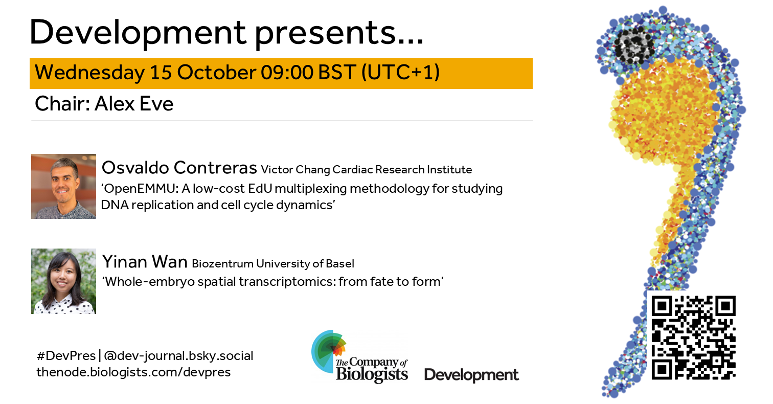
Join us in mid-October to hear from two early-career researchers working on development across scales. Chaired by Development’s Executive Editor, Alex Eve.
Wednesday 15 October – 09:00 BST (UTC+1)
Osvaldo Contreras (Victor Chang Cardiac Research Institute and UNSW) ‘OpenEMMU: A low-cost EdU multiplexing methodology for studying DNA replication and cell cycle dynamics’
Yinan Wan (Biozentrum University of Basel) ‘Whole-embryo spatial transcriptomics: from fate to form’
At the speakers’ discretion, the webinar will be recorded to view on demand. To see the other webinars scheduled in our series, and to catch up on previous talks, please visit: thenode.biologists.com/devpres
I’m a big fan of podcasts, and one of my favorites is Tim Harford’s “Cautionary Tales.” It tells true stories about disasters and what we can learn from them. One episode particularly stuck with me—the story of Tenerife.
On March 27, 1977, two Boeing 747s collided on a foggy runway in Tenerife, killing 583 people. This wasn’t just about miscommunication, mechanical failure or bad weather. The investigation revealed something more profound: the captain was the airline’s Chief Flying Instructor, thus creating a steep “gradient” that prevented his first officer from challenging a fatal mistake.
When the first officer realized they didn’t have takeoff clearance, he saw disaster coming but couldn’t bring himself to forcefully challenge his superior.
This got me thinking: when power dynamics prevent people from speaking up, disaster follows. Does this also apply to academia?
A Pattern Worth Noticing
Tim Harford’s podcast reveals a disturbing pattern: many disasters across different fields stem from the same problem—people being unable to challenge authority when they see danger ahead. From naval catastrophes to medical errors, from financial crashes to engineering failures, a common thread is often authority gradients that silence dissenting voices.
To be clear, most academic labs aren’t disaster zones. Most PIs, including my own, are thoughtful mentors who genuinely care about their students’ growth and scientific development. Many labs operate with healthy dynamics where ideas flow freely and disagreement is welcomed.
But here’s a learning from other fields: even well-intentioned leaders can unknowingly create subtle power imbalances. And in science, our “disasters” aren’t plane crashes—they’re missed discoveries, delayed projects, unexplored hypotheses, and brilliant ideas that never see daylight.
The Academic Context
In academia, unlike most corporate environments, one person—your PI—has enormous influence over your career trajectory. As a PhD student or Post-doc, you commit years to one supervisor’s lab. They guide your research direction, allow your access to resources, and significantly influence your future opportunities.
This isn’t inherently problematic. Expertise matters, and experienced scientists rightfully guide newcomers. The challenge is when this necessary hierarchy inadvertently creates barriers to open scientific dialogue.
Even in the best labs, there might be subtle versions of this dynamic. A student hesitating to present data that contradicts the PI’s hypothesis. A postdoc avoiding questions that might seem to challenge established lab protocols. These aren’t dramatic confrontations—they’re quiet moments where respect for authority might overshadow respect for scientific inquiry.
The Free Resource We Maybe Missing
Of all the things science needs—expensive equipment, ample funding, and reagents—respect, costs nothing. Yet, it might be our most powerful tool. Every carefully planned experiment and every piece of expensive equipment depends on people thriving in an environment where they feel safe, heard, and valued.
Science thrives on disagreement. The best discoveries often come from questioning prevailing wisdom and challenging assumptions. But when subtle power dynamics make people hesitate to speak up, we miss out on breakthrough ideas.
The most productive labs may be doing something simple: they separate intellectual discussion from hierarchy. In these labs, everyone responds to contradictory data with curiosity, not defensiveness. Unexpected results are seen as learning opportunities, not failures.
A Quick Self-Check for the Lab
As an opportunity for reflection, PIs and mentees can ask themselves:How often do mentees feel comfortable disagreeing with an idea? If it’s rare, it may be worth examining why. Perhaps even create a “disagreement board” to make the act of questioning a hypothesis more salient and celebrated. What’s the atmosphere like when someone presents data that contradicts an expectation? Do people feel comfortable sharing results that go against the grain?
These aren’t accusations; they’re simply opportunities for growth and improvement. The goal isn’t to flatten hierarchies but to ensure that authority serves discovery, not ego. Sometimes, the most junior person in the lab has a game-changing insight. But they can only share it if they feel safe to do so.
The bottom line
Listening to cautionary tales from other fields reminded me that power dynamics are everywhere, often subtle, and worth examining. In science, where truth-seeking is our highest goal, creating space for respectful disagreement isn’t just good mentorship—it’s essential for discovery.
Sameer Thukral is a post doc in the lab of Yu-Chiun Wang at RIKEN-BDR, Kobe, Japan, where he loves discussing science in a healthy and respectful environment. He is developmental biologist with a focus on mechanics of yolk-blastoderm interactions. He is also the co-founder of BDR-Launchpad, a post-doc network for supporting ECRs with the hidden curriculum of science.
The observations made here are his own and do not reflect the opinions of the employer. This article was written by Sameer Thukral, with formatting, structuring and framing support of Claude AI.
Our ‘Featured resource’ series aims to shine a light on the resources that support our research – the unsung heroes of the science world. In this post, we learn about the data and functionalities available at Facebase, and hear about new initiatives they are developing.
What is FaceBase?
FaceBase is a public data resource and repository dedicated to advancing basic and clinical research spanning the translational spectrum of dental, oral, and craniofacial (DOC) biology, as well as related systemic health and disease models throughout the data lifecycle. FaceBase realizes this mission by recruiting, transforming, and publicly sharing research and clinical data.
This freely available and public resource currently hosts over 1,100 datasets, approximately 3,000 experiments, over 210,000 images, and more than 8,000 genomics files. FaceBase exemplifies FAIR (Findability, Accessibility, Interoperability and Reusability) and TRUST (Transparency, Responsibility, User focused, Sustainability, and Technology) principles of scientific data sharing, ensuring that its clean, well-structured datasets are not only easy to find and reuse, but are also inherently AI-ready for integration into modern computational workflows.
FaceBase hosts data from both human subjects and animal models, encompassing a wide array of experimental approaches, including multiple omics and imaging data types. This platform welcomes contributions of data from the community after going through a careful review process and quality assurance.
Human subjects and animal model data (Current animal models include mouse, zebrafish, chimp and chick)
Controlled-access and public data
Genomic and phenotypic data from multiple species
Most known types of genomics and imaging data
Resources and strategies to enhance data reproducibility
State-of-the-art data science methods to support cutting edge research
Standards and educational resources for improving data management and sharing practices across the community
FaceBase demonstrated itself as a credible resource for the DOC research community through its CoreTrustSeal accreditation after a two-year approval process, as well as becoming one of a select number of NIH approved Controlled Access Data Repositories (CADRs) handling genomics and other sensitive data.
What inspired the development of FaceBase?
In 2009, National Institute of Dental and Craniofacial Research, National Institutes of Health (NIDCR, NIH) launched FaceBase in response to the need for more comprehensive analysis of craniofacial development. With the immense amount of craniofacial data being generated, there is a danger of relevant datasets being buried in the avalanche of genomic and other data.
The first five years (known as FaceBase 1) started with a spoke-and-hub of 10 spoke projects and resulted in almost 600 datasets and over 100 publications. The next phase of FaceBase (FaceBase 2) began in August 2014 with 10 spoke projects and a new hub that developed an updated data model allowing for more data integration and faceted searches with a new server interface. The third phase (FaceBase 3) dismantled the spoke-and-hub model in favor of a community-based model that opened submissions to any contributor. We also promoted the idea of self-curation which allowed us to scale up considerably: since opening up to community contributions, we have more than doubled the number of contributors and our dataset growth has kept pace with prior years.
How can scientists use FaceBase in their research?
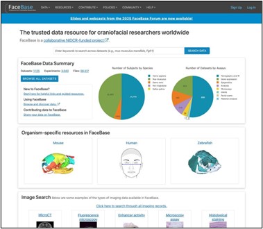
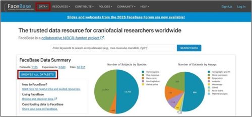
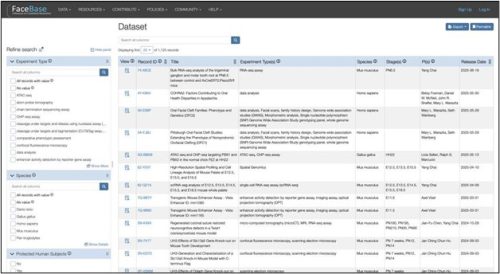
For researchers and clinicians seeking to generate a hypothesis for a new grant, validate their own data by comparing with controls, or examine phenotypes in mutant models, the FaceBase Data Browser provides an intuitive interface. Data are represented as filtered records, with sidebar attributes that function similarly to filters on an online shopping site.
Find Data
You may begin your search with the BROWSE ALL DATASETS button on the homepage or you can use the DATA tab in the top navigation menu bar (available on all pages) to start with a particular model.
When you start searching the data browser you will see:
Search results based on filters
Faceted navigation sidebar on the left
Search bar above the results
By default, the data is sorted to display the most recently released data first. On the left side is the faceted navigation based on characteristics of the data and experiments. Scroll down to see all the categories of filters available to narrow down your search.
Export Data
All open access data can be downloaded directly from the browser without requiring login. If you want to download a large amount of data, you can use our BDBag protocol-derived tool, which allows for reliable transfer of a “bag” of digital content – in this context, a group of files that you want to export in bulk. It is available as a GUI client and a command-line client.
We also offer resources to help you include FaceBase in your Data Management Sharing (DMS) plan, including template text that you can copy and paste into your plan. You can find guidance on how to fill out the various fields here: https://www.facebase.org/contributing/dms/.
Who are the people behind the resource?
FaceBase is run by University of Southern California’s Center for Craniofacial Molecular Biology (CCMB) and Information Sciences Institute (ISI) in Los Angeles.
Our current leadership and staff include:
• Principal Investigators – Yang Chai (CCMB) and Carl Kesselman (ISI)
• Co-Investigators – Robert Schuler (ISI, technical lead) and Parish P. Sedghizadeh (Herman Ostrow School of Dentistry of USC)
• Scientific Curators – Jifan Feng, Tingwei Guo, and Thach Vu Ho (CCMB)
• Data Management Lead – Alejandro Bugacov (ISI)
• Collaborations and Communications Coordinator – Cris Williams (ISI)
• Project Manager – VyVy Nguyen (CCMB)
How can researchers help and contribute to the resource?
The most effective ways to support FaceBase are two pronged: 1) contribute data to improve the breadth and depth of our offerings and 2) cite any data you deposit or reuse by using the citation tools embedded in the platform.
Contribute data
FaceBase welcomes biomedical basic and clinical research across the translational spectrum related to the DOC domains as well as those from related systems. We are also an approved repository for the HEAL Initiative, an NIH-wide effort to speed scientific solutions to stem the national opioid public health crisis.
Our current funding phase expands our focus to accept research and data on relevant anatomical and biological health and disease models beyond DOC domains, for example the ear and eye or biomarkers that overlap with those found in DOC regions.
After a review process from the FaceBase team and NIH program staff, approved projects will receive a one-hour one-to-one tutorial to learn how to curate their data using the online metadata forms and how to upload data. You can find more information about the process here: https://docs.facebase.org/docs/Data-Submission-Key-Concepts/.
Note that our focus is on high quality data that conforms to FAIR initiatives that bolster or expand existing data. Find more detailed descriptions of the types of data we are especially interested in here: https://www.facebase.org/contributing/data-priorities/. If you have any questions about whether your data is a good fit, please contact us at help@facebase.org.
Cite FaceBase data
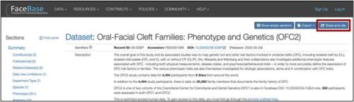
FaceBase has been leading the charge on effective and transparent citation of data for many years. Every data record has its own unique, permanent identifier. In addition, every Dataset and Project page has a registered Digital Object Identifier (DOI) and a “Share and cite” button that provides citation text that you can simply copy and paste into your publication.
EarBase: As part of our new focus to include research and data from relevant anatomical and biological health and disease models, FaceBase is collaborating with the National Institute on Deafness and Other Communication Disorders (NIDCD) to migrate 3D images of the temporal bone that were previously held in a private enclave.
CranioRate: Another new development is our collaboration with CranioRate, a user interface that is being launched in late 2025 to help surgeons and clinicians manage metopic craniosynostosis cases, a birth defect that affects the structure of the skull. In particular, FaceBase is supporting their open access human craniosynostosis image bank and working towards standardized vocabularies and ontologies to ensure the data’s FAIR-ness.
Integrating clinical elements from Electronic Health Records (EHR)
We are collaborating with clinician-scientists on a pilot project to integrate clinical data from patients with temporomandibular disorders (TMD) into FaceBase. Important directives of this pilot include ensuring clear patient consent for repository use (that specifically permit the use of identifiable health information for research without requiring re-authorization) and exploring the potential of AI/ML methods to analyze clinical notes and improve diagnostic accuracy.
Advanced computation and AI-ready analytics
By definition, aligning the data in the FaceBase repository with FAIR principles means that our data, which is clean, well-formatted, with structured metadata and provenance, is ready for a data scientist to pull into analytics platforms. In the future, we plan to continue to enhance the AI-readiness of our data, provide curated collections of “reference datasets” for training purposes, and enable interoperability with LLMs and lab notebooks and develop an AI-assisted curation bot for data contributors.
Interoperability with external data resources
We are also developing a pipeline to transform raw FaceBase data into a processed format that can be ingested by external resources, for example a cloud-based analytics platform.
We are all stepping into a story where evolution, development, and regeneration converge in the eye of a snail.
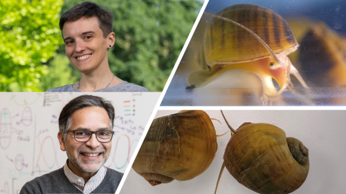
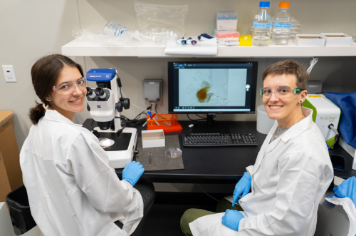
Portraits of Dr. Alice Accorsi and Dr. Alejandro Sánchez Alvarado, shown alongside the apple snail, Pomacea canaliculata. Image source : Alice Accorsi and Joaquin Benitez, College of Biological-Sciences, UC Davis and Stowers Institute for Medical Research.
Throughout their lives, organisms encounter injuries and stresses that threaten the integrity of their bodies and have evolved remarkable ways to restore lost or damaged tissues. This ability to replace body parts, which can range from reorganizing existing structures to generating entirely new ones—is known as regeneration.
Among many forms of regeneration, the ability to rebuild eyes is especially striking. Eyes are among the most intricate organs, requiring precise anatomical organization and highly ordered neural wiring to restore function. Across the animal kingdom, eyes vary widely, reflecting adaptation to different ecological demands. While regeneration of simpler structures, such as planarian pigmented eye cups, and partial regeneration of camera-type eyes in vertebrates has been described, the idea that complete adult camera-type eyes could regenerate has long seemed improbable. These highly specialized organs, capable of high-resolution vision, present unique challenges that extend beyond conventional models.
In a recent groundbreaking Nature Communications study, Alice Accorsi, Alejandro Sánchez Alvarado, and colleagues demonstrate that the apple snail, Pomacea canaliculata can completely regenerate its camera-type eyes. By coupling this discovery with CRISPR–Cas9 genome editing, they establish a new genetically tractable model to probe regeneration of complex sensory organs. Here are behind the scene stories from the corresponding authors – Dr. Alice Accorsi and Dr. Alejandro Sánchez Alvarado.
First we have behind the science stories from Dr Alice Accorsi !
How did you first get introduced to apple snails, and what drew you to them? Tell us about your PhD work.
Throughout my career I have worked with several invertebrate species, such as snails, leeches and planarians. These apple snails are originally from South America, particularly Brazil and Argentina, but have now spread to parts of Asia, Europe, and North America, where they pose a serious threat to local ecosystems. The same traits that make them invasive, such as resilience, rapid growth and prolific reproduction, also make them easy to care for. And it turns out this also makes them excellent laboratory models. My PhD mentor, Dr. Enzo Ottaviani, once purchased some apple snails from a pet shop and had them in his office. It was during one of our meetings that we wondered if we could use them as another invertebrate in my research! During my graduate studies, I was interested in studying their immune system to understand what makes them so resilient and to explore ways to affect their survival without using environmentally harmful compounds. I was also intrigued by the possibility that their immune and nervous systems might communicate with each other, as we see in vertebrates. My research uncovered evidence of this crosstalk, offering a new evolutionary perspective on neuroimmune interactions.
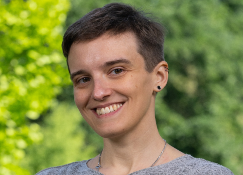
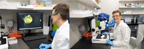
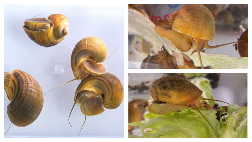
Pictures featuring Dr. Accorsi visualizing the apple snail. Image source : Joaquin Benitez, College of Biological-Sciences, UC Davis.
What convinced you to keep working with snails in your research – even during post doc and now in your independent research program as a faculty? What led you to the Sánchez Alvarado lab?
This journey began with a conversation between Dr. Alejandro Sánchez Alvarado (Stowers Institute for Medical research, Kansas City, MO) and me at the Marine Biological Laboratory in Woods Hole, MA. I was still a graduate student at the time, studying the immune system of apple snails, while Alejandro’s laboratory was focused on regeneration in planarians. Although snails have been known for their regenerative abilities since the 1700s, no one had explored their biology using modern molecular tools. That conversation sparked my interest in applying these approaches to snails to see what we could uncover. We already have several model systems that excel at regenerating different body parts, such as planarians, hydras, and axolotls. I began to wonder whether these snails could regenerate an organ that the others could not, making them unique and even more relevant to study. That is when I discovered that apple snails possess complex camera-type eyes, the same kind of eyes found in humans. This opened up a unique opportunity to explore regenerative biology in a new way, with potential implications for human health. That is what convinced me to continue working with snails, even as I transitioned into postdoctoral and now independent research.
How was your transition from Italy to US for postdoctoral work?
Moving abroad for my postdoctoral studies was a major life change. I left my family behind and immersed myself in a new culture and scientific environment. I moved from a small lab with limited resources where I was the most senior member to the Stowers Institute for Medical Research, a place with nearly unlimited possibilities and a large, diverse team of scientists, including many senior researchers. Despite the challenges, I never regretted the move. I learned more than I ever imagined and had the chance to connect with scientists across the country and the world. The Technology Centers at Stowers supported my work and introduced me to techniques I had only read about before. I am deeply grateful for the preparation I received through the Italian educational system, which gave me the foundation to take this leap.
What was it like to take on eye regeneration in snails – a phenomenon that hadn’t really been studied in them before?
Taking on a project about complete eye regeneration in snails was both exciting and challenging. Since this phenomenon had not been studied before and this was a relatively novel model system, we had to start from scratch. We began by characterizing the morphology of apple snail eyes using microscopy and histological techniques to understand their structure and cellular composition. Then, we performed genomic and transcriptomic analyses to identify the genes involved in eye development and regeneration. Finally, we developed techniques to manipulate their genome to test gene function. This multi-approach research allowed us to build a comprehensive picture of apple snail eye anatomy, gene expression and regeneration, laying the groundwork for deeper investigations into the molecular mechanisms behind this process.
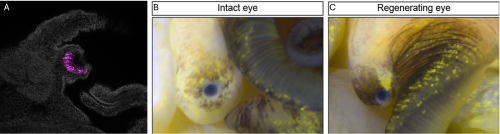
Images showing A) embryonic snail eye with fluorescent photoreceptor cells in magenta, B) and C) showing intact and regenerating adult eye respectively. Picture credits : Alice Accorsi, College of Biological-Sciences, UC Davis.
Your genomic analyses revealed genes shared between apple snails, humans, and Drosophila, particularly related to eye development and photoreceptor formation. What does this shared genetic toolkit tell us about the evolution of complex eyes across distant lineages?
Our molecular studies revealed that many genes are involved in forming both snail and human eyes, even though these eyes evolved independently. This suggests that, while there may be many ways to build an eye, the fundamental genetic building blocks are conserved between very different species (humans and snails). These findings have important implications for evolutionary biology. By comparing the development of camera-type eyes in snails, cephalopods, and humans we can shed light on how these complex structures evolved multiple times independently. This helps us identify both conserved mechanisms and evolutionary novelties across species.
Can you describe the moment you first saw a regenerated camera type eye?
Seeing the regenerated eye for the first time was exciting, but in that moment, I was not even close to fully grasp the importance of that one piece of data. It was later on, reading literature and looking through old papers and I started appreciating how this unconventional system could reveal something truly profound about regeneration. That realization was the real turning point that deepened my commitment to this research.
Your experiments showed eye regeneration unfolded in defined stages—wound healing, blastema formation, tissue emergence, and maturation. Did any of these phases surprise you ?
One of the most remarkable aspects of apple snail eye regeneration is how fast, precise, and reproducible it is. After complete eye removal, early signs of regrowth appear in less than two weeks, and a fully reconstructed eye, with all its components, is restored in under a month. What surprised me most was the efficiency and consistency of this process. The speed at which regeneration unfolds, and the minimal variability between individuals, suggest a tightly regulated mechanism. Just as striking was the discovery that many of the genes active during regeneration are also involved in vertebrate eye development. This points to a shared genetic toolkit and opens exciting possibilities for comparative studies that could inform regenerative medicine.
Your pax6 studies reaffirmed its conserved function, do you think the role of pax 6 is binary ?
In our system, pax6 appears to play a binary role. When pax6 is knocked out, eye development is completely abolished. We did not observe any eye-related structures or any intermediate phenotypes, which underscores how essential this gene is. It is astonishing to see such a conserved function across species.
Do you plan to test the behavioral capabilities of regenerated eyes?
Absolutely. One of our main goals moving forward is to study the behavior and visual capabilities of apple snails. We are planning to collaborate with labs that specialize in behavioral neuroscience and vision to explore what snails can see in their environment and how well regenerated eyes can function.
What challenges did you face developing CRISPR lines?
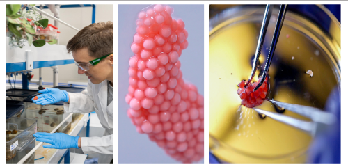
Establishing stable CRISPR/Cas9 mutant lines in snails was a major technical challenge. A few steps were quite difficult. The first was collecting and injecting the zygotes, as they are very small! The next difficult step was ensuring their survival to adulthood after we removed them from the eggs. It took a lot of trial and error. Each step required patience and precision, but eventually, we developed a reliable workflow that allowed us to generate reproducible mutant phenotypes.
How do snails complement fly eye development in other model systems like Drosophila?
While Drosophila has been a powerhouse for studying eye development, its compound eyes are anatomically different from human eyes. Moreover, adult fruit flies do not regenerate their eyes after injury. Apple snails, on the other hand, have camera-type eyes, just like us, and can regenerate them completely. This makes apple snails a powerful complementary model. Their regenerative abilities, combined with shared genetic pathways, offer a unique window into how complex organs can be rebuilt. Studying molecular pathways involved in eye formation and function across such diverse species helps us identify conserved mechanisms and evolutionary innovations, expanding our understanding of how regeneration evolved.
What was your most validating moment in this project?
The most emotional moment of this project was when I obtained pax6 homozygous mutants. I looked in the microscope without daring to hope for anything special. But after getting the embryos in focus, I saw that some of them did not develop eyes. That was the moment I knew CRISPR/Cas9 was working and the function of the gene pax6 was conserved in apple snails. It was incredibly validating and empowering. That was the moment when I truly felt I could start thinking about “the rest of my scientific career” as the leader of a lab using apple snails to study eye regeneration.
Can you share some challenging moments from the project. What were your ways to reset/unwind ?
One of the biggest challenges of this research was figuring out how to collect, inject and raise snail embryos to adults. This was a long, slow and meticulous process. I spent hours carefully observing embryos trying to pinpoint what was not working and letting the biology guide the adjustments. I for sure learnt patience and resilience through this process. Outside the lab, I love to do yoga, listen to audiobooks and spend time with the people I love. These moments help me recharge and return to the lab with fresh energy.
Were there any quirky moments that shaped the trajectory of the study?
A quirky moment that shaped not just this study but of my entire career happened during graduate school. I was so excited about regeneration after attending the MBL Embryology Course in Woods Hole that I immediately wanted to test if the apple snails I was working on were able to survive injuries and regenerate. I got dissection scissors and… well, luckily for me and for them, they regenerated!
How did your team coordinate such a complex study?
At Stowers, I had incredible support from the Technology Centers, which helped optimize protocols, run experiments and maintain the snails. At UC Davis, we also have excellent core facilities for imaging and sequencing, but the members of my lab play a central role in all the work that we do. I encourage everybody on my team to learn all aspects of research, from animal husbandry, to sample processing and data analysis. Through this approach I aim to foster collaboration and independence.
What big questions are you excited to explore next?
Pictures featuring Dr Accorsi alongside graduate student Annika Patel. Check out the Accorsi lab web page to know more about the lab and the exciting ongoing research. Image source : Joaquin Benitez, College of Biological-Sciences, UC Davis
Some of the key questions I hope to answer about apple snail eye regeneration revolve around uncovering the fundamental biological mechanisms behind this remarkable process. One major area of interest is identifying the specific cell types responsible for regenerating all the eye components: the retina, lens, and cornea. Understanding whether these structures arise from a shared pool of cells or from distinct cell populations is essential to understanding how such complex tissues are rebuilt. Equally important is exploring the genes involved in the regeneration process and how they are regulated. Dissecting these molecular circuits could reveal conserved pathways and highlight potential targets for biomedical applications. Another critical question is how neural connections between the regenerated eye and the brain are re-established. While regenerating the physical structure of the eye is impressive, full functional recovery requires precise reintegration into the central nervous system. Studying how apple snails accomplish this could provide valuable insights into nervous system regeneration. Finally, one of the most exciting prospects is the potential to identify specific genes or regulatory elements that can be tested in species lacking natural regenerative capacity. By comparing regenerative and non-regenerative systems, we may uncover key factors that could one day be harnessed to promote regeneration in humans.
Anything you’d like to highlight about your lab?
We are always interested in hearing from people who are excited about development, regeneration and snails and who would be interested in joining our team or collaborate with us! We highly value basic science, curiosity, creativity and community.
Now we have behind the science stories from Dr Alejandro Sánchez Alvarado !
You’ve pioneered much of what we know about planarian regeneration. What motivated you to pivot toward the apple snail? What were your initial plans when you and Alice started the project?
Curiosity has always driven my research. After years delving into planarian regeneration, I wanted to take the lessons learned and test their validity in other systems. I knew from the work of Charles Bonnet (Observations sur la Physique, sur l’ Histoire Naturelle et sur les Arts, vol. 10, Paris, 1777, in Tracts on the Natural History of Animals and Vegetables, 2nd, ed., vol. II, Edinburgh, 1803, plate 8, p. 360) that some snails could regenerate their heads after decapitation. Given that such a head included complex sensory organs such as camera type eyes, I was intrigued to see how much regeneration was possible in snails and thought of it as a great opportunity to test how far fundamental principles of regeneration extend beyond our favorite models. When Alice and I initiated the project, we aimed to develop the apple snail into a powerful system, one where we could explore not only eye regeneration but new rules for organ complexity and repair.
Having studied planarians extensively, what similarities and differences strike you most between their eye regeneration and what you observed in Pomacea canaliculata?
In planarians, eye regeneration is fairly direct, that is, the structure is simple, and the set of participating cells is relatively constrained. Apple snail eyes, in contrast, are much more anatomically elaborate: they possess a lens, cornea, and a retina. Despite these differences, we observed the employment of a surprisingly conserved genetic toolkit, yet the deployment is tailored to the organism’s needs and eye architecture. While planarians offer lessons in simplicity and robustness, snails challenge us to understand regeneration in complex, multi-tissue architectures.
As you said, snail eyes are highly organized with a lens, cornea, and retina. How did you approach regeneration of a complex organ? What were your reactions when Alice and the team showed you the eye regeneration phenotype? How did you celebrate?
We approached snail eye regeneration with a mix of excitement and humility. Knowing the added complexity, our first step was to characterize the anatomy and developmental processes in exquisite detail, as we’d done in planarians. When Alice showed me the early phenotypes (eyes regrowing with partial or complete restoration of layers) it was exhilarating. There was a sense of witnessing something extraordinary, something no one had seriously documented in this way before. We asked ourselves: if this is the wild type (eye regeneration) imagine what phenotypes will we get once we can begin to genetically perturb this process? We celebrated in true lab fashion: with data, good coffee, and a shared sense of purpose.
For the broader scientific community, how important it is to move beyond conventional systems towards models which are more “problem suited”?
Snail images. Image source : Stowers Institute of Medical research and Alice Accorsi, College of Biological-Sciences, UC Davis.
I believe science advances most meaningfully when we select models tailored to address questions, not just because they’re easy or fashionable. Apple snails forced us to reconsider mechanisms dogmatically ascribed to “higher” animals. For example, we unexpectedly found developmental modules acting outside canonical developmental windows, hinting at a flexibility in the animal’s response to injury or loss. Integrating these observations required both developmental and regenerative frameworks to be more plastic and open to revision. In essence, exploring unconventional systems not only expands our sense of what is possible in biology, but also reminds us, quite humbly, that we have yet to discover the full scope of what biology is already capable of achieving.
Across planarians, snails, and vertebrates, pax6 seems to act as a unifying thread in eye development. How do you see your work helping to connect these very different models into a broader evolutionary framework?
Pax6 is a beautiful example of deep homology: one gene at the crux of eye development in organisms as disparate as worms, snails, and humans. Our work allows us to chart the variations on a theme: the “melody” played by pax6, for example, shifts based on the “instrument.” This comparative approach helps trace evolutionary logic in how complex traits are built, lost, or re-invented, and fosters a more unified evolutionary understanding.
Was there a moment in this project that reminded you of your early planarian work—perhaps seeing the first signs of tissue re-emergence or recognizing a familiar gene playing a role in an unexpected context?
Absolutely. Seeing the initial re-emergence of eye tissue in snails, especially with familiar candidates like pax6 lighting up, evoked the earliest days in our planaria research. There’s a special thrill in spotting a familiar genetic face performing in a new “play.” These moments reinforce just how interconnected biology’s solutions really are. Perhaps more importantly, it presses us to recognize that, among countless possible outcomes, biology did not have to unfold in precisely this way, yet it did. The question, then, is why? What fundamental principles have shaped these solutions over evolutionary time, and might there be yet-undiscovered rules underlying these phenomena that the study of regeneration could help us uncover?
Do you imagine a comparative roadmap, linking regeneration in planarians, snails, and vertebrates, that might one day illuminate how regenerative capacity has been gained or lost across the tree of life?
One of my greatest hopes is for the field to embrace genuine comparative biology across multiple scales and levels of resolution—a comprehensive roadmap that interweaves regeneration in planarians, snails, vertebrates, and beyond. By charting where regenerative capacity is retained or lost, and probing the underlying reasons, we may finally decode the molecular signatures and constraints that shape these outcomes. This is an ambitious, long-term vision that traces its roots back to my earliest work (BioEssays, 22:578–590, 2000).
Read the paper to learn about a new protocol that enables collection of P. canaliculata zygotes and their ex ovo culture in perivitelline fluid extract — making it possible, for the first time, to observe embryonic development in real time. Images shown contain Alice showing the clutch collection process (the pink granular spheres forming a distinct speckled structure). Image source : UC Davis and Stowers Institute for Medical Research.
How do you look at processes of regeneration and development – where do they overlap, and where do they diverge?
Regeneration recapitulates development, sometimes literally, often figuratively. There are clear overlaps in gene regulatory networks and cell behaviors, but crucial divergences arise: injury response, aged tissue, functional integration of new tissues with old, and organismal context all shape outcomes. Examining both processes in parallel ensures our interpretations remain grounded and discerning, fostering an appreciation for both their commonalities and their distinctions.
You’ve mentored so many students and postdocs who have gone on to start their own labs and do incredible science. What is your mentoring philosophy?
Mentoring is, without question, the most rewarding aspect of this work. Science is inherently a human pursuit, and watching students and postdocs mature into independent thinkers is the ultimate measure of success. My approach centers on fostering autonomy, intellectual rigor, and genuine kindness. My greatest hope is that everyone who passes through my lab carries forward a deep sense of curiosity, confidence, and thoughtful skepticism wherever their careers take them. To me, choosing to mentor means embracing the responsibility to help cultivate scientists who will one day surpass us and, in doing so, move the field forward in ways we have yet to imagine.
Experiments don’t always work, and science can be frustrating. How do you help your students and trainees stay curious, motivated, and resilient during unfavorable circumstances?
I frequently remind my lab that failed experiments are the tuition we pay for discovery. I encourage tenacity by fostering a culture in which failures are shared, analyzed, and celebrated as learning opportunities. Curiosity is self-sustaining if it’s nurtured, and joy in small wins (finding a new phenotype, seeing cells behave unexpectedly) is kept front and center. It is important to emphasize that both true innovation and robust, lasting knowledge are built bit by bit, through careful testing, iterative refinement and the willingness to work patiently in the face of complexity, particularly when the prevailing winds conspire against such efforts. Our job as scientists is to contribute and continue to build a legacy of discovery that is as relevant tomorrow as it is today.
What do you find most awe-inspiring about nature’s capacity to regenerate, and how does that influence the way you think about biology?
To witness a fragment of an animal regenerate into a complex, living structure is to brush up against the truly profound. These moments evoke a sense of philosophical awe, as life reasserts itself with ancient, elegantly orchestrated mechanisms. Nature’s answers to damage and loss inspire both humility and an unshakable urge to understand how such feats are possible. In this light, every act of regeneration becomes a fresh retelling of an ancient narrative, one that has unfolded, again and again, across the history of life on Earth.
What continues to drive your curiosity and excitement about regeneration after all these years?
It’s the interplay of questions, the unexpected twists, and the pure delight in discovering something genuinely new. Regeneration is a frontier: every answer spawns new mysteries, and the joy of discovery, whether majestic or subtle, never fades.
A note from Shefali : I came across this beautiful research paper by Accorsi et al on bluesky and it literally blew my mind. It’s one of the rare times in the year when you stumble upon a piece of science that reminds you why you chose this path in the first place. As a grad student who is in the last leg of their PhD, it’s easy to to lose sight of the bigger picture – this paper brought it all back. I urge you all to read it—it’s rare and remarkable.Check out the Accorsi lab webpage and reach out if you’re interested in studying development and regeneration in snails.
 (No Ratings Yet)
 (11 votes)