After a pandemic related hiatus the MBL Gene regulatory networks (GRNs) course at Woods Hole is back. It’s been refreshed with new course directors and faculty and is better than ever. It covers experimental and computational methods used to study GRNs, through highly interactive lectures, discussions, group projects, and practical tutorials.
Gene regulatory networks (GRNs) are key to the genomic control of development in animals and plants. To study GRNs requires insights from various research fields, including systems biology, developmental and evolutionary biology, as well as functional genomics, and provides an integrative approach to fundamental research questions in biology. This course introduces the concepts of GRNs, and teaches experimental and computational methods used to study them, through highly interactive lectures, discussions, group projects, and practical tutorials. We will cover a broad range of topics, including transcriptional control systems, the structural organization of hierarchical networks, developmental functions of GRN circuit modules, GRN evolution, and computational modelling using BioTapestry as well as Boolean and quantitative mathematical approaches. Students will learn how to generate GRN models based on data extracted from the literature, and will generate computational models to analyze dynamic circuit behavior. We will present and discuss a broad range of experimental approaches and how they are effectively used for studying gene regulation and developmental GRNs. Examples of experimentally solved developmental GRNs from a variety of organisms, such as flies, sea urchins, frogs, chicken, and mice, will be explored. Students are encouraged to share their research projects in a poster session, and to discuss with course faculty how to apply the approaches taught in the course to their own research questions. The course is intended for advanced graduate students, postdoctoral scholars, and faculty.
Course Faculty and Lecturers Leslie Babonis, Cornell Sarah Tulin, Canisius College Crystal Rogers, UC Davis Scott Barolo, University of Muchigan Doug Erwin, Smithsonian Mark Rebeiz, University of Pittsburgh William Longabaugh, Institute for Systems Biology Paola Oliveri, University College London Roberto Feuda, University of Leicester Nipam Patel, Marine Biological Laboratory Zeba Wunderlich, Boston University Hui-Chun Lu (grace), University College London James Briscoe, Crick Institute Andrea Streit, King’s College London Megan Martik, UC Berkeley
In this latest SciArt profile, we spoke with Kimberley Snowden, who has a background in plant molecular and developmental biology and makes science-inspired polymer clay artwork.
Can you tell us about your background and what you work on now?
I have a background in plant molecular and developmental biology. I enjoy working out at a mechanistic level how plants get their very complex forms. In particular, I have worked on plant shape or architecture, and the molecular genetic controls of plant branching for a while now. While we understand a lot about the hormones and signals that contribute to the development of branches, there is still a lot to learn, particularly at the level of the whole plant.
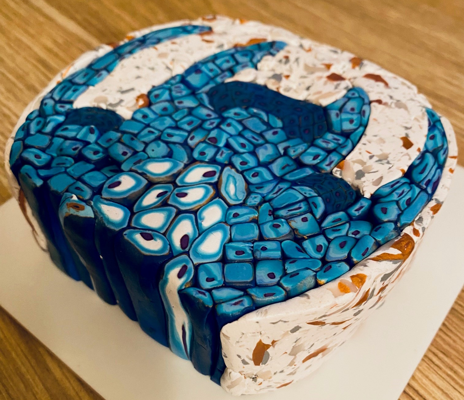
Leaf cross section used on a tea-light holder. A complex polymer clay cane was made to represent the cellular organisation of a leaf, including the epidermis, mesophyll, vascular bundles and guard cells. Slices of the cane were used here to cover a tea-light holder. The air spaces of the leaf were made using translucent clay.
Were you always going to be a scientist?
Yes I think so. From a reasonably early age I enjoyed the idea that we can understand the world around us using science. That if we collected data and used some logic, then perhaps we could figure out how things work. The idea that science can help us solve problems and help advance the human race is very appealing to me. Although as I’ve gotten older I’ve come to appreciate our problems require so much more than science to give us enduring solutions.
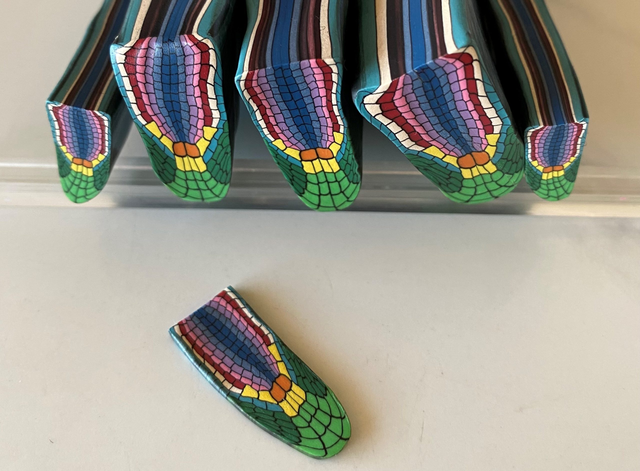
Root tip This cane is a stylised representation of the cellular organisation of a plant root tip.
And what about art – have you always enjoyed it?
I never felt I had much aptitude for art for much of my life. During my postdoc years I lived in a few very beige apartments and became very drawn to environments and art that were full of colour. I had dabbled in pottery on and off for many years, but had been frustrated that I couldn’t get the detail or colour that I wanted to. And then by chance in 2017 I stumbled into the world of polymer clay, being captivated by the colours and possibilities of this medium. Along the way I learned cane making, and have been hooked ever since.
Meristem Cane This is a full-sized cane before reduction (approximately 12 cm wide). The subject is a plant shoot tip, with the darker cells portraying the shoot apical meristem along with two axillary meristems in the developing leaf axils. This one is probably the closest to the science I do!
What or who are your most important artistic influences?
I’ve always been drawn to tactile types of art, including ceramics, glass, and sculpture. In general I like colour and abstract art, but actually many different art styles and forms appeal to me, so it’s hard to narrow that down. In the polymer clay world I am inspired by many artists, and would suggest to those unfamiliar with this medium to check out the works of people like Kathleen Dustin, Georg Dinkel, and Adam Thomas Rees. On a more personal level, a lot of the skills I learned to make canes came from the generosity of artists like Fiona Abel-Smith.
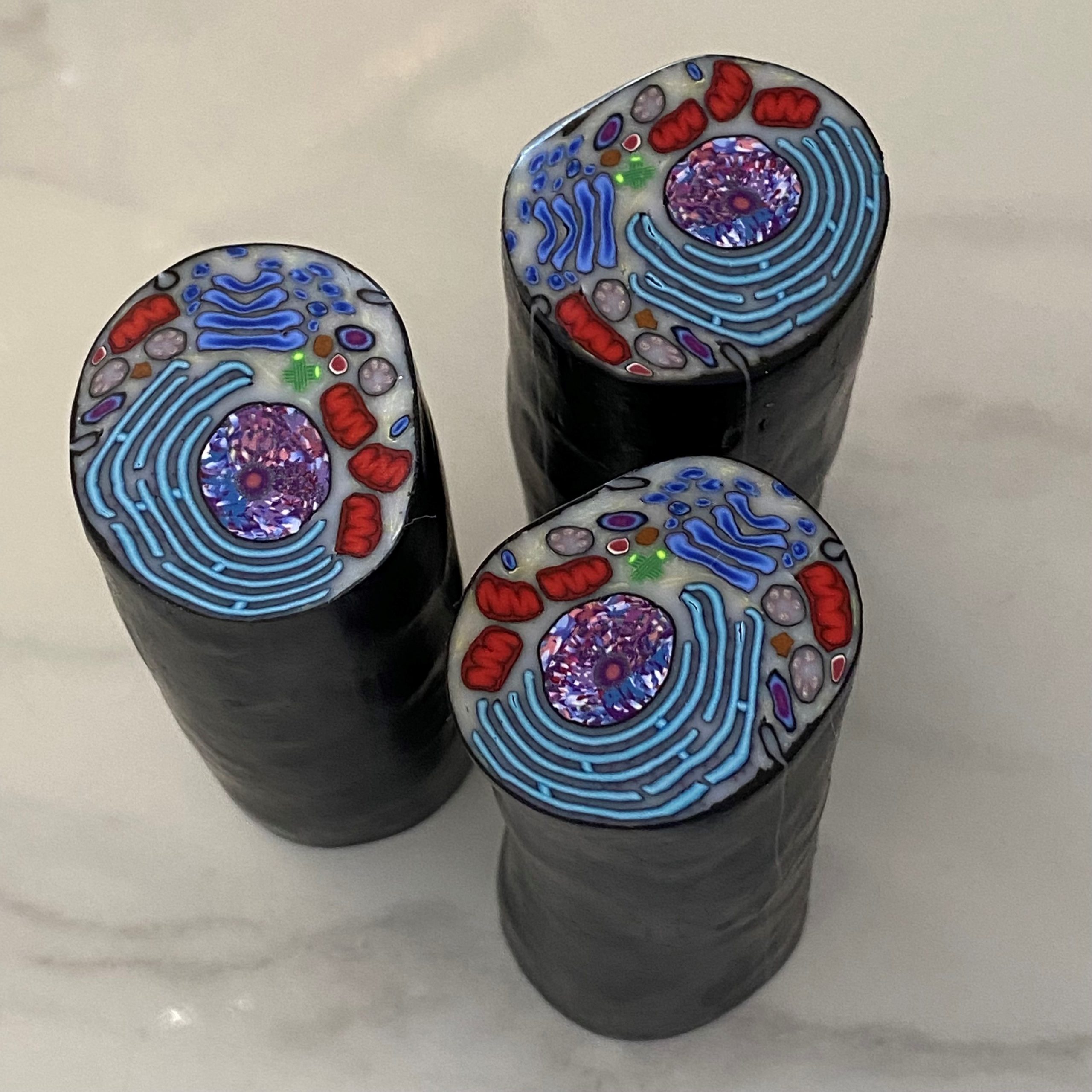
Animal cell cane This is a polymer clay cane portraying an animal cell. This cane has been reduced and is about 3 cm wide, while the original cane started out about 11 cm wide.
How do you make your art?
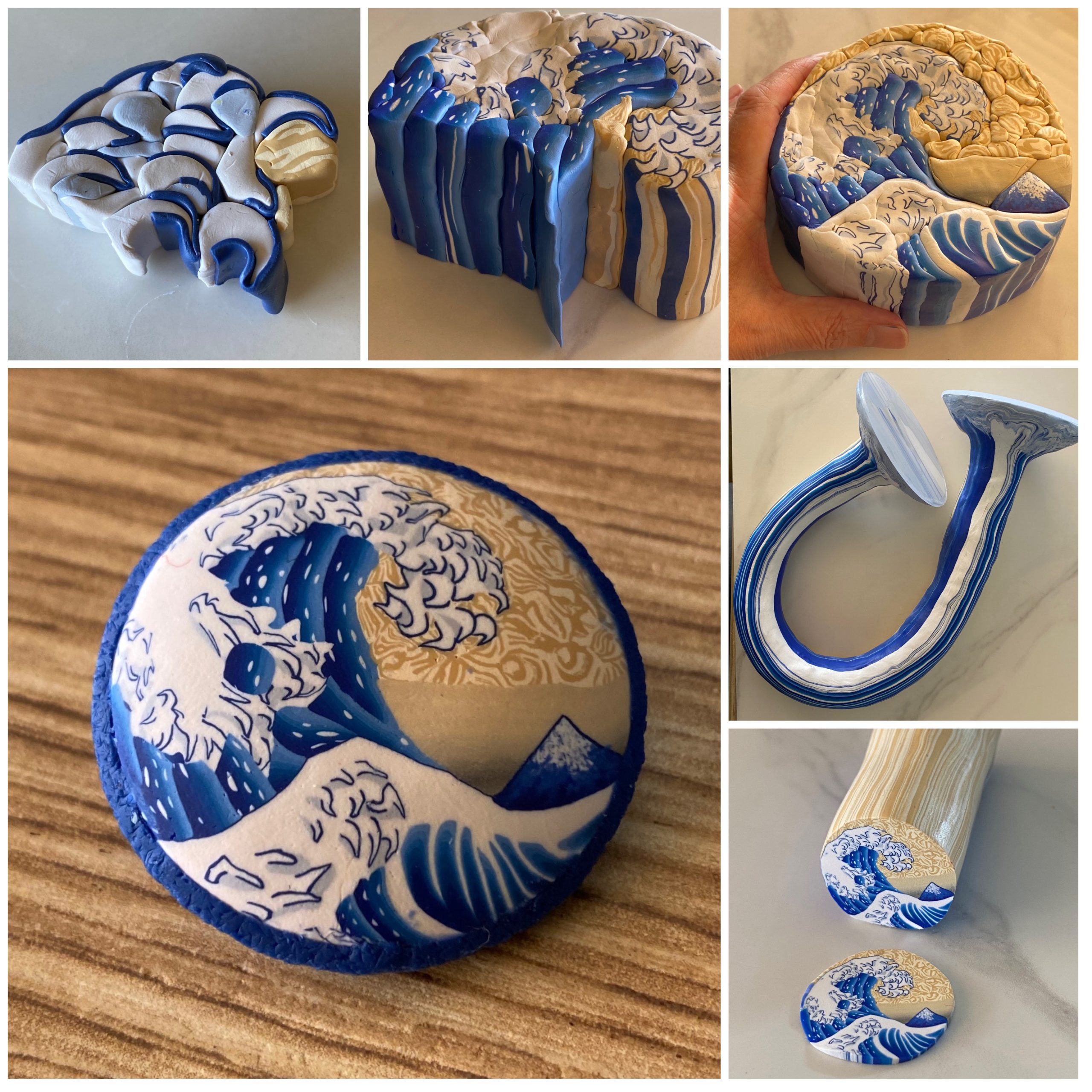
I start with a rough sketch of my subject, often based on looking at many images of whatever that might be. Sometimes I spend time learning about the subject as well, so that I feel I know a bit more than what I might see in photographs. Then I need to work out the level of detail needed, and spend time thinking about how to achieve that, or if it is achievable at all at the scale I work at. Some things that might be easy to draw can be very difficult to convert to a cane format. An example of that is when I recently made a version of Hokusai’s Great Wave off Kanagawa in cane form – there the small details of the wavelets took me a few attempts and approaches to get something I was happy with. Consideration of contrast and colour combinations is also very important, and not something I feel I have mastered yet. Then I usually build the components of the cane, incorporating blends, and a few other different methods before reducing those to a size that can be used in the cane. Assembly of the components into the cane happens next and requires components to be sized appropriately and for everything to be fitted well together. Rushing that aspect usually causes a lot of problems in the final cane, as the physics of reduction mean that clay will move into any gaps causing distortion. My complex canes are often built around 12 cm in diameter and 4 or 5 cm in height, and once finished the whole cane is reduced down to something more like 3 cm in diameter. Final items are made with slices of my canes, which are then cured by baking in a domestic oven.
Diatom picture. This one is based on a photograph by Anatoly Mikhaltsov of a fossil diatom from New Zealand.
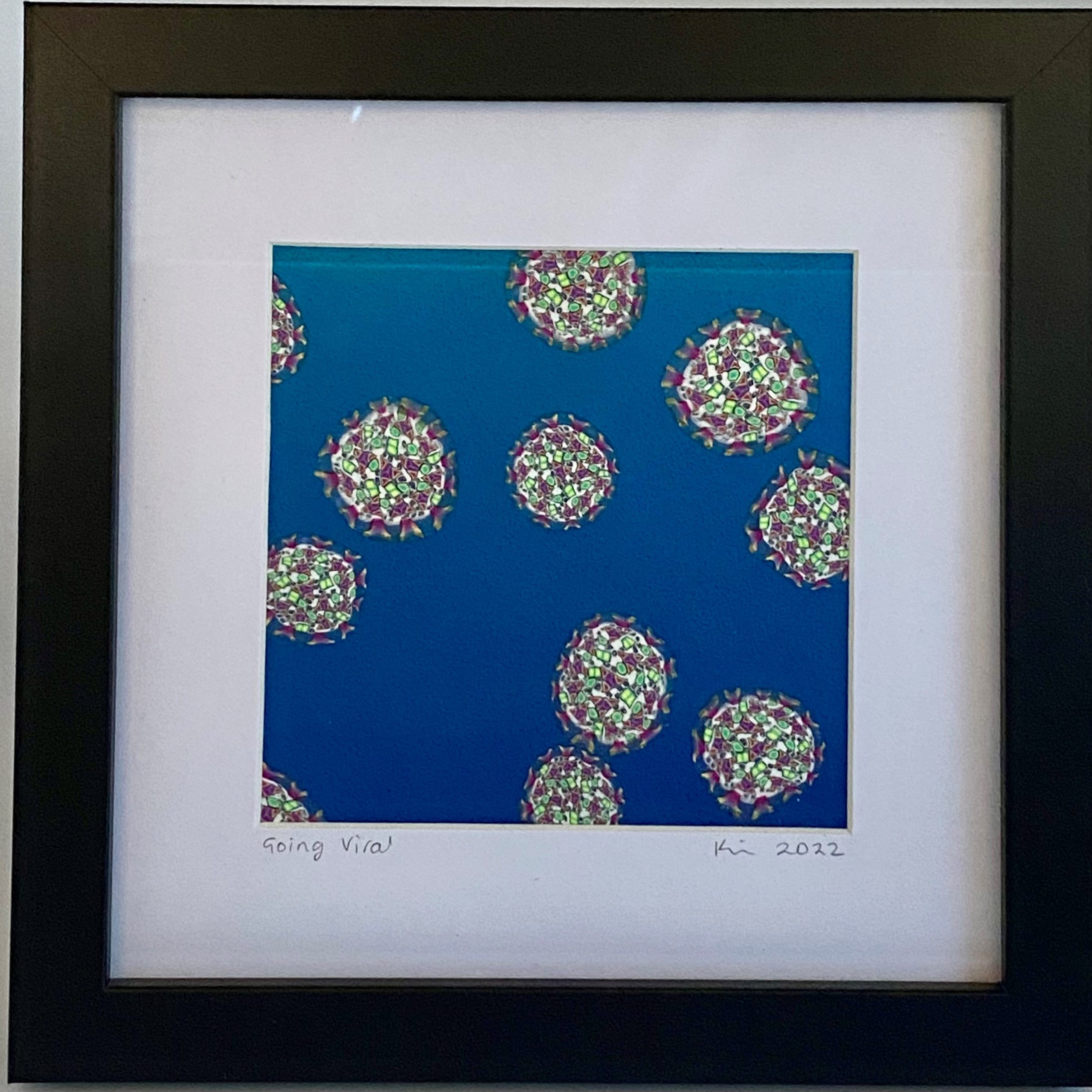
Going viral. While in lockdown in 2020, I made a cane that was my version of a SARS-CoV-2 virus. A couple of years later I used slices to assemble this piece.
Does your art influence your science at all, or are they separate worlds?
I think it is more that my science influences my art. My science background has taught me to be detail oriented which often results in me putting too much detail into my art. I have to balance portraying what is real versus what I think could convey the essence of a subject. An example of that would be my animal cell cane. There are no animal cells that look exactly like that, and the scale of different organelles is all over the place, which was necessary so that they could still be recognisable in the finished piece. In spite of that most people would still recognise it as being a cell.
What are you thinking of working on next?
I haven’t decided, and I’m always open to ideas! A few of my science canes have come from suggestions by other scientists. Some science subjects don’t lend themselves easily to a cane design, and so I sometimes think about a possible subject a long time before I start on it. Currently I have another native New Zealand flower in mind as one possibility. In the meantime, I often make kaleidoscope canes when I’m between complex cane projects, which I enjoy because of the combination of symmetry and often quite organic designs.
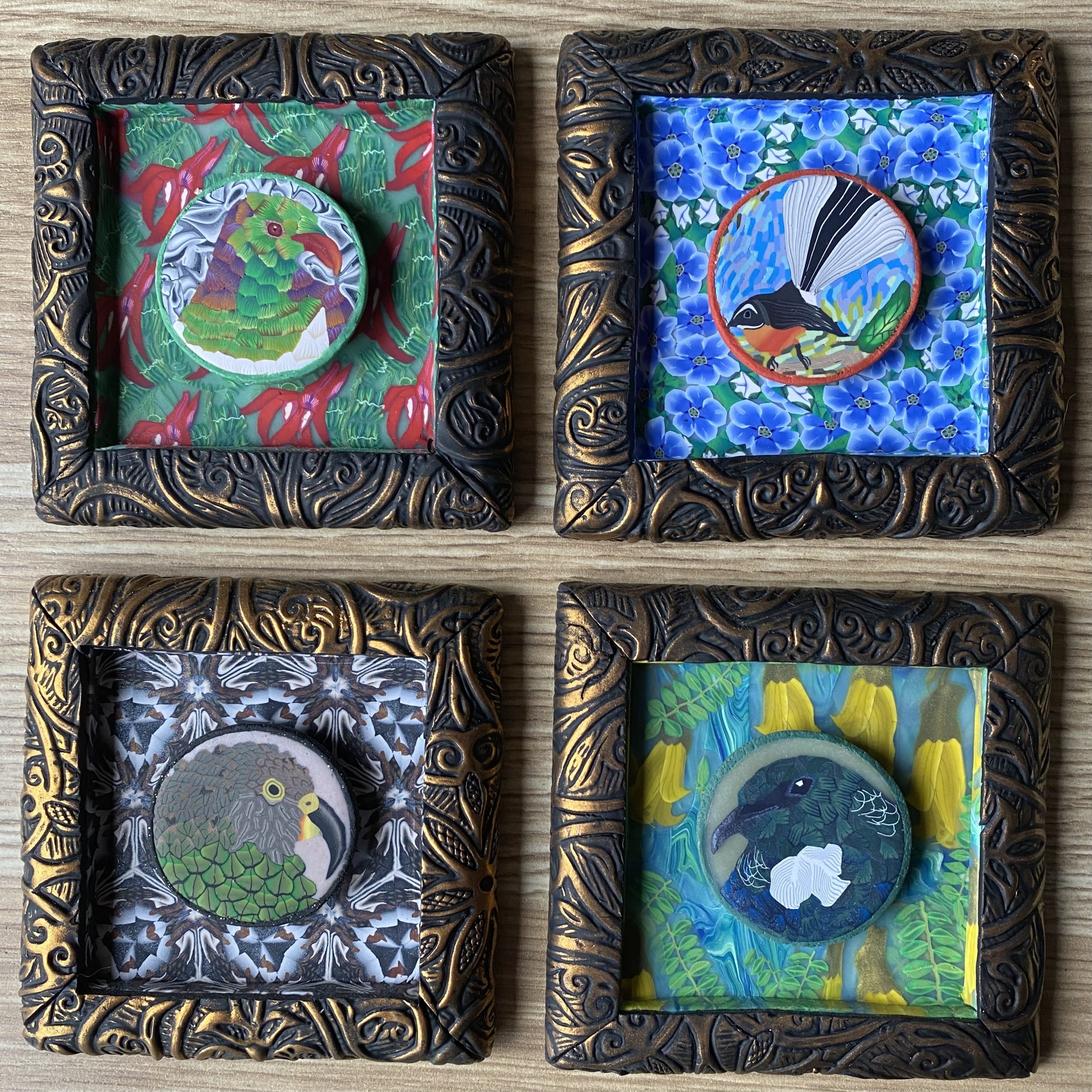
Native birds and plants of New Zealand. These small artworks are each approximately 10 cm square. They are made entirely of polymer clay (except a small bit of embedded wire for hanging). They each show a native New Zealand bird with a background appropriate to where these birds are found. Top left is a kererū (native wood pigeon) with a background of ngutu kākā flowers and foliage. Top right shows a pīwakawaka (also known as a fantail) with a background of kōpukapuka (also known as Chathan Islands forget-me-not, although it is not a true forget-me-not). On the bottom left is a kea (a very smart alpine parrot), with a more abstract background (working with alpine colours in a kaleidoscope format), and on the bottom right is a tūī with a background of kōwhai flowers and foliage. In the wild our kea, ngutu kākā and kōpukapuka are threatened from a conservation stand-point.Making of a complex cane. This montage shows a few of the steps involved in making a complex polymer clay cane. The top left panel shows the beginnings of the wave construction, the top middle shows construction about half way through and the top right shows the completed cane at full size (approximately 11 cm in diameter). On the middle right, you can see the cane during reduction, and the bottom right image shows the completed cane with the interior showing (here the cane is approximately 3 cm in diameter). The large image on the left shows a completed pin constructed from a slice of this cane.
Alessandra M. Norris, Ambili Bai Appu, Connor D. Johnson, Lylybell Y. Zhou, David W. McKellar, Marie-Ange Renault, David Hammers, Benjamin D. Cosgrove, Daniel Kopinke
Nikola Sekulovski, Jenna C. Wettstein, Amber E. Carleton, Linnea E. Taniguchi, Xiaolong Ma, Sridhar Rao, Jenna K. Schmidt, Thaddeus G. Golos, Chien-Wei Lin, Kenichiro Taniguchi
Eduardo D. Gigante, Katarzyna M. Piekarz, Alexandra Gurgis, Leslie Cohen, Florian Razy-Krajka, Sydney Popsuj, Hussan S. Ali, Shruthi Mohana Sundaram, Alberto Stolfi
Marta Moreno-Oñate, Lourdes Gallardo-Fuentes, Pedro M. Martínez-García, Silvia Naranjo, Sandra Jiménez-Gancedo, José L. Gómez-Skarmeta, Juan J. Tena, José M. Santos-Pereira
John R Klem, Tae-Hwi Schwantes-An, Marco Abreu, Michael Suttie, Raeden Gray, Hieu Vo, Grace Conley, Tatiana M Foroud, Leah Wetherill, CIFASD, charles B lovely
Teresa Krammer, Hannah T. Stuart, Elena Gromberg, Keisuke Ishihara, Manuela Melchionda, Jingkui Wang, Elena Costantini, Stefanie Lehr, Dillon Cislo, Laura Arbanas, Alexandra Hörmann, Ralph A. Neumüller, Nicola Elvassore, Eric Siggia, James Briscoe, Anna Kicheva, Elly M. Tanaak
Wilma Tixi, Maricela Maldonado, Ya-Ting Chang, Amy Chiu, Wilson Yeung, Nazia Parveen, Michael Nelson, Ryan Hart, Shihao Wang, Wu Jih Hsu, Patrick Fueger, Janel L. Kopp, Mark O. Huising, Sangeeta Dhawan, Hung-Ping Shih
An Chengrui, Alison M. Farley, Sam Palmer, Dong Liu, Anastasia I. Kousa, Paul Rouse, Viktoria Major, Joanna Sweetman, Jan Morys, Andrea Corsinotti, Jennifer Nichols, Jan Ure, Renee McLay, Luke Boulter, S. Jon Chapman, Simon R. Tomlinson, C. Clare Blackburn
Tania L Gonzalez, Sahar Wertheimer, Amy E Flowers, Yizhou Wang, Chintda Santiskulvong, Ekaterina L Clark, Caroline A Jefferies, Kate Lawrenson, Jessica L Chan, Nikhil V Joshi, Yazhen Zhu, Hsian-Rong Tseng, S. Ananth Karumanchi, John Williams III, Margareta D Pisarska
Ricardo Fuentes, Florence Marlow, Elliott Abrams, Hong Zhang, Manami Kobayashi, TriptiGupta, Lee Kapp, Zachary DiNardo, Felipe Montecinos-Franjola, William Vought, Charles Vejnar, Antonio E Giraldez, Mary C Mullins
Sudha Sunil Rajderkar, Kitt Paraiso, Maria Luisa Amaral, Michael Kosicki, Laura E. Cook, Fabrice Darbellay, Cailyn H. Spurrell, Marco Osterwalder, Yiwen Zhu, Han Wu, Sarah Yasmeen Afzal, Matthew J. Blow, Guy Kelman, Iros Barozzi, Yoko Fukuda-Yuzawa, Jennifer A. Akiyama, Veena Afzal, Stella Tran, Ingrid Plajzer-Frick, Catherine S. Novak, Momoe Kato, Riana D. Hunter, Kianna von Maydell, Allen Wang, Lin Lin, Sebastian Preissl, Steven Lisgo, Bing Ren, Diane E. Dickel, Len A. Pennacchio, Axel Visel
Rinaldo Catta-Preta, Susan Lindtner, Athena Ypsilanti, James Price, Armen Abnousi, Linda Su-Feher, Yurong Wang, Ivan Juric, Ian R. Jones, Jennifer A. Akiyama, Ming Hu, Yin Shen, Axel Visel, Len A. Pennacchio, Diane Dickel, John L R Rubenstein, Alex S Nord
Andrew J. Aman, Lauren M. Saunders, August A. Carr, Sanjay R. Srivatsan, Colten D. Eberhard, Blake Carrington, Dawn Watkins-Chow, William J. Pavan, Cole Trapnell, David M. Parichy
Ricardo Fuentes, Florence L. Marlow, Elliott W. Abrams, Hong Zhang, Manami Kobayashi, Tripti Gupta, Lee D. Kapp, Zachary DiNardo, Ronald Heller, Ruth Cisternas, Felipe Montecinos-Franjola, William Vought, Mary C. Mullins
Richard Francis, Jovenal T San Agustin, Heather L. Szabo Rogers, Cheng Cui, Julie A. Jonassen, Thibaut Eguether, John A. Follit, Cecilia W. Lo, Gregory J. Pazour
Laura Massoz, David Bergemann, Arnaud Lavergne, Célia Reynders, Caroline Désiront, Chiara Goossens, Lydie Flasse, Bernard Peers, Marianne L. Voz, Isabelle Manfroid
Georgios Tsissios, Anthony Sallese, J. Raul Perez-Estrada, Jared A. Tangeman, Weihao Chen, Byran Smucker, Sophia C. Ratvasky, Erika Grajales-Esquivel, Arielle Martinez, Kimberly J. Visser, Alberto Joven Araus, Hui Wang, Andras Simon, Maximina H. Yun, Katia Del Rio-Tsonis
Fabrizio E. Mancini, Paul Evelyn Alexander Humphreys, Steven Woods, Nicola Bates, Sara Cuvertino, Julieta O’Flaherty, Leela Biant, Marco A.N. Domingos, Susan J. Kimber
Joshua Hislop, Amir Alavi, Qi Song, Rayna Schoenberger, Kamyar Keshavarz F., Ryan LeGraw, Jeremy Velazquez, Tahere Mokhtari, Mohammad Nasser Taheri, Matthew Rytel, Susana M Chuva de Sousa Lopes, Simon Watkins, Donna Stolz, Samira Kiani, Berna Sozen, Ziv Bar-Joseph, Mo R. Ebrahimkhani
Kiryu K. Yap, Jan Schröder, Yi-Wen Gerrand, Anne M. Kong, Adrian M. Fox, Brett Knowles, Simon W. Banting, Andrew G. Elefanty, Eduoard G. Stanley, George C. Yeoh, Glen P. Lockwood, Victoria C. Cogger, Wayne A. Morrison, Jose M. Polo, Geraldine M. Mitchell
Christian Wiese, Miriam Abele, Benjamin Al, Melina Altmann, Alexander Steiner, Nils Kalbfuss, Alexander Strohmayr, Raksha Ravikumar, Chan Ho Park, Barbara Brunschweiger, Chen Meng, Eva Facher, David W. Ehrhardt, Pascal Falter-Braun, Zhi-Yong Wang, Christina Ludwig, Farhah F. Assaad
Facundo Romani, Susanna Sauret-Güeto, Marius Rebmann, Davide Annese, Ignacy Bonter, Marta Tomaselli, Tom Dierschke, Mihails Delmans, Eftychios Frangedakis, Linda Silvestri, Jenna Rever, John L Bowman, Ignacio Romani, Jim Haseloff
Martina Jablonski, Guillermina M Luque, Matias Gomez-Elias, Claudia Sanchez-Cardenas, Xinran Xu, Jose L de la Vega-Beltran, Gabriel Corkidi, Alejandro Linares, Victor Abonza, Dario Krapf, Diego Krapf, Alberto Darszon, Adan Guerrero, Mariano G Buffone
Anna E. Williamson, Sanuri Liyanage, Mohammadhossein Hassanshahi, Malathi S.I. Dona, Deborah Toledo-Flores, Dang X.A. Tran, Catherine Dimasi, Nisha Schwarz, Sanuja Fernando, Thalia Salagaras, Aaron Long, Jan Kazenwadel, Natasha L. Harvey, Grant R. Drummond, Antony Vinh, Vashe Chandrakanthan, Ashish Misra, Zoltan Neufeld, Joanne T.M. Tan, Luciano Martelotto, Jose M. Polo, Claudine S. Bonder, Alexander R. Pinto, Shiwani Sharma, Stephen J. Nicholls, Christina A. Bursill, Peter J. Psaltis
The conference will for the first time since COVID-19 bring together the research community using avian models in research. We have an exciting line-up of top international speakers covering topics like stem cells, developmental biology, new technologies, immunology, comparative genomics and evolution, and the use of avian models for medical research. Besides the keynote speakers, the programme features short and flash talks selected from the abstracts, poster sessions, a plenary discussion on avian models in research, and a PI meeting to discuss the development of resources for the international avian research community.
Outside the conference Portsmouth and the South coast of England provide a range of attractive sights to visit. Portsmouth can easily be reached by train and from London Heathrow and Gatwick airports and Southampton airport. It also has ferry links with the Channel Islands, France and Spain.
We are very much looking forward to welcoming the avian model research community in Portsmouth. Please spread the word!
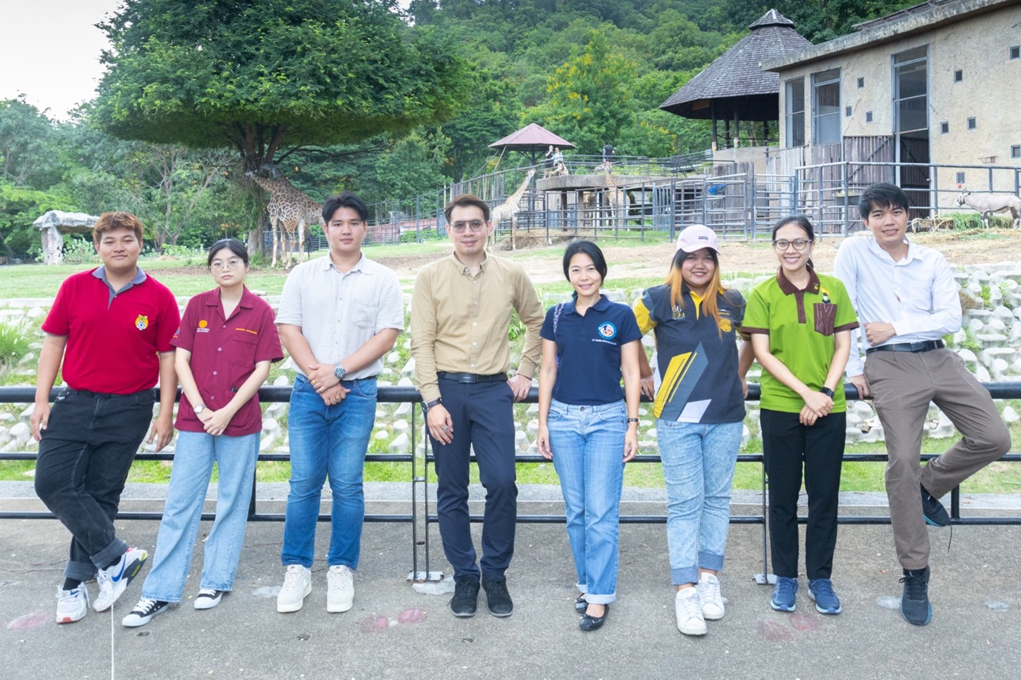
Excellent Unit of Wildlife Stem Cells (eWIS) is housed in two remarkable locations: Wildlife Reproductive Innovation Center (WRIC) atKhao Kheow Open Zoo (as part of Zoological Part Organization of Thailand) and Department of Biology, Faculty of Science, Burapha University. Both reside in Chonburi Province, Thailand.
Research summary
The eWIS initiative brings together a team of stem cell scientists and veterinarians, forging connections between universities and zoos. The primary objective is to explore the immense potential of stem cell technology in establishing a biobank comprised of stem cells derived from endangered mammalian and avian species. Our research focuses on species that require urgent preservation efforts to safeguard genetic diversity through the collection of viable cells capable of generating entire animal bodies, known as induced pluripotent stem cells (iPSCs). Additionally, we are striving to generate wild felid/ domestic and non-domestic cat embryonic stem cells.
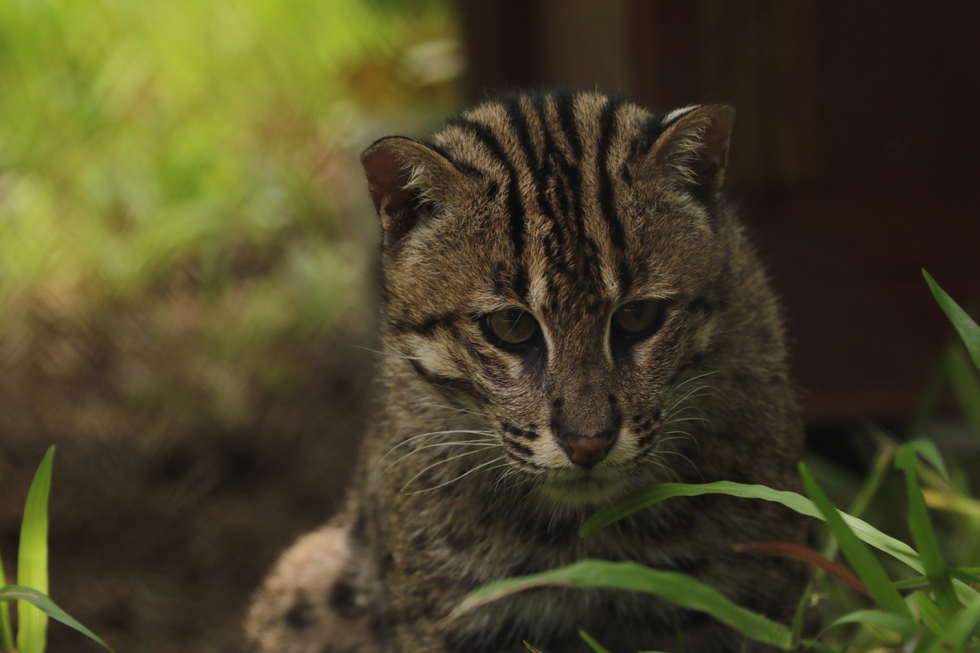
Our primary focus lies in wild felid species, including fishing cat (Prionailurus viverrinus) and clouded leopard (Neofelis nebulosa), and domestic cat (Felis catus) as a model. Our goal is to generate iPSCs from these wild cats, unravelling the possibilities of utilizing iPSCs for conservation purposes through in vitro gametogenesis, animal cloning, and assisted reproductive technology (ART). We are also devoted to establishing wild avian iPSCs, with specific attention given to hornbills and peafowl.
Furthermore, we employ a range of evolutionary tools to delve into the conservation and divergence of pluripotency networks across various vertebrate species. In particular, our research involves studying Oct4 homologues to comprehend the effects of evolutionary changes on conserved and diverged Oct4 functions. The profound insights gained from understanding the Oct4 network and early developmental programs contribute to translational approaches, aimed at overcoming reprogramming barriers in wild species.
The eWIS research team primarily works within one of the largest zoos in the world, “Khao Kheow Open Zoo,” and Burapha University in Chonburi, Thailand. From left to right: Pawares, Chichakon, Pitiwut, Woranop, Ampika, Rattanaporn, Santhita, Nattakorn. (Photo credit : Nattawut Dueanjam).
Lab roll call
Santhita: Researcher – Managing routine lab works/paper works at WRIC, bridging zoo units to collect wild animal tissues (mostly dead) to eWIS; Organizing biobank at the zoo
Pathira : Lab Manager/Researcher – Managing routine lab works/paper works/ordering at eWIS lab at Department of Biology; Generating barnacle collection
Pawares: Pre-doc student – searching cat ovaries and testes around the city!, IVF of cat embryos, cat pluripotency explorer
Nattakorn: Master student – primary culture of tissues from live and dead mammals and birds; RNA-based reprogramming; bioinformatics on pluripotency network
Ratchapon: Master student – Wild avian/chicken reprogramming and understanding the network within avian pluripotency
Pitiwut & Rattanaporn & Phanpimon: BSc students – characterization of felid pluripotency and cat IVF/IVM
Chichakon & Phornchanok: BSc students – avian reprogramming: testing various sets of reprogramming factors
Ampika: PI – advice the team, developing ART research, searching research grant, building capacity of the team and strengthen research network
Woranop: PI – teaching, admin jobs, pipetting, being Head (meeting job) & Biology program director, making connections between organizations
Favourite technique, and why
Woranop: Cellular reprogramming is my favourite tool. It is fascinating of how these felid cells can uptake reprogramming factors and transform the morphology and reverse cell fate! I love immunostaining of pre-implantation embryos from IVF- this is stunning how a single cell can become diversified in forms and functions! Recently, I discover myself to enjoy working on structural models predicted from AlphaFold2 which helps understand the evolution of proteins orchestrating the development.
Ampika: ARTs including AI, IVF and cloning are my favourite techniques for enhancing breeding conservation program of important species such as fishing cat, clouded leopard, flat-headed cat (Prionailurus planiceps), Asian golden cat (Catopuma temminckii), Eld’s deer (Rucervus eldii) and Asian elephant (Elephas maximus). Developing cryopreservation for establishment of “Viable Cell Bank” is crucial as it is important as insurance against loss or extinct of endangered species. These techniques are valuable tools that we use to sustain the genetic diversity of our endemic and global important species.
Fishing cat, one of the wild felid species, is currently in a vulnerable status due to wetland destruction. The Zoological Park Organization of Thailand (ZPOT) prioritizes research efforts with universities to save fishing cats via assisted reproductive technology, particularly artificial insemination. We are utilizing stem cell technology to establish a biobank and perform cellular reprogramming in the Excellent Unit of Wildlife Stem Cells (eWIS). (Photo credit: Piyapong Chinnadate)
Apart from your own research, what are you most excited about in developmental and stem cell biology?
Woranop: It is amazing how cells know how to reorganize themselves into specific shapes and transform into particular lineages. The technology to generate synthetic embryos, based on our understanding of cell potency and differentiation, is incredibly exciting. I am curious to see how this technology will merge with our understanding of wild stem cells and eventually contribute to saving wild animals.
Ampika: It is very exciting to see how we can develop stable stem cell colonies and differentiate them into various cell types that can be applied for medical treatments and reproductive purposes. This capability is incredibly useful for sustaining wildlife populations in the future!
How do you approach managing your group and all the different tasks required in your job?
Woranop: Everything is all about team work and communication! Regular meet-up helps a lot. We also frequently hang out for lunch or dinner, creating a comfortable environment for students to freely talk and share ideas.
Ampika: Update and discuss in anytime and set up a small meeting at least once a month.
What is the best thing about where you work?
Woranop : I enjoy working multidisciplinary fields of biology at our department and visiting the zoo every time I go to the eWIS lab. It allows me to appreciate animals and nature.
Ampika : The best part of our work is that we apply all our knowledge to prevent our wildlife from facing the brink of extinction! Our work supports not only ex situ management but also in situ conservation.
Santhita: At the zoo, we have access to a diverse range of wild animal species, which enables us to quickly collect samples for our work after the animals die or during health checks.
Pathira: The department admin staffs – we can ask for almost everything from lab materials to animal/plant specimens.
Pawares: Working with developing embryos often fills my heart with boundless excitement.
Nattakorn: I love being a TA and teaching undergraduate students! It provides me with opportunities to help other young students develop a passion for science through activities like science camps. It also allows me to support them in navigating university courses and life.
Ratchapon: The teachers in our department are very friendly, and we can contact them to get advice anytime.
Pitiwut: Everyone here is a bundle of joy- we can engage in conversations about anything, no matter how unrelated to work it may be!
Rattanaporn: Learning new things all the time
Phanpimon: Our advisors are the best! They always provide great support.
Chichakon:There are plenty of opportunities to engage in biology research starting from my third year! I love the open exchange of ideas we have in the lab.
Phornchanok: The great friendships in the lab make everything easier.
Purachet: Enjoy Sukiyaki parties with admin staffs in the department lunch breaks!
eWIS research team
What is there to do outside of the lab?
Woranop : I teach undergraduate and graduate students, and it brings me joy to share the fascinating stories of developmental biology with them outside the lab. We are also located near Bangsaen Beach! I often hang out with our team and visit other beaches along the eastern coast of Thailand.
Ampika : Since we work with 6 zoos nationwide and the Elephant Kingdom Project, it is very enjoyable to explore the places and talk to local people. And of course, Bangsaen is my favourite place to R&R!
Santhita: Sometimes in the evening, we take walks around the zoo to be close to nature. Nearby, there are tourist attractions such as Chan Ta Then waterfall and Bang Pra reservoir.
Pathira: Hangout at the zoo, Capybara watching! & definitely, Bangsaen is a right place for seafood, in particular grilled shrimp/squid
Pawares: I help organize department events and take care of our small colonies of chickens!
Nattakorn: I enjoy playing online games while waiting for my phylogenetic analyses! The beautiful Bangsaen beach also provides great help during exam or lab meeting crises
Ratchapon: I assist the department’s administrative staffs for specific events and serve as a TA to help Woranop teach the Developmental Biology Lab.
Pitiwut:I keep myself fit and healthy by doing cardio exercises at the nearby gym! I think it helps clear my thoughts a lot after a long day of studying and working in the lab.
Rattanaporn: Moo-gata (Traditional Thai BBQ!) is a must here.
Phanpimon: Jet skiing, running on the beach, or even feeding the monkeys on Khao Sam Muk hill.
Chichakon: I love designing art works for my lab and also others in the department.
Phornchanok: We can do a lot of things in Bangsaen – shopping, visiting cafes, having dinner, and enjoying the beach!
Purachet: Outside of class and the lab, I spend most of my time in the department studio, where I assist with photography and collect biological samples for departmental teaching.
Browse through other ‘Lab meeting’ posts featuring developmental and stem cell biology labs around the world.
The Company of Biologists has launched a new server on Mastodon: biologists.social. It is a space for biologists to discuss science, research, teaching, life and more.
“Richard III was King of England from 1483 to 1485. There are some contemporary records. One talks about him having one shoulder higher than the other. So when we were excavating, we thought maybe we’ll find somebody with a spinal abnormality.“
Prof. Turi King
In the latest episode of the Genetics Unzipped podcast, we’re venturing into ancient territory and archaeological digs, excavating the complex ethics of extracting and sequencing DNA from human remains. We chat to Prof. Turi King about her role uncovering the body King Richard III and who decides which research gets done.
On 21 June 2023, Development hosted a webinar featuring talks on the topic of growth and morphogenesis from three early career researchers, chaired by Development’s Editor-in-Chief, James Briscoe. Below are the recordings of the talks.
Patricio Pérez-Henríquez (University of California, Riverside)
Talk and Q&A by Patricio Pérez-Henríquez
Stefania Tavano (MedUni Vienna)
Talk and Q&A by Stefania Tavano
Stefan Harmansa (IBDM and Turing Center for Living Systems)
Talk and Q&A by Stefan Harmansa (No Ratings Yet) Loading...
Looking forward to welcome you to our EMBO Workshop – Developmental metabolism: flows of energy, matter, and information
Recent work revealed the intricate relationship between energy flux, spatial and temporal control of metabolites, the environment, and embryogenesis🤯. #EMBODevMet
Hydrozoan cnidarians have a population of stem cells, known as i-cells. The developmental potential of only one other hydrozoan i-cell has been described before this paper. Hydra‘s i-cells can give rise to the neuroglandular lineage and to germ cells. Their ectodermal and endodermal epithelial cells are self-renewing, constituting separate lineages. i-cells multipotent but not pluripotent (Bosch & David, 1987).
In terms of Hydractinia, it was established that a population of stem cells (known as i-cells) were able to give rise to every single lineage (Müller, et al., 2004). This showed that as a population, i-cells can give rise to the somatic and germ cell lineages. The potential of a single i-cell, however, was still unknown. Either i-cells had their own distinct lineage restricted sub-populations or they were pluripotent, and one i-cell could give rise to all cell types.
The aim of my research was to transplant a single i-cell and observe it in vivo. For this purpose, we had an extremely useful fluorescent reporter animal at our disposal. This animal had two fluorescent transgenes, the first transgene was Piwi1- driven GFP which is expressed in only i-cells and germ cells. It is downregulated in differentiated cells. The other transgene was Beta-tubulin- driven mScarlet. This transgene is expressed by all differentiated cells but not i-cells. This allowed us to identify i-cells by their GFP expression and lack of mScarlet expression and identify the i-cells progeny by their mScarlet expression and dim GFP fluorescence due to the half-life of the GFP. The aim of the project was to transplant a single GFP positive i-cell into a wild type recipient and identify the resulting progeny that would be identifiable by their mScarlet expression.
Bottlenecks on bottlenecks
Transplanting a single cell was my first objective when I started this project as a 1st year PhD student. I figured that it should take me 3-4 months maximum to get it sorted, optimised and then I could press on with the actual experiments and start generating some (hopefully decent) data. I began by looking at what single cell transplantations had been performed before in comparable models and hoped that it would be just as successful in Hydractinia.
Firstly, I tried incorporation of a cell via aggregation. This is something that was established in Hydra (Murphy & David, 1977). It required dissociation of full Hydractinia polyps. I could then identify a single i-cell by its GFP fluorescence and its lack of mScarlet fluorescence. This would then be picked up by a needle attached to a micro-injector and ejected into a tube full of dissociated wild type cells. These would then be centrifuged into an aggregate and cultured till, ideally, a polyp with a single transplanted i-cell would be established. Turns out, betting on a single cell surviving aspiration and/or centrifugation is a long shot. In meetings with my PI, Uri, I frustratedly referred to it as “a bottleneck on a bottleneck”.
After, countless amounts of failed transplantations via aggregation I was reinspired by the planarian community’s favourite method of single cell transplantation: direct injection into the animal (Wagner, et al., 2011). Perhaps by directly delivering a single cell I could finally cross off my first objective. This required a steady hand to ensure you didn’t overshoot the piercing of the epidermis and inject a cell into the gastric cavity. Unfortunately, after injecting into adult polyps and larvae from all angles I realised that this method wasn’t going to be feasible either. I was just not seeing any i-cell engraftment. It may have been a combination of mechanical issues on my part, maybe unhappy cells or also Hydractinia having a much more confined stem cell niche area than planarians do. Ultimately, we never found out why. We just knew it wasn’t a way forward to achieve our aims. At this point, I was almost 2 years into my PhD and that first objective was still not completed. The pressure was mounting, and I started considering alternative projects in the worst-case scenario. Whatever came next would be my last attempt at getting this project off the ground.
Keeping it in the niche
Finally, we had our own form of a eureka moment. We had been dissociating the animals to isolate one cell (like the planarian and Hydra communities had previously done) but we had overlooked something major and wonderful about Hydractinia. We had overlooked what sets them apart from both Hydra and planarians. Hydractinia is a colonial animal that can fuse and swap cells with a histocompatible relative via stolonal networks. I had been working to establish a method of transplanting cells when our model already had one itself. We could keep our i-cells within their own niche! I just had to find a way to effectively leverage it to my aims.
This began with me setting up Hydractinia “neighbourhoods”: a donor colony settled next to a recipient colony. The donor colony would grow and then fuse with the recipient by extended stolons and I could patiently observe i-cells migrating into the recipient tissue, waiting for a perfect moment to strike and isolate one cell (Figure 1). I had noticed that when the colonies were fed less, they were more likely to grow long branches of stolons rather than extending their mat. A long branch was preferable as it meant the i-cells could be transplanted “in single file” rather than like an encroaching army that was more unpredictable and difficult to follow a single cell. After numerous fusions of donor and recipient I isolated a single donor i-cell in a piece of recipient stolon tissue. The next day there were two i-cells. Then four. Then there were mScarlet expressing somatic cells AND i-cells. My transplanted cell was proliferating and differentiating. This new chimeric piece of stolon grew into a small colony with both wild type cells and cells derived from the one i-cell. Extraordinarily relieved, I checked off that first objective.
At the early stages there was a split between recipient and donor derived cells in the colony. To ensure that the donor cells were not outcompeted I performed “selective pruning”. This involved excising large portions of tissue that had only/mainly wild type recipient cells. This gave us a lot of peace of mind as we were more confident that the donor cells wouldn’t be outcompeted before they had developed sexual polyps.
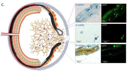
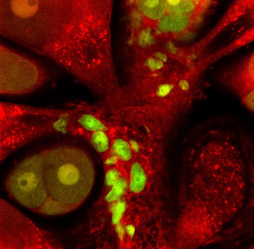
Figure 1: Colony grafting via parabiosis. A wild-type colony is positioned close to a transgenic fluorescent colony. Fusion between the histocompatible relatives allows i-cells to move from one colony to the other. A small section of wild-type stolon with a single donor fluorescent i-cell is isolated by excising the surrounding donor and recipient tissues.(Varley, et al., 2023)
When the animal was sexually mature, we were excited to see sexual polyps that were male (from the recipient) but also fluorescent and female (from the donor). Some sexual polyps were even both. These donor derived GFP+ and mScarlet+ oocytes were able to be fertilised and gave rise to a new primary polyp that inherited the transgenes from the single donor i-cell (Figure 2). From this we knew that one i-cell can contribute to the next generation of Hydractinia.
Using live confocal microscopy on polyps cut from the colony, we were able to identify mScarlet+ neurons, epithelial cells, nematocytes and i-cells by their characteristic morphologies. Gland cells, however, were more difficult to identify. We didn’t have any antibodies that could identify them and even by morphology they often look like more vesicular epithelial cells. To address this, we had to look at it from a different perspective. At this later stage of the colony, we were able to confirm by flow cytometry that all cells present were transgenic and there were no recipient cells remaining. This meant that all cells, including gland cells, came from the single donor i-cell. This was the ultimate proof of pluripotency for us. There was not a single cell in the animal that one i-cell did not give rise to.
Don’t doubt your model
In humans, pluripotency occurs for a brief snippet of time pre-gastrulation. This paper established that Hydractinia stem cells are pluripotent for the full extent of their life. When starting, I didn’t expect for one single cell to be as globally influential as it was. In retrospect, I should have never doubted our model and its ability to impress.
Figure 2: Donor derived oocyte and germ cells (green) in a chimeric sexual polyp (Varley, et al., 2023)
References
Bosch, T. & David, C., 1987. Stem cells of Hydra magnipapillata can differentiate into somatic cells and germ line cells. Developmental Biology, pp. 182-191.
Müller, W. A., Teo, R. & Frank, U., 2004. Totipotent migratory stem cells in a hydroid. Developmental Biology, Volume 275, pp. 215-224.
Murphy, S. & David, C. N., 1977. Characterisation of interstitial stem cells in hydra by cloning. Developmental Biology, 58(2), pp. 372-383.
Varley, Á. et al., 2023. Pluripotent, germ cell competent adult stem cells underlie cnidarian regenerative ability and clonal growth. Current Biology, 33(10), pp. 1883-1892.
Wagner, D. E., Wang, I. E. & Reddien, P. W., 2011. Clonogenic neoblasts are pluripotent adult stem cells that underlie planarian regeneration. Science, 332(6031), pp. 811-816.
 (No Ratings Yet)
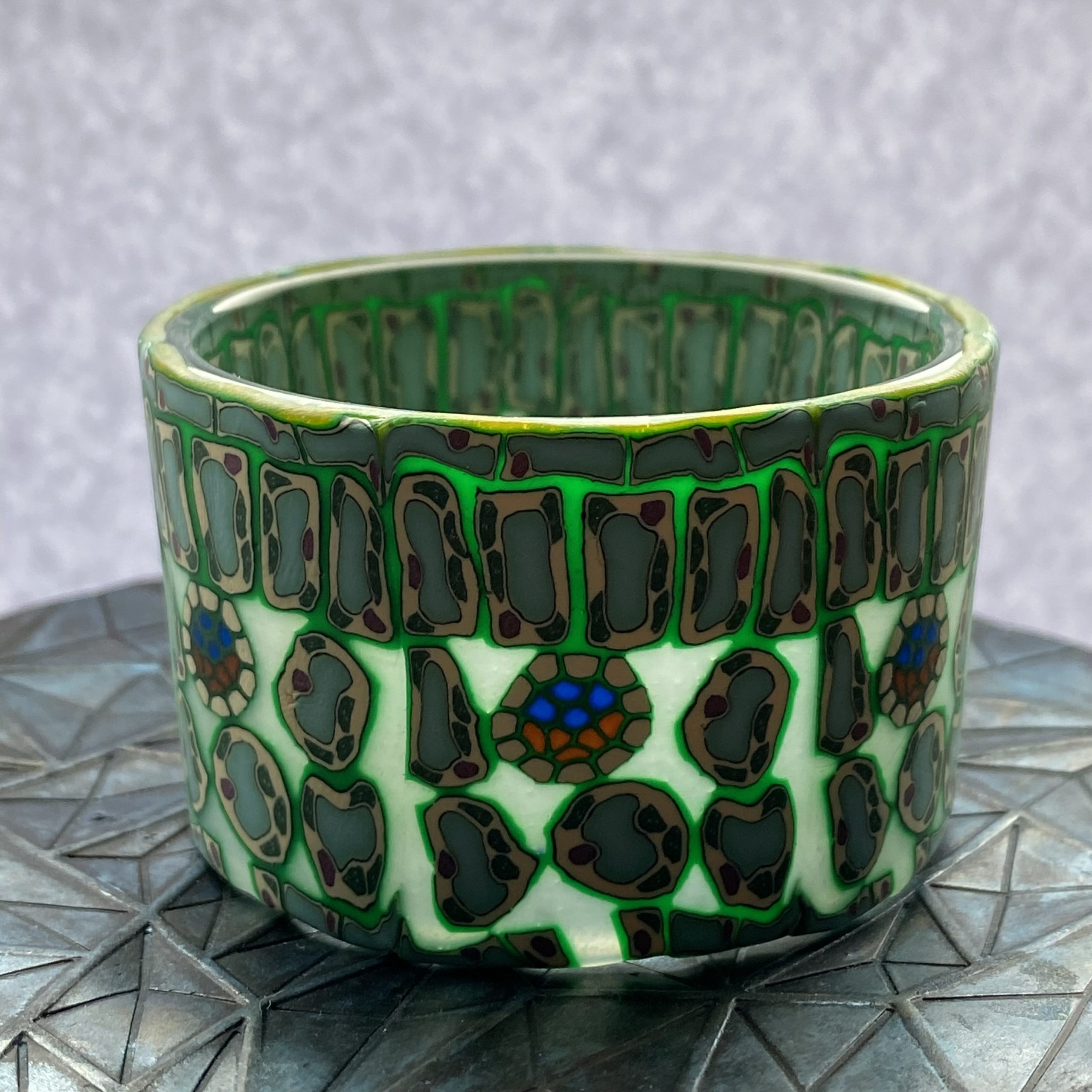

 (2 votes)