Welcome to the spring edition of our ‘Occasional writing ideas’ newsletter. With the world opening up again, we have focussed our ideas on getting back to ‘normal’ or finding our new normal. However, we are not limiting our posts to these topics, and you are welcome to post on any subject relevant to the developmental and stem cell biology community. If you would like to discuss any ideas for new content, or would like help with editing or posting, please get in contact at thenode@biologists.com.
Helen Zenner Community Manager, the Node The Company of Biologists
Writing Ideas
Meetings or symposia
Are you about to attend your first in-person meeting, or just your first for two years? You could share your experiences in a meeting report. See our blog on writing meeting reports here for guidance on what you can include in your post.
Will you be taking advantage of the fact that so many meetings now have a virtual component? Is this for public health, accessibility or sustainability reasons? What do you think conference providers can do to improve your experience?
Webinar series/online communities
Are you involved with a webinar series that you would like to highlight? Do you have any tips and tricks to overcome Zoom fatigue and keep your audience engaged?
Did you start, or become part of, an online community during the pandemic? How has this helped your mental health, your research, or your career?
Lab life
What are the positive and negative effects of the pandemic on your research? Has the pandemic led to any behavioural changes that are set to stay?
“We’ve tamed the toxicity of this cancer drug by attaching it to an exosome. Previously the side effects prevented it from becoming a treatment, but because it’s associated with an exosome, it stays in the lesion and we get all of the drug effects locally without the tolerability issues”
Dr Doug Williams, CEO of Codiak Biosciences
In the latest episode of the Genetics Unzipped podcast, sponsored by Lonza, unpacking the science behind exosomes: one of the hottest new areas of research for both diagnosing and treating diseases. Once thought to be little more than ‘dust’, exosomes are tiny biological mailbags that travel around the body, as Kat Arney hears from Rossella Crescitelli. Sally Le Page talks to Doug Williams to learn how exosomes can be used to reduce the toxicity of cancer treatments by targeting drugs directly to tumours. And Davide Zocco tells us how they are scaling up manufacturing to take exosome treatments from the lab to clinics.
In our latest SciArt profile, we meet Maria Abou Chakra. Maria is a theoretical biologist who introduces us to her sci-sketchnotes and highlights the importance of creativity in science.
Where are you originally from and what do you work on now?
I am from Canada. I am a theoretical biologist, with a focus on complex biological phenomena using evidence linked modelling. For my PhD thesis, I was trained in evolutionary biology and theoretical morphology, in the lab of Jon Stone at McMaster University, Canada. I developed a mathematical model that explores both growth and form of sea urchin skeletons. After graduating, I moved to Germany to work at the Max Planck Institute for Evolutionary Biology, where I was trained, under the supervision of Arne Traulsen, in evolutionary game theory. I developed models that capture and predict behaviours in complex social dilemmas such as climate change negotiations and host parasite interactions. Since 2016, I have been working as research associate, in the lab of Gary Bader, creating 3D mathematical models that explore cell development. My current research tries to understand how cell decisions happen and use mathematical modelling to predict how that affects cell fates during development.
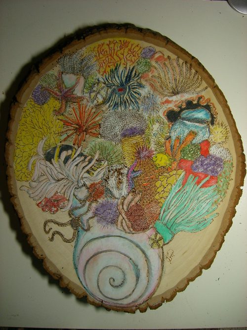
Sea bloom, using wood burning
Were you always going to be a scientist?
No, my plan throughout high school was to become an engineer. I loved programming and I thought that engineering would be a good path, but then I discovered biology in my last term which led to a sudden change of heart and I switched to science!
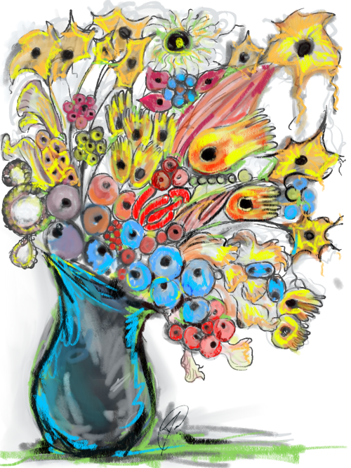
Stem cells in bloom is a bouquet illustrating my first interpretation of the diversity stem cells in a developing system.
And what about art – have you always enjoyed it?
Yes, art has been a part of my life for as long as I can remember. My dad is an architect, so he sketched all the time and as soon I could hold a pencil, I sat next to him and sketched. During my teens, I learned to paint and that became my medium of choice. As an undergrad, I tried to take art classes in parallel to my bio degree, but all the classes conflicted with lab time so I couldn’t manage both and painting became a hobby.
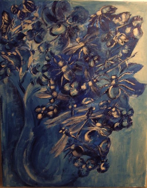
Bouquet of bugs was a gift to an entomologist friend of mine, it is an acrylic painting and up close you will see it is not flowers but a collection of different insects
When did you started using science in your artwork?
As an undergrad, sketching my notes helped me retain information longer. Then, as a grad student, I helped develop an anatomy class that introduced students to sketching notes to learn and memorise information. Their incentive was that they were allowed to use their sketches during the infamous ‘bell-ringer’ exam. I now use my sci-sketchnotes as electronic summaries of research papers or talks.
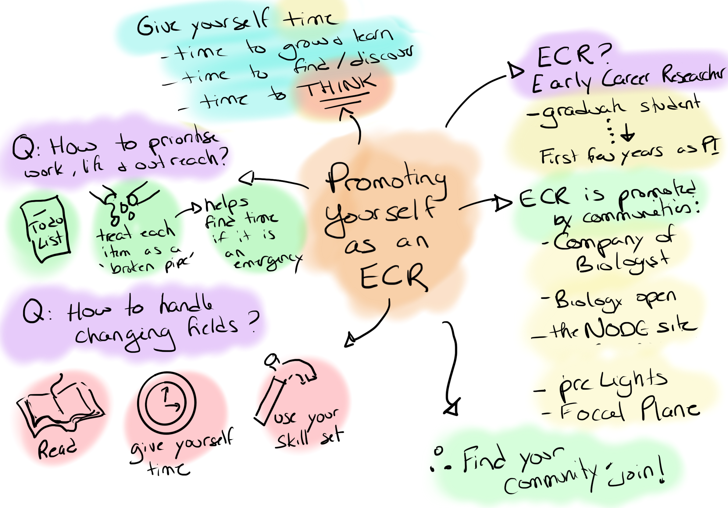
sci-sketchnotes from ECR advocacy event with The Company of Biologists
At the same time, it was hard to resist sketching the beautiful skeletons of the sea urchins that I studied during my PhD. My art then evolved into painting model organisms and other marine organisms. My supervisor and another professor appreciated them and asked to use them as the covers on the yearly reports for the department.
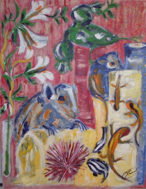
Acrylic painting for the biology department at McMaster (2005), I drew the model organisms (Arabidopsis, mouse, rat, frog, fruit fly, zebrafish and axolotl) trying to escape.
Does your art influence your science at all, or are they separate worlds?
I cannot imagine that it is possible to separate them. I think my creativity is one of the sources of my success in my research. I think having the ability to see things from a different perspective is an asset in science, and art helps me do that. A while back I read a piece by Sydney Brenner and a line stuck with me, ‘Science is the product of human minds, and the essence of research is creative innovation’ (Brenner, 1994). To me this quote says that you need both art and science, and that creativity is essential for discovery and progress in research.
SciArt poster that I created for a women in science meeting. For the central figure I chose Libby Hyman, a biologist who wrote the massive and beautiful echinoderm volumes which were an asset during my PhD.
You can find out more about Maria’s science and artwork on Twitter and on her webpage.
Thanks to Maria and all the other SciArtists we have featured so far.You can find the full list here. We’re always on the lookout for new people to feature in this series – whatever kind of art you do, from sculpture to embroidery to music to drawing, if you want to share it with the community just email thenode@biologists.com (nominations are also welcome!)
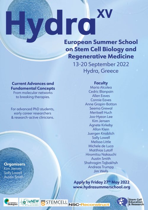
Registration is open for the 2022 Hydra Summer School in Stem Cell Biology and Regenerative Medicine.
This is a stimulating high-level course for postdocs, advanced PhD students (usually 2nd year & above), junior PIs and research-active clinicians. Places are limited and will be allocated on a competitive basis.
Leaders in the field will join you on the beautiful Greek island of Hydra to discuss topics ranging from recent advances in fundamental developmental and stem cell biology through to the latest breakthroughs in the clinic. There will also be sessions on ethics, research culture, careers, publishing, and outreach.
The Hydra summer school has been running since 2005 and has an outstanding reputation. Check out the testimonials from previous delegates: https://www.hydrasummerschool.org/testimonials
Often in scientific writing, we are up against strict limits, whether it be the number of words for a conference abstract, the number of pages for a grant proposal or even just the number of characters in a Tweet. Below, I’ve summarised a list of writing tricks to help you reduce the number of words in your scientific writing purely by changing the grammar and without sacrificing key information, clarity or accuracy. If you are not trying to fit within a specific word limit, then these tricks need not apply and instead write however you like. I do, however, think there is a case that concise writing can be clearer and more accessible, but that’s a matter of style and preference.
I’ve organised these tips in tables, in order from the easiest changes to do (remove and replace) to those changes that require more thought (rephrase). Within each table, tricks are organised in descending order with the most effective (i.e. most words chopped) at the top.
Take a look at the Twitter thread and #wordcountchop, for the list and other contributions from the community and add your own tips using the comments below.
1. Remove
First, aim to remove all unnecessary or redundant words and phrases to streamline your text and focus on the key points.
It has been shown that X interacts with… (8) X interacts with… (3)
X and colleagues show X and colleagues have shown In a study by X and colleagues
(or any of the above with ‘co-workers’ or ‘et al’)
Unless you are directly comparing two or more studies (e.g. to reconcile conflicting results) it is not necessary to mention studies by name. If you do choose to do so, make sure you mention all studies by name otherwise it appears biased.
To be able (3)
To be able to determine X… (5) To determine X… (3)
In this article (3)
In this article, we discuss… (5) Here, we discuss… (3) We discuss… (2)
In order (2)
In order to determine X…. (5) To determine X… (3)
Which is (2)
X, which is a transcription factor, … (6) X, a transcription factor, … (4)
Here, we will discuss… (4) Here, we discuss… (3) We discuss… (2)
X will then translocate… (4) X then translocates… (3) X translocates… (2)
2. Replace
Don’t be overzealous with cuts, sometimes it is necessary to re-orientate the reader or relate various concepts with each other. Here, you can replace some lengthy phrases or unweidly grammar with shorter, or more precise, alternatives.
Original
Alternative(s)
Due to the fact that (5)
As (1) Due to the fact that we could not… (8) As we could not… (4)
Because (1) Possibly due to the fact that X can… (8) Possibly because X can… (4)
It had been previously thought that X (8)
Historically, X was thought to… (5) It was thought that X… (5)
Imprecise: Has an effect on (4) (Positively/negatively) regulates (2) (Positively/negatively) modulates (2) Controls (1)
It is possible that X (4) One possibility is that X (5)
X may (2) One possibility is that X induces Y. (7) X may induce Y. (4)
It is thought that X (5) It is believed that X (5)
X might (2) X could (2) X may (2) It is thought that X interacts with Y. (8) X might interact with Y. (5)
At the same time (4)
Concurrently (1) Simultaneously (1)
Sometimes appropriate (can also refer to space as well as time) In parallel (2) Coincident (1)
On the other hand (4)
Alternatively (1) Conversely (1) In contrast (2)
To start off with (4) To begin with (3) First of all (3)
First (1)
A number of (3)
Many (1) Several (1)
The majority of (3)
Most (1)
In regards to (3) With regards to (3)
Regarding (1)
As well as (3)
And (1) Together with (2)
In particular (2)
Specifically (1)
The most important (3) The most significant (3)
The primary (2) The key (2)
As (1) Because (1) Since (1)
; (0) X activates Y because (the) loss of X reduces Y. (10) X activates Y; loss of X reduces Y. (8)
[…]. This X… (2)
‘This’, when used alone, is imprecise and unclear. It’s better to immediately qualify what ‘this’ refers to; for example, ‘X’ here could mean ‘this experiment’, ‘this approach’, ‘this protein’, ‘this cell’, etc.
, (0)
[…]. This result indicates that… (4) […], indicating that… (2)
, which (1) […], which indicates that… (3)
Such as (2)
Like (1) Including (1)
During which (2)
When (1)
As opposed to (3)
Rather than (2)
Time period (2)
Time (1) Period (1)
For example, …. (2) For instance, …. (2) Such as, …. (2)
(e.g. …) (1) X interacts with co-factors, such as Y, Z and A. (10) X interacts with co-factors (e.g. Y, Z and A). (9)
3. Rephrase
Finally, use grammar to your advantage. These tricks require a bit more conscious editing to rephrase the whole sentence or clause properly but, generally, you can convey the same message just as clearly.
Technique
Example
Abbreviations and acronyms
Abbreviate but don’t use more than one or two unfamiliar abbreviations and make sure you define them all on first use. Generally, don’t abbreviate single words or words that you use only once to help readability.
Familiar to developmental biologists: ChIP, ECM, EMT, ESC, NCC, PSC, HSC, SVZ, RA, PBS, TF etc.
Tense
Try to use a consistent tense throughout your article. If you say in the first paragraph, ‘one study has shown…’ do not then switch to, ‘one study showed’.
Bear in mind that tense also changes the tone. Present tense makes the article feel timely and recent, whereas past tense does the opposite.
Present-perfect to present simple: X has been shown to be Y. (6) X is Y. (3)
It has been shown that X interacts with Y. (9) X interacts with Y. (4)
Present perfect to past simple: Using X they have shown Y. (6) Using X they showed Y. (5)
Future simple to present simple: X will determine… (3) X determines… (2)
Possessive nouns
The X domain of Y… (5) Y’s X domain… (3)
Overexpression of Y in Z cells… (6) Y overexpressionin Z cells… (5)
US English grammar
Using the US English em dash without spaces joins up words that the UK en dash would not.
X interacts with Y – but not Z – in Q cells. (12) X interacts with Y—but not Z— in Q cells. (9) or X interacts with Y, but not Z, in Q cells. (10)
Hyphenateadjectives
(See also possessive nouns)
The domain of X that binds to DNA… (8) X’s DNA-binding domain… (3)
Allows X to be specifically expressed in Q cells. (9) Allows Q cell-specific expression of X. (6)
Plurals
In the X… (3) In Xs… (2)
Phylogeny
Phylogeny is your friend but use it with caution. It is better to be as specific as possible. Most scientists, however, usually consider that if something is true/conserved in mice and humans, it is ‘conserved in mammals’ etc.
Mice and rats Rodents
Rodents and humans (or other mammals) Mammals/mammalian
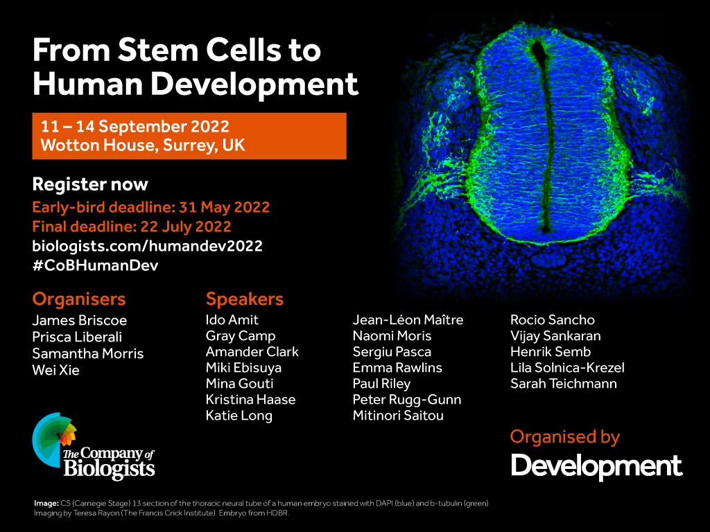
September 2022 sees the return of the popular Development Journal Meeting focussed on human development. After the successful virtual event in 2020, this year’s meeting will be hosted at Wotton House in the heart of the English countryside. The organisers, James Briscoe (The Francis Crick Institute, UK), Prisca Liberali (Friedrich Miescher Institute for Biomedical Research, Switzerland), Samantha Morris Washington (University School of Medicine, USA) and Wei Xie (Tsinghua University, China) have put together an exciting programme covering topics from early cell fate choices to tissue organogenesis and from stem cell pluripotency to techniques to visualise the entire human embryo. As well as the outstanding list of invited speakers, 15 short talks will be selected from the submitted abstracts. Early-career researchers are particularly encouraged to put themselves forward for a short talk.
Places at the meeting are limited, so we encourage you to apply as soon as possible, and we look forward to welcoming you to Wotton House in September!
preLights is a preprint highlighting service that is centered around a community of early-career researchers. Launched in 2018, this initiative has gained significant attention from researchers as well as the publishing industry, being nominated for an ALPSP Award for Innovation in Publishing in 2019. We are now looking for the right person to join us for the next phase of community building and the site’s growth and development.
Joining an experienced and successful publishing team, this is an exciting opportunity for an enthusiastic and motivated team player to take a step into publishing or for someone already working in publishing to extend their interest in online communities.
Applicants will have relevant research experience, ideally a PhD in a field that features in preLights’ coverage. They should have a good understanding of the needs of scientists and the growing impact of preprints in biomedical research.
Core responsibilities include: • maintaining an active team of community contributors • providing feedback to the community team on their posts • conducting interviews and ensuring a healthy flow of preprint highlight content • smooth running of the preprint highlighting service (a WordPress site) • working with the technical team on new features and improvements • identifying opportunities that allow us to evolve this community initiative • promoting the service e.g. through an active social media presence • networking in the preprint space, acting as an ambassador for preLights
Essential requirements for the job are excellent communication skills, a good writing style, plus confidence in networking and building relationships. The successful candidate will have a diplomatic style, enthusiasm, judgement and integrity.
The position has an attractive salary and benefits and represents a unique career opportunity within a highly successful not-for-profit publisher. Currently, staff are working remotely, but post-COVID the role will be based in our modern offices on the outskirts of Cambridge, UK.
The Company of Biologists (biologists.com) exists to support biologists and inspire advances in biology. At the heart of what we do are our five specialist journals – Development, Journal of Cell Science, Journal of Experimental Biology, Disease Models & Mechanisms and Biology Open. We take great pride in the experience of our editorial team and the quality of the work we publish. We believe that the profits from publishing the hard work of biologists should support scientific discovery and help develop future scientists. Our community sites provide researchers with opportunities for networking and the sharing of research and related information. Our grants help support societies, meetings and individuals. Our workshops and meetings give the opportunity to network and collaborate.
To apply, please send your CV by email to recruitment@biologists.com along with a covering letter that states your current salary, summarises your relevant experience and why you would be suitable for this role, and explains why you are enthusiastic about this opportunity and preprints in general. Applicants should be eligible to work in the UK.
On Wednesday 23 February, in collaboration with our sister sites FocalPlane and preLights, we welcomed three fantastic panellists, Maria Abou Chakra, Pablo Sáez and Sarvenaz Sarabipour, to our webinar focussed on promoting yourself as an early-career researcher. It was great to have so many questions coming from the audience and we hope that you enjoyed the event as much as we did. You can find a recording of the panel discussion below, or on The Company of Biologists YouTube channel.
The Company of Biologists has a number of initiatives aimed to help ECRs that we discussed in the webinar:
There were a few questions that we didn’t have time to address during the discussion, which Maria and Sarvenaz have answered here:
How advantageous/disadvantageous is having interdisciplinary training at different stages of career when one wants to apply for a faculty position?
MAC: Interdisciplinary training can be advantageous when trying to communicate across disciplines/field, it gave me a broader point of view, and helped me switch topics and methods more easily. That said, it is not always perceived as a good feature in research, it was placed as a weakness when I applied for grad school, it was seen as being dispersed and lacking focus.
SS: In the long run (the duration of a scientific career), interdisciplinary training is very advantageous. It broadens one’s perspective and widens an ECR’s network.
What are your favourite ways to build meaningful connections with researchers (especially outside your field and internationally) aside from in-person conferences?
MAC: My prefered way is to have virtual coffee/tea meetings with casual conversations.
SS: There are valuable books written on this topic and this stage of career development. Seeking good mentors that can help you navigate the ECR stage is critical. Peer support can help too. Online support communities such as GradSlack and the Future PI Slack can help.
What advice or tips would you give to people looking for a laboratory to do the PhD in?
SS: PIs typically describe their desired characteristics in new colleagues in the job advertisement. Every researcher also brings unique qualities to a lab and these are valuable. Other aspects can be discussed with the PI during job interview and lab onboarding.
Advocating for equitable access (conference/parents/underrepresented students) seems to fall on the ECRs who are affected by the lack of access. It can be a large side job. How do you balance advocacy with career advancement?
MAC: Everything comes at a cost, so when I’m deciding whether to help/volunteer my time I make sure I will be able to fulfil my commitment (you can be left with very little free time). I usually step up to advocate because I feel it will help others and I don’t want to work in a field/place that is ignoring systemic inequities. I ensure that my work and research is always on track, but as a parent/caretaker I am already at a disadvantage in terms of career advancement whether I stay silent or advocate.
What steps should I take as a PhD or Postdoc to prepare myself to be a PI?
SS: Early discussions with advisors/PIs/mentors can be very helpful to shape your way of thinking regarding research and career development. Conversations with fellow graduate and postdoctoral researchers and shared wisdom can also be helpful.
How do you identify collaborators for on-going research and future grant applications?
SS: It is not easy to find collaborators since connections develop over time and interests may not always match. Initiating discussions after departmental seminars, other online webinars, at conferences or at local events and retreats can help. Online networking and career development platforms can also provide access to a larger pool of academics. Scientific societies also often fund discussion groups, ECR committees or workshops where junior and senior academics meet and discuss research.
You can also check out Pablo twitter thread for his additional answers:
Finally, we would like to thank everyone who was involved in making the event a success, especially our panellists, Maria, Pablo and Sarvenaz. Please let us know if you have any suggestions for events you would like to see from our community sites in the future.
The 2011 Vision and Change document called for undergraduate STEM majors to have an authentic research experience during their undergraduate careers(Holm, Carter, & Woodin, 2011). There is evidence that research experiences help to increase retention and persistence in STEM (Estrada et al., 2016). One of the biggest challenges of this charge at a PUI (Primarily Undergraduate Institution) is capacity: there are often too few faculty and too many majors, making it impossible to provide traditional mentored research experiences for every student. One way to address this issue is through the implementation of Course-based Undergraduate Research Experiences (CUREs), which embed authentic hypothesis-driven research projects within a credit-bearing undergraduate (usually laboratory) course. This provides a mechanism for one faculty member to provide authentic research experiences for many (20-30) students all at once.
Students who participate in CUREs show gains in psychosocial metrics similar to those students who participated in traditional undergraduate summer research experiences (Shaffer et al., 2014). Additionally, CUREs can reduce the entrance barrier to research for first-generation, historically minoritized, and non-traditional students. Despite all these advantages, there are barriers of entry to CURE implementation:
Designing a project that can address an authentic research hypothesis and be collaboratively investigated by a large group of undergraduate researchers within the confines of a teaching laboratory course (typically once a week for 3 hours).
Ensuring that students who take the CURE have experiences and exposures to topics and techniques typically covered in more traditionally taught laboratory courses.
The facilities, equipment, and funding available to undertake a large-scale research endeavor.
A significant time investment and skills required to develop an effective CURE that can be broadly implemented.
Institutional support for the implementation of CUREs.
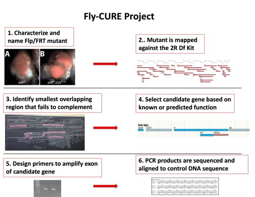
The Fly-CURE
The Fly-CURE is a national CURE that simultaneously addresses many of these issues. Fly-CURE was started at the University of Detroit Mercy in 2012 and is based on a genetic screen in Drosophila looking for conditional regulators of developmental signaling, cell growth control, and cell division (Kagey, Brown, & Moberg, 2012). In each class section of the Fly-CURE, students work on a novel mutant from the genetic screen. Throughout the semester the students name their mutant; characterize the mosaic phenotype; genetically map the location of the mutation; sequence potential candidate genes; and present their work in written and oral formats.
Overview of Fly-CURE Curriculum
In 2014, the Fly-CURE began to expand to the other institutions. The pilot implementation of the Fly-CURE at the University of Evansville provided evidence that this project could be successfully expanded across the country, dramatically increasing the number of undergraduate STEM majors impacted by this project. Initially, 7 institutions participated in an expanded pilot and in 2021, the Fly-CURE was awarded an NSF IUSE grant to expand the project to 20 institutions. The goals of this grant are to assess the impact of this experience on student attitudes towards science and on key learning objectives in genetics.
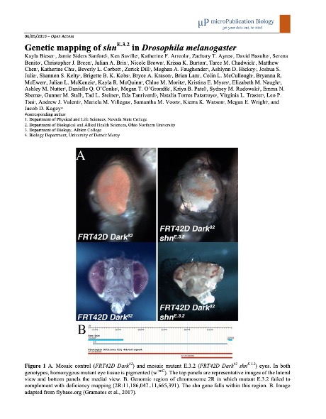
Currently, the Fly-CURE is being taught at sixteen institutions including PUI’s, a CC, and MSI’s. Faculty implementers have a wide range of previous fly experience: some run their own Drosophila research lab, while others had no prior experience with Drosophila before this project. To date, over 500 undergraduate researchers have contributed to the mapping of 14 novel Drosophila mutants, and 6 research papers describing this work have been published with 358 student co-authors across institutions (i.e. (Bieser et al., 2019)).
Bieser K, et al. 2019 Micropublications
Adapting to the COVID-19 Pandemic
When traditionally delivered college courses and labs became disrupted during the COVID-19 pandemic, Fly-CURE pivoted to create materials that allowed for different modes of implementation. Faculty taught the Fly-CURE in person, as a hybrid course, or as a fully online virtual CURE. To accommodate hybrid and virtual courses, Fly-CURE faculty collaborated to create virtual datasets of fly crosses typically viewed in person for students to analyze virtually. For other methods, some faculty created videos performing wet-lab protocols for students to continue participation in the project from home. Our assessment data found that students who analyzed virtual datasets were able to map and characterize a novel Drosophila mutant as successfully as those in traditional in-person labs. Our adaptation provides a model for other CURE and inquiry-based undergraduate laboratory courses to generate virtual/online modules of data analysis. The use of virtual datasets not only accommodated differential instruction during the COVID-19 pandemic, but also provides a model for providing virtual research experiences for under-resourced institutions and training materials for new faculty implementers.
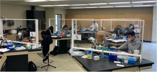
Fly-CURE being taught during the COVID-19 pandemic
Looking Forward
As the Fly-CURE project expands to additional institutions, we have incorporated the expertise and experiences of Fly-CURE faculty to continue improving and expanding the scope of the Fly-CURE project. For example, faculty have added whole genome sequencing and bioinformatics analysis, behavioral assays, and CRISPR analysis of the original mutants found in the genetic screen. We hope to continue to utilize these mutants to bring new techniques and ask new research questions within the classroom setting. Faculty are also conducting new genetic screens and collaborating with the Drosophila community to establish a large pool of mutants for future study by Fly-CURE students.
If you’re interested in learning more about the Fly-CURE, please reach out to one of the Co-PIs:
Kayla Bieser, Nevada State College, kayla.bieser@nsc.edu
Jacob Kagey, University of Detroit Mercy, kageyja@udmercy.edu
Joyce Stamm, University of Evansville, js383@evansville.edu
Alysia Vrailas-Mortimer, Illinois State University, admorti@ilstu.edu
Reference:
Bieser, K., Sanford, J., Saville, K., Arreola, K., Ayres, Z., Basulto, D., . . . Kagey, J. (2019). Genetic mapping of shn(E.3.2) in Drosophila melanogaster. MicroPubl Biol, 2019. doi:10.17912/micropub.biology.000118
Estrada, M., Burnett, M., Campbell, A. G., Campbell, P. B., Denetclaw, W. F., Gutierrez, C. G., . . . Zavala, M. (2016). Improving Underrepresented Minority Student Persistence in STEM. CBE Life Sci Educ, 15(3). doi:10.1187/cbe.16-01-0038
Holm, B., Carter, V. C., & Woodin, T. (2011). Vision and change in biology undergraduate education: Vision and change from the funding front. Biochem Mol Biol Educ, 39(2), 87-90. doi:10.1002/bmb.20502
Kagey, J. D., Brown, J. A., & Moberg, K. H. (2012). Regulation of Yorkie activity in Drosophila imaginal discs by the Hedgehog receptor gene patched. Mech Dev, 129(9-12), 339-349. doi:10.1016/j.mod.2012.05.007
Shaffer, C. D., Alvarez, C. J., Bednarski, A. E., Dunbar, D., Goodman, A. L., Reinke, C., . . . Elgin, S. C. (2014). A course-based research experience: how benefits change with increased investment in instructional time. CBE Life Sci Educ, 13(1), 111-130. doi:10.1187/cbe-13-08-0152
I would occasionally hear hints of something so transgressively bizarre that it made my head spin: cancer cells were having sex
Dr Kat Arney
In the latest episode of the Genetics Unzipped podcast, we’re exploring groundbreaking discoveries about the secret sex lives of cancer cells, and what it means for our understanding of tumour growth, evolution and treatment. Dr Kat Arney tells the story of how we discovered cancer cells were having sex, plus we look into why female tumours and male tumours act so differently.
If you enjoy the show, please do rate and review on Apple podcasts and help to spread the word on social media. And you can always send feedback and suggestions for future episodes and guests to podcast@geneticsunzipped.com Follow us on Twitter – @geneticsunzip

 (No Ratings Yet)
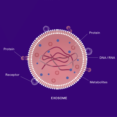

 (4 votes)