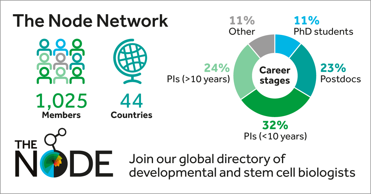
In January 2020 we launched the Node Network and now, two years later, we are delighted to announce that we have over 1000 members from 44 different countries. Read on to find out more about how our global directory of developmental and stem cell biologists can help you.
What is the Node Network?
The Node Network is a directory of developmental and stem cell biologists and is designed to help those organising meetings, assembly committees, seeking speakers or looking for referees to identify individuals who would not otherwise come to mind.
Who is the Network for?
The Node Network is entirely inclusive – anyone from the developmental and stem cell biology community, at any career stage, can add themselves to the directory to become members of the Network. Likewise, anyone searching for reviewers, speakers or committee members can register to access the directory. Users can perform searches based on scientific field, model organism and place of work, but also on aspects of diversity such as gender, race/ethnicity, LGBTQ+ identity and disability status that members can choose to share if they wish.
Why should you sign up?
We caught up with some of our members and users to find out how and why they use the network:
“I often turn to the Node Network to help me identify referees for papers I handle at Development. It’s allowed me to select referees that I wouldn’t normally think of and to find really great ECR referees.” – James Briscoe, Editor-in-chief at Development
“Having a directory of colleagues is useful, whether you are looking for someone in particular or searching for specific keywords/discipline. In combination with the Node, it is a way for junior scientists to share their views and open discussions with peers, which is a precious way of keeping in touch in those times.” – Gaëlle Recher, Permanent Researcher, Biof lab, CNRS
“Postdocs are some of the most reliable reviewers around, but they are not always easy to spot online. The Node Network makes it easy to find and contact postdocs with appropriate expertise, providing a diverse pool of potential paper referees.” – Miguel Branco, QMUL, Editor for PLoS ONE and Epigenetics Communications
“I frequently use the Node Network to expand my referee pool so that the opportunities to peer review are spread more equally between all members of the research community. It’s also a great resource to find potential authors from whom I could commission articles.” – Alex Eve, Reviews Editor at Development
As part of the birthday celebrations, we are running a ‘Promoting yourself as an ECR’ event hosted by FocalPlane, the Node Network and preLights on Wednesday 23 February at 3pm GMT. Come along and listen to the discussions with our panellists and find out more about the Node Network from our Community Manager and Development Editors. If you can’t make the event and have any feedback or suggestions of how we can improve the Node Network send us an email at thenode@biologists.com
Developmental biologists might be interested in a just-published article about Lewis Wolpert’s famous saying, ‘“Not birth, marriage or death but gastrulation”: the life of a quotation in biology’. The piece reconstructs the rich and surprising history of Wolpert’s dictum—including a conference dinner in Antwerp, Jonathan Slack’s From Egg to Embryo and a poster derived from an undergraduate project in Florida—and discusses its uses in teaching, research and public engagement. This uncovers some little-known history of developmental biology, and makes a case for the importance of quotations in communicating recent science.
Howdy all, As promised, here’s a post with book recommendations for developmental biologists.
~First, there are two books that together provide a simply exceptional entry into the field. They will be entertaining for developmental biologists looking for a broader view, but also represent good recommendations to friends and relatives hoping to grasp what the heck we are doing:
Coming to Life: How Genes Drive Development, by Christiane Nüsslein-Volhard. Written by one of our own Nobel Laureates, this book presents all you really need to know about developmental biology in a brisk and lively 150 pages, accompanied by exceptionally cogent illustrations. Undeniably my favorite in the genre.
Life Unfolding: How the Human Body Creates Itself, by Jaime Davies. This very readable, if somewhat dense, book focuses more on human development and gets into many of the gritty details passed over by Coming to Life.
~Two additional books that do the same thing, but from the vantage point of the early 1990s and early 1960s, respectively. Both are written by absolute giants in the field, people whose ideas very much molded our modern practice, These provide a very entertaining glance back in time, and both are geared to a popular audience.
~Next are two books that offer a glimpse into the minds of working developmental biologists.
Egg & Ego: An Almost True Story of Life in the Biology Lab, by Jonathan Slack focuses on his work that of others in the race to link growth factors to embryonic induction and the Spemann-Mangold Organizer in Xenopus. Find out which Xenopus hot-shot is “tall and bluff.”
A History of Embryology, by Joseph Needham is a prolix tome that leaves no corner of European thought on embryos unturned from ancient times to the close of the 18th Century. He concludes with a plea for “a theoretical embryology suited in magnitude and spaciousness to the wealth of facts which contemporary investigators are accumulating day by day.” Guess we all gotta keep our day jobs.
~Two books for thinking about developmental biology as it relates to human form:
Mutants: On Genetic Variety and the Human Body, by Armand Leroi is an exceptionally well-written book explaining developmental biology through discussion of human structural variation. The title has aged poorly, but the book is spectacular.
~Two great books about evolution that nonetheless brought developmental biology to the masses.
The Human Embryo, Aristotle and the European Tradition, Ed. G.R. Dunstan. This collection of essays edited by a central figure in 20th Century medical ethics provides an excellent introduction to ancient European thought about embryos.
Black Apollo of Science: The Life of Earnest Everett Just, by Kenneth Manning is surely the most important (and best) biography of a developmental biologist. Charting Just’s experiences as a Black man in science in the early 20th Century, the book is heartbreaking in its humanity. But the science will be exhilarating to developmental biologists.
The Heritage of Experimental Embryology: Hans Spemann and the Organizer, by Viktor Hamburger is an excellent first-person account of the heady days of experimental embryology. We can thank his narrative of Hilda Mangold’s life in the Appendix of this book for her finding her rightful place in the minds of modern developmental biologists.
From Egg to Embryo: Regional Specification in Early Development (Second Edition), by Jonathan Slack is written for specialists but provides an outstanding overview of the state of the art in about 1990, by which time the major themes of modern developmental biology were squarely in focus. Provides and exceptional primer on the big picture of our field; great for new students.
On Growth and Form, by D’Arcy Wentworth Thompson is a book that Steven Jay Gould describes as the “greatest work of prose in twentieth-century science.” A heralded masterpiece, its quantitative approach was prescient to say the least!
The Dog Stars, by Peter Heller is a novel that has nothing whatsoever to do with developmental biology. But it reads like a mashup of A River Runs Through It and Mad Max and I really liked it and you might want to read it.
The Stowers Institute for Medical Research (Kansas City, Missouri) is now accepting applications for Stowers Research Scholars, a mentored 1-year postbaccalaureate research fellowship. Through guided research experience and academic career mentoring, this program aims to increase the number of NIH-defined underrepresented students in fundamental biological research. This year we are accepting up to three students.
This opportunity is open to US citizen or permanent residents from diverse backgrounds with a BA or BS in a STEM field who are interested in pursuing graduate education in basic biological sciences. Compensation is highly competitive and prior laboratory experience is not required.
1. The worst aspect of working as a scientist is the never ending & all consuming need to secure funds. But there is a simple solution. It will work, it will increase diversity, empower scientists, & improve research outcomes. But the science power structure is arrayed against it
Our application period is open. We have the best REU mentors and offer an amazing opportunity to grow as a young scientist, in a very special place @MBLScience! pic.twitter.com/l2RnwfqJnj
— Veronica G Martinez Acosta (@VeronicaGMarti2) January 14, 2022
On-the-job training?:
If I hadn't packed up and moved my postdoc lab to a new institution in 2020 I don't think I would know what needs to be done to build a lab, or how to do those things and prioritize certain tasks. Why is this not a part of postdoc training?
Mine: – always have a presentation ready – update your CV as you go along – write it down – you WON'T remember – take calculated risks – be true to yourself#science#WednesdayMotivation .@AcademicChatter
A beautiful story on the impact an individual can make on a child’s life by engaging with them:
Thinking about biological collections lately I'm reminded of the time that somehow at age 4, I got a personal tour of the @UCBerkeley entomology collection (Essig?) from a kind scientist (anybody know who?!). Asked my parents how on earth THAT happened, and it turns out…🤯
So thrilled to see @Edutab_Africa bring Foldscope to remote communities in Kenya. Start of a bigger resolution to focus on scale up in Africa this year for us! “Life long love for science” sums it up very well. https://t.co/F0ATI9CMin
If you would like to contribute to our ‘Developing news’ blog, please get in touch at thenode@biologists.com. If you are interested in writing preLights, you can find more information here.
In our first SciArt profile of 2022, we hear from Diego Galagovsky, a postdoc with a passion for using cartoons to communicate science.
Where are you originally from and what do you work on now?
I am originally from Salta, Argentina. After moving to Buenos Aires for my PhD, then Dijon, France for a postdoc, I now live in Jena, Germany where I am working on a second postdoc.
Were you always going to be a scientist?
Since I was very young, I have been curious and interested in understanding how things work. As far as I remember, I got interested in dinosaurs when I was 4 or 5 years old and that became the gateway into science. I started reading books, increasing in complexity and getting interested in all areas of science, reading about animals, ecology, evolution, anthropology and history, physics and astronomy. In school and later in high school, I adored all classes linked to science, and especially biology. And as a kid I would always say that I wanted to be a scientist.
And what about art – have you always enjoyed it?
At the same time, I always enjoyed artistic expression. As a kid I took crafts classes and learned the basics for drawing. It was fun. I was always combining it with my love for science, drawing animals, dinosaurs and planets. I also loved cartoons and learned to draw by copying my favourite characters. Later, I became interested in comic books and started trying to emulate the style and make my own.
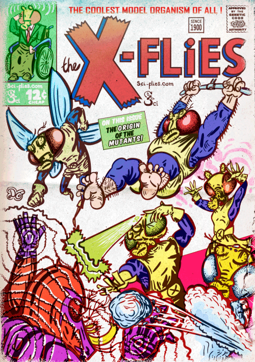
The X-Flies I like X-Men comics a lot. For an article about mutations, I did this homage to the cover of 1961 X-Men #1. I liked the characters and would love to continue using them for more articles.
What or who are your most important artistic influences?
My love for cartoons and comic books has had the greatest impact on my style. I take a lot of my inspiration from cartoons I loved throughout my life and from my favourite comic books. I also like visual arts and design in general, so I try to mix in whatever I find nice and appealing. My main interest is in experimenting and learning something new every time I decide to make a drawing.
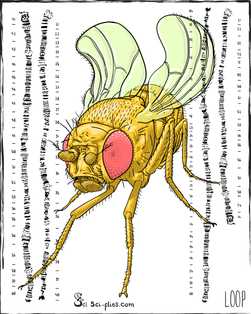
Loop I participated in Inktober 2021. It was an excuse to work hard on drawing and try new things. Here I paid tribute to a beloved fly line, the Curly-O balancer line. Balancer lines have rearranged chromosomes which I depict here next to the fly.
How do you make your art?
I work mainly digitally. I like the results I obtain, and I also find it comfortable because I can work almost anywhere, it takes up little space and allows me to work on several projects at the same time. I have been interested in digital art since I was very little. As a kid I never had access to fancy software or someone to teach me. I remember using basic software and pushing it as far as I could. With the popularization of the internet, I finally started learning to use more professional software design. I learned enough to then start self-teaching and experimenting. Regarding the process of my work, it usually involves a lot of thinking until I get the final idea for the subject and the composition. Then as I work it starts evolving and the initial idea starts changing. It is a very rewarding and stimulating process.
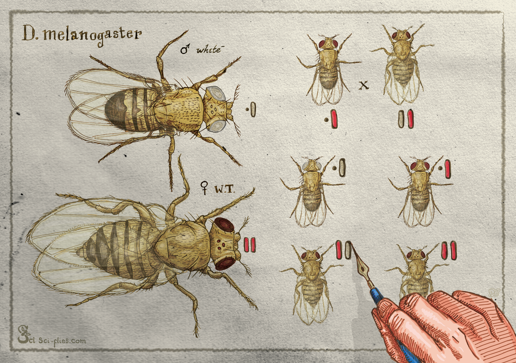
Morgan Genetics One of the first articles I wrote on our website was about genetics. I wrote about Mendel and Morgan. To illustrate it I drew the crosses Morgan performed with his white-eyed mutant, as if he was drawing them with ink and watercolours.
Does your art influence your science at all, or are they separate worlds?
Now I am working mainly on drawings and illustrations that connect directly to my scientific interests. I am doing works that are aimed at not only being aesthetically pleasing but also educational. I want them to be interesting enough to attract people to learn about science.
My art also influences my science in the sense that it has helped me better communicate my projects and my results. I like visual storytelling and understanding this language has helped me a lot with the communication aspect of my scientific work. I’ve also done figures and graphical abstracts for others in which I infused a bit of the artistic interests I had at the time.
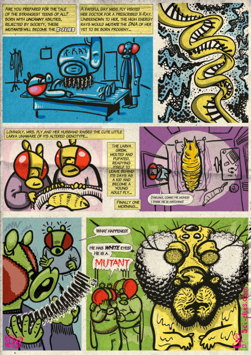
Mutant On our website, we did an article about mutations and I illustrated the use of x-rays to generate mutations in the style of horror/super-hero over-the-top origin story comic books.
What are you thinking of working on next?
I have been working on projects that involved thinking fast and working on a short schedule. As part of trying to work on science education and dissemination, I have been trying to take on challenges online that involve producing drawings every day, like Inktober or now an Advent Calendar featuring Drosophila flies and the science we do with them. I have also made a sticker app for WhatsApp with the same objective, and now I plan to explore that further: can we use stickers to interest people in science? Next, I want to experiment with the visual storytelling format of comic books, and how to use it to tell science online in a more engaging way. I have many ideas I want to explore in those areas.
Thanks to Diego and all the other SciArtists we have featured so far.You can find the full list here. We’re always on the lookout for new people to feature in this series – whatever kind of art you do, from sculpture to embroidery to music to drawing, if you want to share it with the community just email thenode@biologists.com (nominations are also welcome!)
Do not miss the Weinstein Cardiovascular Development and Regeneration Conference in Marseille (France) on 12-14th May, our first time in the South of France.
We are grateful to all our sponsors and specifically the Company of Biologists
To get information about the program, registration, and housing, visit the website:
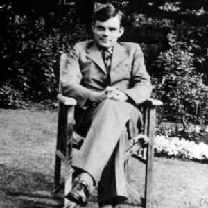
In the first episode of a new season of Genetics Unzipped, the Genetics Society podcast, presenter Kat takes a trip back to the 1950s to find out how one of the world’s greatest minds – Alan Turing – revealed the maths behind some of the deepest mysteries of life, from the patterning of stripes on a zebra to the spots on a leopard, and even the formation of bones in your own body.
If you enjoy the show, please do rate and review on Apple podcasts and help to spread the word on social media. And you can always send feedback and suggestions for future episodes and guests to podcast@geneticsunzipped.com Follow us on Twitter – @geneticsunzip
The Turing Centre for Living Systems (CENTURI) wishes to attract talented PhD students to the Luminy campus. To do so, CENTURI will fund up to 10 PhD positions to start in 2021. PhD students will work in an interdisciplinary life science environment, and have backgrounds in any of the following fields: cell or developmental biology, immunology, neurobiology, biophysics, theoretical physics, computer science, bioinformatics, applied mathematics, engineering.
Candidates can either apply to one of the advertised CENTURI projects or submit their own project, providing that they meet the application criteria and that their application is supported by at least 2 host labs.
PhD students will be co-supervised by two or three supervisors from our community. Candidates can apply to a maximum of three projects.
Do not hesitate to contact the projects’ supervisors for more information.
Applications must be submitted via the project’s application form and must be written in english.
Deadline for application: February 16, 2022
Duration: 3 years
On-site or video interviews: April 26 to April 27, 2022
Expected profile – selection criteria
Candidates will be evaluated based on the following criteria:
Academic achievements
Past research experience (internships, master thesis)
Interest to work in a multidisciplinary research environment
Enthusiasm and communication skills
To apply please fill the form associated to each project. Applications must include the following documents, in English (compiled into a single PDF file):
CV
cover letter
transcript of your MSc’s grades (M1 and M2 if available)
2 letters of recommendation must also be sent by your references. Please note that an automatic email will be sent to them so that they can upload their recommendation letter. We invite you to contact them to make sure that they have received the notification email.
Who are we?
CENTURI brings together leading institutes in biology, physics, mathematics, computer science and engineering to decipher the complexity and dynamics of living systems. CENTURI offers an exceptional international environment for the development of interdisciplinary projects, in developmental biology, immunology and neurosciences.
CENTURI is mainly located on the Luminy campus of Aix-Marseille University and is affiliated to Aix- Marseille University, CNRS, INSERM and École Centrale Marseille.
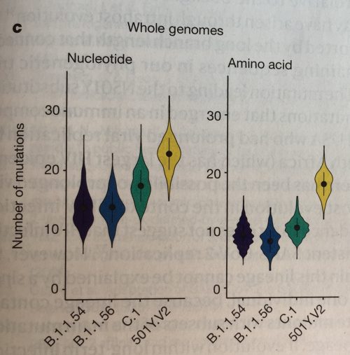
At the end of the year people take stock and reminisce. This also applies to data visualization scientists, who like to review the best visualizations of 2021. I enjoy Maarten Lambrechts summary of all the “Best of 2021” posts [note added: by now this post is included in his list!].
While musing whether to keep or toss my Nature print issues (still undecided!), I browsed through all the science visualizations of 2021. Well, I mainly checked the biology and medicine figures as I really do not understand enough of physics to comment on their charts and plots. Some visualizations and themes really stood out to me – so I decided to summarize the top 10 science visualization trends of 2021 for you.
[Notes: (1) all photos show anonymous excerpts for educational purposes from Nature articles 2021. (2) because of technical difficulties this article was already posted on my personal site.]
Viva viridis!
The viridis color-scheme is now omnipresent. Viridis was developed by Stefan van der Walt and Nathaniel Smith in 2015 as the default sequential color map for matplotlib. Their goal was to create a color scheme in which color changes are perceptually uniform, and to replace the non-linear Jet/Rainbow-color schemes used previously for sequential data.
Viridis quickly gained popularity as we can see in the many examples below. By now viridis is however no longer only used for sequential data. Instead we also see it being applied to diverging and categorical data, which may be not exactly ideal at times. But for now, let’s celebrate the end of rainbow color schemes!
Charts with viridis color-scheme colors
Various charts with the viridis-color scheme.
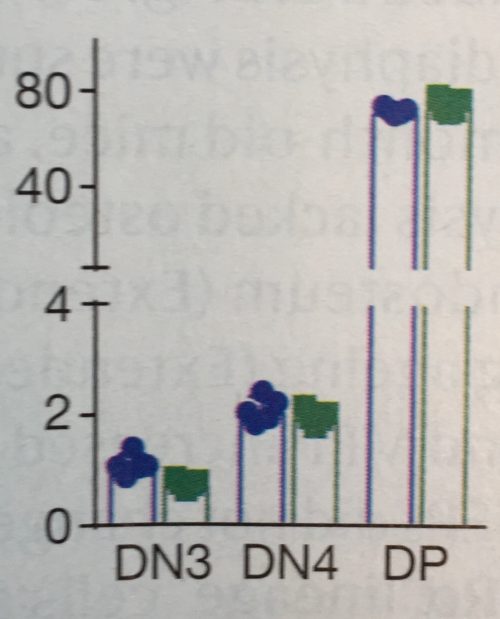
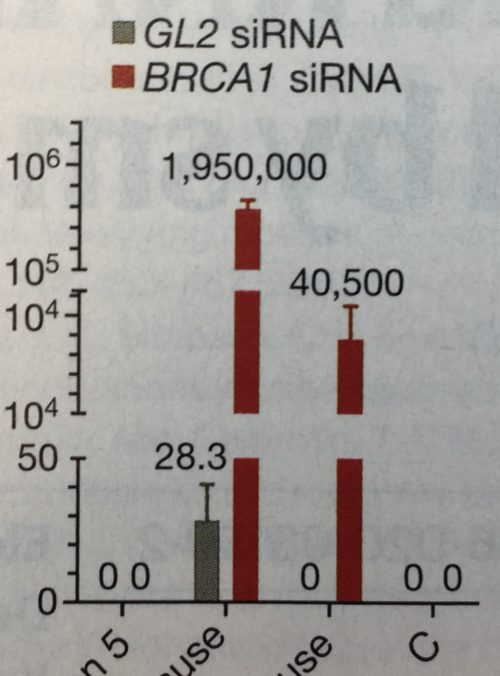
2. Still trending: axis breaks
Yes, axis breaks, necessary or not, are unfortunately still a thing in 2021. And so far, no end in sight. Every time I teach a Data Viz Course, I challenge the students that all axis breaks are avoidable – I have yet to see one where the break really was necessary. Send me yours!
Maybe necessary, unnecessary, and a really-really unnecessary axis break.
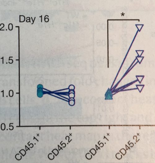
3. Hello slope-charts
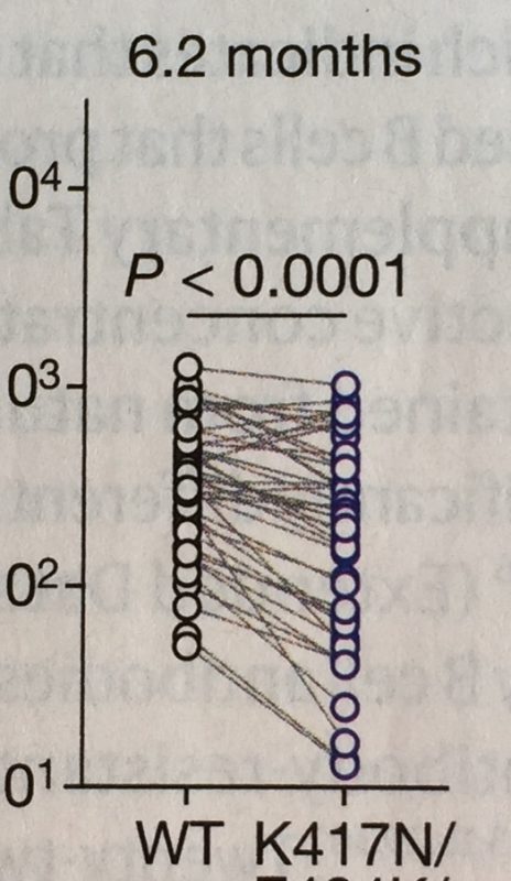
In 2016 the #BarBarChart initiative called for a ban of bar charts for data distributions. They were successful and I have not any in 2021. At that time, Tracey Weissgerber published a notable paper encouraging the use of a slope chart for dependent observations. It seems the science world listened again: numerous papers now use slope charts to illustrate dependent measurements of specimens, for example mice before and after treatment, responders and non-responders in cohorts etc.
Slope-charts for dependent measurements.
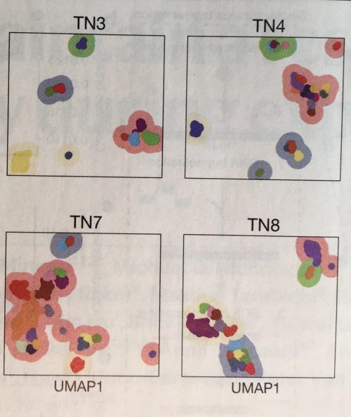
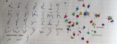
4. tSNE, UMAP, PCA
There don’t seem to be any papers that don’t have at least one dimension reduction plot. t-SNE, UMAP and PCA are omnipresent. These plots are however rarely explained in articles and figure legends, and I suspect even more rarely understood by audiences. In my courses participants are often not familiar with them – not surprising given that they are a recent addition in statistics and still heavily researched in the vis community! To educate your students refer them to these resources from Claus O. Wilke and StatQuest.
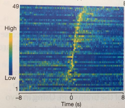
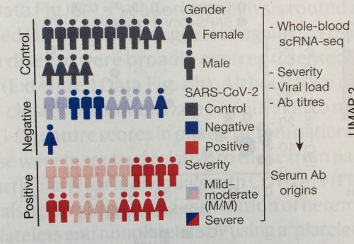
Eye-catching plots like tSNE, UMAP and PCA are useful for genomics, single cell sequencing and even letter-analysis in brain-robots!
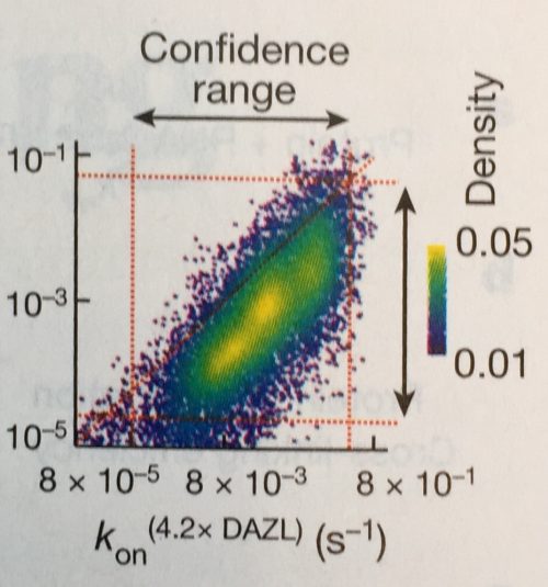
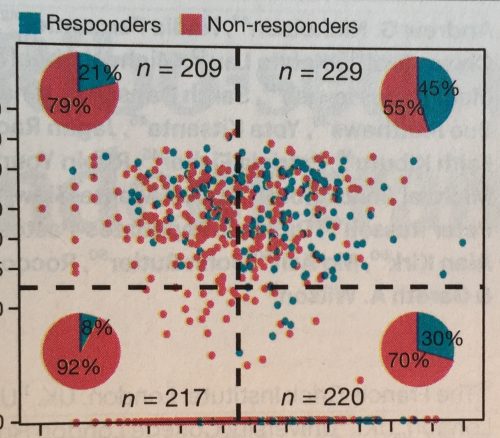
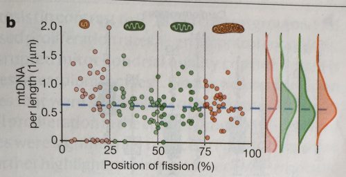
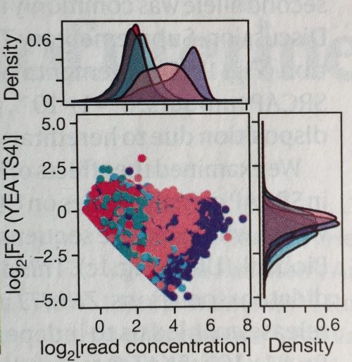
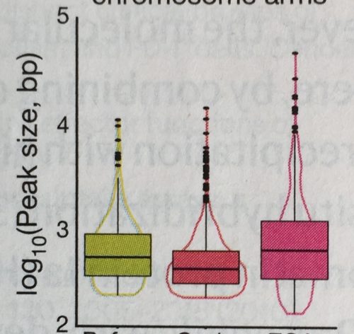
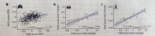
5. Mixing charts
A big fashion trend of 2021 was mixing styles and patterns, and it seems this did spill over to the visualization world. Researchers have become quite experimental with mixing chart types (see also earlier post). I’ve seen a number of scatter plots showing the data distribution summarized along each axis: above for the x-axis and left for the y-axis. I’ve also seen pie charts summarizing the quadrants of a scatter-plot, charts mixing violin and box-plots, of course box-plots with data points, and many more.
Mixed-plots: it has become useful to merge plot types where needed.
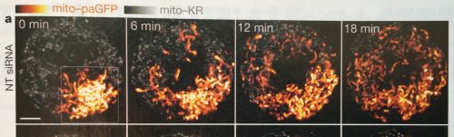
6. Pictograms
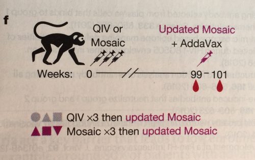
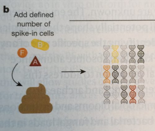
Often novel insights come from a new methodological approach. To familiarize audiences with a new method, papers now regularly include a sketch of the experimental procedure. And, good news, these have begun to look rather nice and clear, whether it’s a stool preparation method, an approach stem cell differentiation, or for a mouse neurobiology set-up.
Pictograms help explain experimental designs.
7. Pictograms for labeling
Pictograms are not only used for explaining procedures, but also often help to quickly orient audiences. Pictograms are for example used instead of text: I’ve seen pictograms as a title, pictogram to name a leaf in a phylogenetic tree, or even a pictogram chart. Overall pictograms seem to be most popular in mouse molecular genetics and evolutionary biology. Go figure.
Pictograms can make titles and labels understandable at first glance.
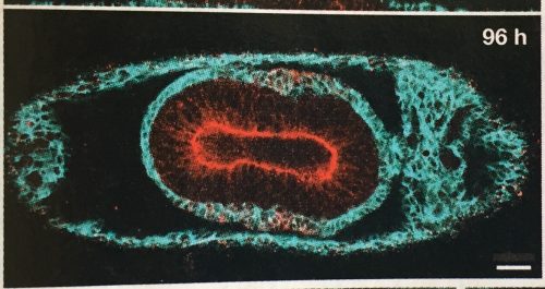
8. Images now come with scale bars!
Scale bars in images are apparently here to stay. A few years ago, I had noticed that scale information was quite often missing in scientific images, even in high profile journals. Eventually this observation prompted a collaborative project to systematically screen the quality of image data in publications.
This year the tide has definitely shifted. Except for a few cases, scale bars are now visibly placed on all images; even in plant and histology images, two fields so far quite notorious for omitting scale information.
Now regularly with scale-bars: images!
9. Colors in images: still problematic
Despite many recent efforts, most recently a technology feature in nature feat. your truly, no end is in sight for images that generously mix red and green, making it illegible for many color-blind people. In addition, often images that show false-color scales do not come with a legend explaining the color intensities.
A glimpse of hope: I’ve seen a color legend once this year (still without quantities in legend), which is already a 100% increase from previous years! And, rescue is near, with QUAREP we have a collaborative consortium addressing quality of light microscopy. Join our working group that develops guidelines for image visualization in publications!
Include legends for intensity values – ideally with quantifications.
10. More space for figures!
Figures get more space in publications. This is quite easy in electronic publications, but also in print editions half or even full-page figures are becoming a sight. This makes it quite enjoyable to read some of the figures, which before were entirely illegible due to being squeezed into a small space.
More space for figures means less space for other sections of a paper. Often materials and methods are now entirely moved to the supplementary materials and citations are limited to a few only.
The figure below for example each took up 2/3 of a page – lovely!
 (No Ratings Yet)
 (1 votes)
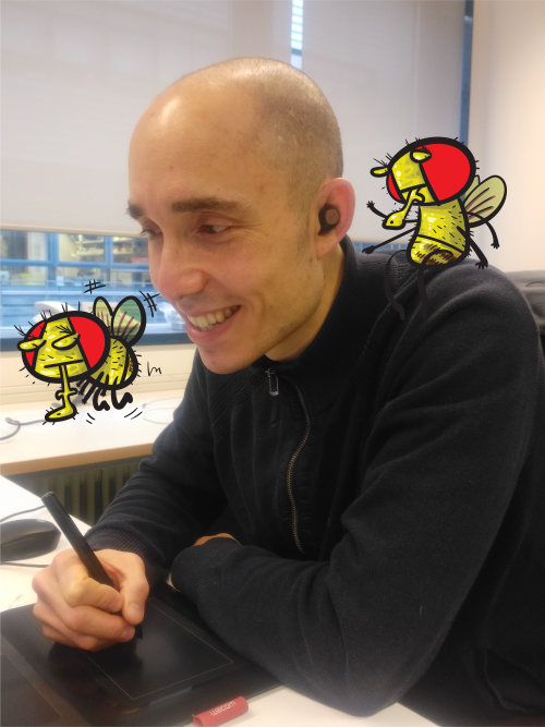


