Looking for contributors
Posted by Patricia Ybot-Gonzalez, on 9 April 2021
Posted by Kat Arney, on 8 April 2021
In this episode we’re taking a look at the history of gene editing, from the early days of restriction enzymes in the 1960s through to the CRISPR revolution and the very latest base editing techniques.
But while these tools are undeniably powerful and hold great promise for treating disease, with great power comes great responsibility: what are the acceptable limits of genome engineering in humans, and will we see more CRISPRd babies in the future?
Genetics Unzipped is the podcast from The Genetics Society. Full transcript, links and references available online at GeneticsUnzipped.com.
Subscribe from Apple podcasts, Spotify, or wherever you get your podcasts.
Head over to GeneticsUnzipped.com to catch up on our extensive back catalogue.
If you enjoy the show, please do rate and review on Apple podcasts and help to spread the word on social media. And you can always send feedback and suggestions for future episodes and guests to podcast@geneticsunzipped.com Follow us on Twitter – @geneticsunzip
Posted by the Node, on 6 April 2021
For last month’s Quintay 2020 Development cover competition, just under a thousand votes were cast from across the world in order to pick the best image taken by students of the 2020 class of the MBL Practical course in Developmental Biology in Quintay, Chile. The votes are in and we can now announce the podium positions:
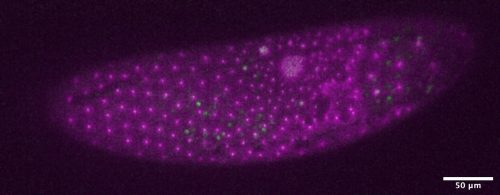
3rd place (12% of the vote) – Parhyale by Diana Carolina Castañeda-Cortés, Nicolas Eduardo Cumplido Salas, Felipe Andres Gajardo Escobar
2nd place (14% of the vote) – Drosophila larval body wall by Pablo Guzman Palma
Congratulations Tonatiuh! The image will appear on the cover of a future issue of Development – look out for it.
The 2022 version of Quintay is hopefully going to take place: check out the homepage for more information.
Posted by the Node, on 1 April 2021
Welcome to our monthly trawl for developmental biology (and related) preprints.
The preprints this month are hosted on bioRxiv, arXiv and preprints.org – use these links to get to the section you want.
Immunoglobulin superfamily receptor Junctional adhesion molecule 3 (Jam3) requirement for melanophore survival and patterning during formation of zebrafish stripes
Dae Seok Eom, Larissa B. Patterson, Raegan R. Bostic, David M. Parichy
Notch signaling represses cone photoreceptor formation through the regulation of retinal progenitor cell states
Xueqing Chen, Mark M. Emerson
TGIF1 is required for chicken ovarian cortical development and generation of the juxtacortical medulla
Martin Andres Estermann, Claire E Hirst, Andrew T Major, Craig A Smith
Human brain organoids assemble functionally integrated bilateral optic vesicles
Elke Gabriel, Walid Albanna, Giovanni Pasquini, Anand Ramani, Natasa Josipovic, Aruljothi Mariappan, Friedrich Schinzel, Celeste M Karch, Guobin Bao, Marco Gottardo, Jürgen Hescheler, Veronica Persico, Silvio O Rizzoli, Janine Altmüller, Giuliano Callaini, Argyris Papantonis, Olivia Goureau, Volker Busskamp, Toni Schneider, Jay Gopalakrishnan
A novel juxtamembrane basolateral targeting motif regulates TGF-β receptor signaling in Drosophila
Aidan J. Peterson, Stephen J. Murphy, Melinda G. Mundt, Maryjane Shimell, Edward B. Leof, Michael B. O’Connor
Strength of interactions in the Notch gene regulatory network determine lateral inhibition patterning
Héctor Sánchez-Iranzo, Aliaksandr Halavatyi, Alba Diz-Muñoz
Duox generated reactive oxygen species activate ATR/Chk1 to induce G2 arrest in Drosophila tracheoblasts
Amrutha Kizhedathu, Piyush Chhajed, Lahari Yeramala, Deblina Sain Basu, Tina Mukherjee, Kutti R. Vinothkumar, Arjun Guha
Interaction of thyroid hormones and gonadotropin inhibitory hormone in the multifactorial control of zebrafish (Danio rerio) spermatogenesis
Maira S. Rodrigues, Hamideh P. Fallah, Maya Zanardini, Hamid R. Habibi, Rafael H. Nóbrega
Retracing Schwann cell developmental transitions in embryonic dissociated DRG/Schwann cell cocultures in mice
Venkat Krishnan Sundaram, Tatiana El Jalkh, Rasha Barakat, Camille Julie Isabelle Fernandez, Charbel Massaad, Julien Grenier
Dual control exerted by dopamine in blood-progenitor cell cycle regulation in Drosophila
Ankita Kapoor, A. Padmavathi, Tina Mukherjee
Pax6 regulates the morphological and electrophysiological development of mouse prethalamic neurons
Tian Tian, Idoia Quintana-Urzainqui, Zrinko Kozić, Thomas Pratt, David J. Price
Ezh2 mediates epigenetic regulation of osteoclastogenesis and bone remodeling in mice
Jin-Ran Chen, Oxana P. Lazarenko, Dongzheng Gai, Can Li, Michael L. Blackburn, Fenghuang Zhan
The feedback regulator Nord controls Dpp/BMP signaling via extracellular interaction with Dally in the Drosophila wing
Takuya Akiyama, Chris W. Seidel, Matthew C. Gibson
Paraxial mesoderm organoids model development of human somites
Christoph Budjan, Shichen Liu, Adrian Ranga, Senjuti Gayen, Olivier Pourquie, Sahand Hormoz
The proteome of remyelination is different from that of developmental myelination
Joana Paes de Faria, Maria M. Azevedo, Damaris Bausch-Fluck, Ana Seixas, Helena S. Domingues, Maria A. Monteiro, Patrick G.A. Pedrioli, Eduarda Lopes, Rui Fernandes, Chao Zhao, Robin J. M. Franklin, Bernd Wollscheid, Joao B. Relvas, Laura Montani
Development of early postnatal inhibitory function in the mouse medial prefrontal and primary somatosensory cortex
Katerina Kalemaki, Xiaxia Xu, Angeliki Velli, Ourania Christodoulou, Myrto Denaxa, Ileana L. Hanganu-Opatz, Domna Karagogeos, Kyriaki Sidiropoulou
A quantitative landscape of cell fate transitions identifies principles of cellular decision-making
M. Sáez, R. Blassberg, E. Camacho-Aguilar, E. D. Siggia, D. Rand, J. Briscoe
Foxq2 determines blue cone identity in zebrafish
Yohey Ogawa, Tomoya Shiraki, Yoshitaka Fukada, Daisuke Kojima
Ciliary Hedgehog signaling regulates cell survival to build the facial midline
Shaun Abrams, Jeremy F. Reiter
Conserved and context-dependent roles for Pdgfrb signaling during zebrafish vascular mural cell development
Koji Ando, Yu-Huan Shih, Lwaki Ebarasi, Ann Grosse, Daneal Portman, Ayano Chiba, Kenny Mattonet, Claudia Gerri, Didier Y.R. Stainier, Naoki Mochizuki, Shigetomo Fukuhara, Christer Betsholtz, Nathan D. Lawson
The enpp4 ectonucleotidase regulates kidney patterning signalling networks in Xenopus embryos
Karine Massé, Surinder Bhamra, Christian Paroissin, Lilly Maneta-Peyret, Eric Boué-Grabot, Elizabeth A. Jones
Modelling the impact of decidual senescence on embryo implantation in human endometrial assembloids
Thomas M. Rawlings, Komal Makwana, Deborah M. Taylor, Matteo A. Molè, Katherine J. Fishwick, Maria Tryfonos, Joshua Odendaal, Amelia Hawkes, Magdalena Zernicka-Goetz, Geraldine M. Hartshorne, Jan J. Brosens, Emma S. Lucas
Embryonic requirements for Tcf12 in the development of the mouse coronal suture
Man-chun Ting, D’Juan T. Farmer, Camilla S. Teng, Jinzhi He, Yang Chai, J. Gage Crump, Robert E. Maxson Jr.
The molecular logic of the self-organization of primitive streak and neuroectoderm fates
Luigi Russo, Hanna L. Sladitschek, Pierre A. Neveu
CA1 pyramidal cell diversity is rooted in the time of neurogenesis
Davide Cavalieri, Alexandra Angelova, Anas Islah, Catherine Lopez, Agnes Baude, Rosa Cossart
Somatic junctions connect microglia and developing neurons
Csaba Cserép, Anett D. Schwarcz, Balázs Pósfai, Zsófia I. László, Anna Kellermayer, Miklós Nyerges, Zsolt Lele, István Katona, Ádám Dénes
Two opposite voltage-dependent currents control the unusual early development pattern of embryonic Renshaw cell electrical activity
Juliette Boeri, Claude Meunier, Hervé Le Corronc, Pascal Branchereau, Yulia Timofeeva, François Xavier Lejeune, Christine Mouffle, Hervé Arulkandarajah, Jean Marie Mangin, Pascal Legendre, Antonny Czarnecki
MRI of Capn15 knockout mice and analysis of Capn 15 distribution reveal possible roles in brain development and plasticity
Congyao Zha, Carole A Farah, Vladimir Fonov, David A Rudko, Wayne S Sossin
Gut regulates brain synaptic assembly through neuroendocrine signaling pathway
Yanjun Shi, Lu Qin, Zhiyong Shao
Early mechanisms of whisker development: Prdm1 and its regulation in whisker development and evolutionary loss
Pierluigi Giuseppe Manti, Fabrice Darbellay, Marion Leleu, Bernard Moret, Julien Cuennet, Frederic Droux, Magali Stoudmann, Gian-Filippo Mancini, Agnès Hautier, Yann Barrandon
Oligodendrocyte Precursor Cells Sculpt the Visual System by Regulating Axonal Remodeling
Yan Xiao, Laura J Hoodless, Luigi Petrucco, Ruben Portugues, Tim Czopka
REST is a major negative regulator of endocrine differentiation during pancreas organogenesis
Meritxell Rovira, Goutham Atla, Miguel Angel Maestro, Vane Grau, Javier García-Hurtado, Maria Maqueda, Jose Luis Mosquera, Julie Kerr-Conte, Francois Pattou, Jorge Ferrer
Ecdysone coordinates plastic growth with robust pattern in the developing wing
André Nogueira Alves, Marisa Mateus Oliveira, Takashi Koyama, Alexander Shingleton, Christen Mirth
The NOTCH3 Downstream Target HEYL Regulates Human Airway Epithelial Club Cell Differentiation
Manish Bodas, Bharathiraja Subramaniyan, Andrew R. Moore, Jordan P. Metcalf, Sarah R. Ocañas, Willard M. Freeman, Constantin Georgescu, Jonathan D. Wren, Matthew S. Walters
A neural progenitor mitotic wave is required for asynchronous axon outgrowth and morphology
Jérôme Lacoste, Hédi Soula, Angélique Burg, Agnès Audibert, Pénélope Darnat, Michel Gho, Sophie Louvet-Vallée
PEG10 viral aspartic protease domain is essential for the maintenance of fetal capillary structure in the mouse placenta
Hirosuke Shiura, Ryuichi Ono, Saori Tachibana, Takashi Kohda, Tomoko Kaneko-Ishino, Fumitoshi Ishino
Persistent ventricle partitioning in the adult zebrafish heart
Catherine Pfefferli, Hannah R. Moran, Anastasia Felker, Christian Mosimann, Anna Jaźwińska
The CATP-8/P5A-type ATPase functions in multiple pathways during neuronal patterning
Leo T.H. Tang, Meera Trivedi, Jenna Freund, Christopher J. Salazar, Nelson J. Ramirez-Suarez, Garrett Lee, Maisha Rahman, Yu Wang, Barth D. Grant, Hannes E. Bülow
Osteoblast-Specific Wnt Secretion is Required for Skeletal Homeostasis and Loading-Induced Bone Formation in Adult Mice
Lisa Y Lawson, Michael D Brodt, Nicole Migotsky, Christopher Chermside-Scabbo, Ramya Palaniappan, Matthew J Silva
Fmrp regulates oligodendrocyte lineage cell specification and differentiation
Caleb A. Doll, Kayt Scott, Bruce Appel
FAM57B is a modulator of ceramide synthesis that regulates sphingolipid homeostasis and synaptic composition in the developing brain
Danielle L. Tomasello, Jiyoon L. Kim, Yara Khodour, Jasmine M. McCammon, Maya Mitalipova, Rudolf Jaenisch, Anthony H. Futerman, Hazel Sive
The cAMP effector PKA mediates Moody GPCR signaling in Drosophila blood-brain barrier formation and maturation
Xiaoling Li, Richard Fetter, Tina Schwabe, Christophe Jung, Hermann Steller, Ulrike Gaul
Cell competition removes segmental aneuploid cells from Drosophila imaginal disc-derived tissues based on ribosomal protein gene dose
Zhejun Ji, Jacky Chuen, Marianthi Kiparaki, Nicholas E. Baker
Vegfr3-tdTomato, a reporter mouse for microscopic visualization of lymphatic vessel by multiple modalities
Esther Redder, Nils Kirschnick, René Hägerling, Nils Hansmeier, Friedemann Kiefer
Pathogenic LRRK2 control of primary cilia and Hedgehog signaling in neurons and astrocytes of mouse brain
Shahzad S. Khan, Yuriko Sobu, Herschel S. Dhekne, Francesca Tonelli, Kerryn Berndsen, Dario R. Alessi, Suzanne R. Pfeffer
An inverse modelling study on the local volume changes during early morphoelastic growth of the fetal human brain
Z. Wang, B. Martin, J. Weickenmeier, K. Garikipati
Expression of Sonic Hedgehog and Pathway Components in the Embryonic Mouse Head: Anatomical Relationships Between Regulators of Positive and Negative Feedback
Crystal L Sigulinsky, Xiadong Li, Edward M Levine
Crosstalk in oxygen homeostasis networks: SKN-1/NRF inhibits the HIF-1 hypoxia-inducible factor in Caenorhabditis elegans
Dingxia Feng, Zhiwei Zhai, Zhiyong Shao, Yi Zhang, Jo Anne Powell-Coffman
Distinct Hypoxia-induced Translational Profiles of Embryonic and Adult-derived Macrophages
Nicholas S. Wilcox, Timur O. Yarovinsky, Prakruti Pandya, Vinod S. Ramgolam, Albertomaria Moro, Yinyu Wu, Stefania Nicoli, Karen K. Hirschi, Jeffrey R. Bender
Insights into in vivo adipocyte differentiation through cell-specific labeling in zebrafish
Paola Lepanto, Florencia Levin, Uriel Koziol, Leonel Malacrida, José L. Badano
YAP1 Regulates the Self-organized Fate Patterning of hESCs-Derived Gastruloids
Servando Giraldez, Eleonora Stronati, Ling Huang, Hui-Ting Hsu, Elizabeth Abraham, Kathy A. Jones, Conchi Estaras
Prolonged development of long-term potentiation at lateral entorhinal cortex synapses onto adult-born neurons
Nicholas P. Vyleta, Jason S Snyder
Svep1 stabilizes developmental vascular anastomosis in reduced flow conditions.
Baptiste Coxam, Yvonne Padberg, Katja Maier, Simone Jung, Eireen Bartels-Klein, Anna Szymborska, Lise Finotto, Christian S.M. Helker, Stefan Schulte-Merker, Didier Y.R. Stainier, Holger Gerhardt
Stress-driven tissue fluidization physically segments vertebrate somites
Elijah R. Shelton, Sangwoo Kim, Ben J. Gross, Ray Wu, Marie Pochitaloff, Irene Lim, Ellen M. Sletten, Otger Campàs
Scribble mutation disrupts convergent extension and apical constriction during mammalian neural tube closure
Alyssa C. Lesko, Raymond Keller, Ping Chen, Ann Sutherland
Epiblast morphogenesis is controlled by selective mRNA decay triggered by LIN28A relocation
Miha Modic, Igor Ruiz de Los Mozos, Sebastian Steinhauser, Emiel van Genderen, Silvia Schirge, Valter Bergant, Joel Ryan, Christopher B Mulholland, Rupert Faraway, Flora C Y Lee, Tajda Klobučar, Juliane Merl-Pham, Stephanie M Hauck, Micha Drukker, Sebastian Bultmann, Heinrich Leonhardt, Heiko Lickert, Nicholas M Luscombe, Derk ten Berge, Jernej Ule
Germ plasm anchors at tight junctions in the early zebrafish embryo
Nadia Rostam, Alexander Goloborodko, Stephan Riemer, Andres Hertel, Sabine Klein, Dietmar Riedel, Gerd Vorbrüggen, Roland Dosch
Slit2 is necessary for optic axon organization in the zebrafish ventral midline
Camila Davison, Flavio R. Zolessi
Actin dependent membrane polarization reveals the mechanical nature of the neuroblast polarity cycle
Bryce LaFoya, Kenneth E. Prehoda
Transthyretin promotes axon growth via regulation of microtubule dynamics and tubulin acetylation
Jessica Eira, Joana Magalhães, Nídia Macedo, Maria Elena Pero, Thomas Misgeld, Mónica M Sousa, Francesca Bartolini, Márcia A Liz
A folder mechanism ensures size uniformity among C. elegans individuals by coupling growth and development
Benjamin D. Towbin, Helge Grosshans
Neurexin and Frizzled signaling intercept axonal-transport at microtubule minus-ends to control synapse formation
Santiago Balseiro-Gómez, Yang Yue, Lin Shao, Selim Ҫetinkaya, Caroline Kuzoian, Kristen J Verhey, Shaul Yogev
The C. elegans PTCHD homolog PTR-4 is required for proper organization of the pre-cuticular apical extracellular matrix
Jennifer D Cohen, Carla E. Cadena del Castillo, Andres Kaech, Anne Spang, Meera V Sundaram
3D Cell Neighbour Dynamics in Growing Pseudostratified Epithelia
Harold F. Gómez, Mathilde S. Dumont, Leonie Hodel, Roman Vetter, Dagmar Iber
Intertissue mechanical interactions shape the olfactory circuit in zebrafish
P Monnot, G Gangatharan, M Baraban, K Pottin, M Cabrera, I Bonnet, MA Breau
Desmosomes polarize mechanical signaling to govern epidermal tissue form and function
Joshua A. Broussard, Jennifer L. Koetsier, Marihan Hegazy, Kathleen J. Green
Vinculin controls endothelial cell junction dynamics during vascular lumen formation
Maria P. Kotini, Miesje M. van der Stoel, Mitchell K. Han, Bettina Kirchmaier, Johan de Rooij, Markus Affolter, Stephan Huveneers, Heinz-Georg Belting
A genetic screen for regulators of muscle morphogenesis in Drosophila
Tiffany Ou, Gary Huang, Beth Wilson, Paul Gontarz, James B. Skeath, Aaron N. Johnson
A Kalirin Missense Mutation Enhances Dendritic RhoA Signaling and Leads to Regression of Cortical Dendritic Arbors Across Development
MJ Grubisha, T Sun, SL Erickson, L Eisenman, S Chou, CD Helmer, MT Trudgen, Y Ding, GE Homanics, P Penzes, ZP Wills, RA Sweet
Lamb1a regulates atrial growth by limiting excessive, contractility-dependent second heart field addition during zebrafish heart development
Christopher J. Derrick, Eric J. G. Pollitt, Ashley Sanchez Sevilla Uruchurtu, Farah Hussein, Emily S. Noёl
The zebrafish presomitic mesoderm elongates through compression-extension
Lewis Thomson, Leila Muresan, Benjamin Steventon
Extracellular vesicles synchronize cellular phenotypes of differentiating cells
Tomohiro Minakawa, Tetsuya Matoba, Jun K Yamashita
Extensive structural remodeling of the axonal arbors of parvalbumin basket cells during development
Kristina D. Micheva, Marianna Kiraly, Marc M. Perez, Daniel V. Madison
Regulation of apical constriction via microtubule- and Rab11-dependent apical transport during tissue invagination
Thao Phuong Le, SeYeon Chung
A cell-ECM mechanism for connecting the ipsilateral eye to the brain
Jianmin Su, Yanping Liang, Ubadah Sabbagh, Lucie Olejníková, Ashley L. Russell, Jiang Chen, Yuchin Albert Pan, Jason W. Triplett, Michael A. Fox
Brazil Nut Effect Drives Pattern Formation in Early Mammalian Embryos
Zheng Guo, Jie Yao, Xu Zheng, Jialing Cao, Zheng Gao, Shuyu Guo, Dandan Qin, Min Tan, Bo Wang, Fanzhe Meng, Jing Zhang, Lei Li, Jing Du, Yubo Fan
Expression and protein sequence analyses of zebrafish impg2a and impg2b, two proteoglycans of the interphotoreceptor matrix
M.E. Castellini, G. Spagnolli, E. Biasini, S. Casarosa, A. Messina
Semaphorin 3C attracts MGE-derived cortical interneurons in the deep migratory stream guiding them into the developing neocortex
Kiara Aiello, Jürgen Bolz
Knockout of zebrafish desmin genes does not cause skeletal muscle degeneration but alters calcium flux
Gulsum Kayman Kurekci, Ecem Kural Mangit, Cansu Koyunlar, Seyda Unsal, Berk Saglam, Bora Ergin, Merve Gizer, Ismail Uyanik, Niloufar Boustanabadimaralan Düz, Petek Korkusuz, Beril Talim, Nuhan Purali, Simon M. Hughes, Pervin R. Dincer
Mechanical bistability enabled by ectodermal compression facilitates Drosophila mesoderm invagination
Hanqing Guo, Michael Swan, Shicheng Huang, Bing He
Mechanically Sensitive HSF1 is a Key Regulator of Left-Right Symmetry Breaking in Zebrafish Embryos
Jing Du, Shu-Kai Li, Liu-Yuan Guan, Zheng Guo, Jiang-Fan Yin, Li Gao, Toru Kawanishi, Atsuko Shimada, Qiu-Ping Zhang, Li-Sha Zheng, Yi-Yao Liu, Xi-Qiao Feng, Dong-Yan Chen, Hiroyuki Takeda, Yu-Bo Fan
Impaired Cx43 gap junction endocytosis causes cardiovascular defects in zebrafish
Caitlin Hyland, Michael Mfarej, Giorgos Hiotis, Sabrina Lancaster, Noelle Novak, M. Kathryn Iovine, Matthias M. Falk
In vivo dissection of Rhoa function in vascular development using zebrafish
Laura M. Pillay, Joseph J. Yano, Andrew E. Davis, Matthew G. Butler, Keith A. Barnes, Vanessa L. Reyes, Daniel Castranova, Aniket V. Gore, Matthew R. Swift, James R. Iben, Amber N. Stratman, Brant M. Weinstein
Global analysis of cell behavior and protein localization dynamics reveals region-specific functions for Shroom3 and N-cadherin during neural tube closure
Austin T. Baldwin, Juliana Kim, John B. Wallingford
Tight junction ZO proteins maintain tissue fluidity, ensuring efficient collective cell migration
Mark Skamrahl, Hongtao Pang, Maximilian Ferle, Jannis Gottwald, Angela Rübeling, Riccardo Maraspini, Alf Honigmann, Tabea A. Oswald, Andreas Janshoff
Collagen polarization provides a structural memory for the elongation of epithelial anlage
Hiroko Katsuno-Kambe, Jessica L. Teo, Robert J. Ju, James E. Hudson, Samantha J. Stehbens, Alpha S. Yap
A feedback mechanism mediated by myosin II-dependent apical targeting of Rab11 vesicles reinforces apical constriction
Wei Chen, Bing He
Semaphorin3F Drives Dendritic Spine Pruning through Rho-GTPase Signaling
Bryce W. Duncan, Vishwa Mohan, Sarah D. Wade, Young Truong, Alexander Kampov-Polevoi, Brenda R. Temple, Patricia F. Maness
Oct1 recruits the histone lysine demethylase Utx to canalize lineage specification
Jelena Perovanovic, Yifan Wu, Zuolian Shen, Mahesh B Chandrasekharan, Dean Tantin
Super interactive promoters provide insight into cell type-specific regulatory networks in blood lineage cell types
Taylor M. Lagler, Yuchen Yang, Yuriko Harigaya, Vijay G. Sankaran, Ming Hu, Alexander P. Reiner, Laura M. Raffield, Jia Wen, Yun Li
Single cell trajectory modeling identifies a primitive trophoblast state defined by BCAM enrichment
Matthew Shannon, Jennet Baltayeva, Barbara Castellana, Jasmin Wächter, Samantha Yoon, Jenna Treissman, Hoa Le, Alexander G. Beristain
Cell-type-specific chromatin occupancy by the pioneer factor Zelda drives key developmental transitions in Drosophila
Elizabeth D. Larson, Hideyuki Komori, Tyler J. Gibson, Cyrina M. Ostgaard, Danielle C. Hamm, Jack M. Schnell, Cheng-Yu Lee, Melissa M. Harrison
Tissue-specific versus pleiotropic enhancers within the bric-a-brac tandem gene duplicates display differential regulatory activity and evolutionary conservation
Henri-Marc G. Bourbon, Mikhail H. Benetah, Emmanuelle Guillou, Luis Humberto Mojica-Vazquez, Aissette Baanannou, Sandra Bernat-Fabre, Vincent Loubiere, Frédéric Bantignies, Giacomo Cavalli, Muriel Boube
The Nestin neural enhancer is essential for normal levels of endogenous Nestin in neuroprogenitors but is not required for embryo development
Ella Thomson, Ruby Dawson, Chee Ho H’ng, Fatwa Adikusuma, Sandra Piltz, Paul Q Thomas
A silencer repressing redundant enhancer activities revealed by deleting endogenous cis-regulatory element of ebony in Drosophila melanogaster
Noriyoshi Akiyama, Shoma Sato, Kentaro M. Tanaka, Takaomi Sakai, Aya Takahashi
Two promoters integrate multiple enhancer inputs to drive wild-type knirps expression in the D. melanogaster embryo
Lily Li, Rachel Waymack, Mario Elabd, Zeba Wunderlich
Piecemeal regulation of convergent neuronal lineages by bHLH transcription factors in C. elegans
Neda Masoudi, Ralf Schnabel, Oliver Hobert
RNA polymerase II depletion from the inactive X chromosome territory is not mediated by physical compartmentalization
Samuel Collombet, Isabell Rall, Claire Dugast-Darzacq, Alec Heckert, Aliaksandr Halavatyi, Agnes Le Saux, Gina Dailey, Xavier Darzacq, Edith Heard
Drosophila primary microRNA-8 encodes a microRNA encoded peptide (miPEP) acting in parallel of miR-8
Audrey Montigny, Patrizia Tavormina, Carine Duboe, Hélène San Clémente, Marielle Aguilar, Philippe Valenti, Dominique Lauressergues, Jean-Philippe Combier, Serge Plaza
Decoding the molecular landscape of the developing spatial processing system and production of entorhinal stellate cell-like cells by a direct programming approach.
Yong Liu, Tobias Bergmann, Leo Mogus, Julie Lee, Ulrich Pfisterer, Louis-Francois Handfield, Andrea Asenjo-Martinez, Irene Lisa-Vargas, Stefan E Seemann, Jimmy Tsz Hang Lee, Nikolaos Patikas, Birgitte Rahbek Kornum, Mark Denham, Poul Hyttel, Menno P Witter, Jan Gorodkin, Tune Pers, Martin Hemberg, Konstantin Khodosevich, Vanessa J Hall
H3K9 methyltransferase EHMT2/G9a controls ERVK-driven non-canonical imprinted genes
Tie-Bo Zeng, Nicholas Pierce, Piroska Szabo
Non-canonical function of the Sex-lethal gene controls the protogyny phenotype in Drosophila melanogaster
Ki-Hyeon Seong, Siu Kang
Unbiased transcriptomic analysis of chondrocyte differentiation in a high-density cell culture model
Claudia Kruger, Aimee Limpach, Claudia Kappen
Somatic piRNAs and Transposons are Differentially Regulated During Skeletal Muscle Atrophy and Programmed Cell Death
Junko Tsuji, Travis Thomson, Christine Brown, Subhanita Ghosh, William E. Theurkauf, Zhiping Weng, Lawrence M. Schwartz
Single cell chromatin accessibility reveals regulatory elements and developmental trajectories in the embryonic forebrain
Christopher T. Rhodes, Apratim Mitra, Dongjin R. Lee, Daniel J. Lee, Yajun Zhang, Joyce J. Thompson, Pedro P. Rocha, Ryan K. Dale, Timothy J. Petros
Widespread translational control regulates retinal development in mouse
Kaining Chen, Congying Chen, Huihui Li, Jiaqi Yang, Mengqing Xiang, Hongwei Wang, Zhi Xie
A pro-endocrine pancreatic transcriptional program established during development is retained in human gallbladder epithelial cells
Mugdha V. Joglekar, Subhshri Sahu, Wilson KM Wong, Sarang N. Satoor, Charlotte X. Dong, Ryan J Farr, Michael D. Williams, Prapti Pandya, Gaurang Jhala, Sundy N.Y. Yang, Yi Vee Chew, Nicola Hetherington, Dhan Thiruchevlam, Sasikala Mitnala, Guduru V Rao, Duvvuru Nageshwar Reddy, Thomas Loudovaris, Wayne J. Hawthorne, Andrew G. Elefanty, Vinay M. Joglekar, Edouard G. Stanley, David Martin, Helen E. Thomas, David Tosh, Louise T. Dalgaard, Anandwardhan A. Hardikar
Transcriptome profiling of embryonic retinal pigment epithelium reprogramming
Jared A Tangeman, Agustín Luz-Madrigal, Sutharzan Sreeskandarajan, Erika Grajales- Esquivel, Lin Liu, Chun Liang, Panagiotis A. Tsonis, Katia Del Rio-Tsonis
Homothorax Controls a Binary Rhodopsin Switch in Drosophila Ocelli
Abhishek Kumar Mishra, Cornelia Fritsch, Ruben Voutev, Richard S. Mann, Simon G. Sprecher
An “individualist” model of an active genome in a developing embryo
Shao-Kuei Huang, Sayantan Dutta, Peter H. Whitney, Stanislav Y. Shvartsman, Christine A. Rushlow
Topaz1, an essential gene for murine spermatogenesis, down-regulates the expression of many testis-specific long non-coding RNAs
Manon Chadourne, Elodie Poumerol, Luc Jouneau, Bruno Passet, Johan Castille, Eli Sellem, Eric Pailhoux, Béatrice Mandon-Pépin
The Disease-Associated Proteins Drosophila Nab2 and Ataxin-2 Interact with Shared RNAs and Coregulate Neuronal Morphology
J. Christopher Rounds, Edwin B. Corgiat, Changtian Ye, Joseph A. Behnke, Seth M. Kelly, Anita H. Corbett, Kenneth H. Moberg
The SWI/SNF chromatin remodeling assemblies BAF and PBAF differentially regulate cell cycle exit and cellular invasion in vivo
Jayson J. Smith, Yutong Xiao, Nithin Parsan, Michael A. Q. Martinez, Frances E. Q. Moore, Nicholas J. Palmisano, Abraham Q. Kohrman, Mana Chaíndhok Delos Reyes, Rebecca C. Adikes, Taylor N. Medwig-Kinney, Simeiyun Liu, Sydney A. Bracht, Wan Zhang, Kailong Wen, Paschalis Kratsios, David Q. Matus
Coordinated Changes in Gene Expression Kinetics Underlie both Mouse and Human Erythroid Maturation
Melania Barile, Ivan Imaz-Rosshandler, Isabella Inzani, Shila Ghazanfar, Jennifer Nichols, John C. Marioni, Carolina Guibentif, Berthold Göttgens
The role of H3K36 methylation and associated methyltransferases in chromosome-specific gene regulation
Henrik Lindehell, Alexander Glotov, Eshagh Dorafshan, Yuri B. Schwartz, Jan Larsson
H3 acetylation selectively promotes basal progenitor proliferation and neocortex expansion by activating TRNP1 expression
Cemil Kerimoglu, Linh Pham, Anton B. Tonchev, M. Sadman Sakib, Yuanbin Xie, Godwin Sokpor, Pauline Antonie Ulmke, Lalit Kaurani, Eman Abbas, Huong Nguyen, Joachim Rosenbusch, Alexandra Michurina, Vincenzo Capece, Meglena Angelova, Miriam Esgleas, Mareike Albert, Radoslav Minkov, Emil Kovachev, Ulrike Teichmann, Rho H. Seong, Wieland Huttner, Magdalena Götz, Huu Phuc Nguyen, Anastassia Stoykova, Jochen F. Staiger, Andre Fischer, Tran Tuoc
PDX1 directs a core developmentally and evolutionarily conserved gene program in the pancreatic islet
Xiaodun Yang, Jeffrey C. Raum, Junil Kim, Reynold Yu, Juxiang Yang, Gabriella Rice, Changhong Li, Kyoung-Jae Won, Doris A. Stoffers, Diana E. Stanescu
The anterior Hox gene ceh-13 and elt-1/GATA activate the posterior Hox genes nob-1 and php-3 to specify posterior lineages in the C. elegans embryo
John Isaac Murray, Elicia Preston, Jeremy P. Crawford, Jonathan D. Rumley, Prativa Amom, Breana D. Anderson, Priya Sivaramakrishnan, Shaili D. Patel, Barrington Alexander Bennett, Teddy D. Lavon, Felicia Peng, Amanda L. Zacharias
Chromatin accessibility and microRNA expression in nephron progenitor cells during kidney development
Andrew Clugston, Andrew Bodnar, Débora Malta Cerqueira, Yu Leng Phua, Alyssa Lawler, Kristy Boggs, Andreas Pfenning, Jacqueline Ho, Dennis Kostka
Unconventional Translation Initiation Factor EIF2A is required for Drosophila spermatogenesis
David D. Lowe, Denise J. Montell
Loss of SETD1B results in the redistribution of genomic H3K4me3 in the oocyte
Courtney W. Hanna, Jiahao Huang, Christian Belton, Susanne Reinhardt, Andreas Dahl, Simon Andrews, A. Francis Stewart, Andrea Kranz, Gavin Kelsey
RNA-silencing induces target gene relocalization toward a specialized nuage domain
Yuchen Yang, David Grunwald, James R. Priess, Craig C. Mello
Cell-specific alterations in Pitx1 regulatory landscape activation caused by the loss of a single enhancer
Raquel Rouco, Olimpia Bompadre, Antonella Rauseo, Olivier Fazio, Fabrizio Thorel, Rodrigue Peraldi, Guillaume Andrey
Epigenetic clocks reveal a rejuvenation event during embryogenesis followed by aging
Csaba Kerepesi, Bohan Zhang, Sang-Goo Lee, Alexandre Trapp, Vadim N. Gladyshev
Endomitosis controls tissue-specific gene expression during development
Lotte M van Rijnberk, Reinier L van der Palen, Erik S Schild, Hendrik C Korswagen, Matilde Galli
Bloom syndrome helicase contributes to germ line development and longevity in zebrafish
Tamás Annus, Dalma Müller, Bálint Jezsó, György Ullaga, Gábor M. Harami, László Orbán, Mihály Kovács, Máté Varga
Polycomb suppresses a female gene regulatory network to ensure testicular differentiation
So Maezawa, Masashi Yukawa, Kazuteru Hasegawa, Ryo Sugiyama, Mengwen Hu, Miguel Vidal, Haruhiko Koseki, Artem Barski, Tony DeFalco, Satoshi H. Namekawa
The arrested state of processing bodies supports mRNA regulation in early development
M. Sankaranarayanan, Ryan J. Emenecker, Marcus Jahnel, Irmela R. E. A. Trussina, Matt Wayland, Simon Alberti, Alex S. Holehouse, Timothy T. Weil
DNA methylation clocks show slower progression of aging in naked mole-rat queens
Steve Horvath, Amin Haghani, Nicholas Macoretta, Julia Ablaeva, Joseph A. Zoller, Caesar Z. Li, Joshua Zhang, Masaki Takasugi, Yang Zhao, Elena Rydkina, Zhihui Zhang, Stephan Emmrich, Ken Raj, Andrei Seluanov, Chris G. Faulkes, Vera Gorbunova
Differential condensation of sister chromatids coordinates with Cdc6 to ensure distinct cell cycle progression in Drosophila male germline stem cell lineage
Rajesh Ranjan, Jonathan Snedeker, Matthew Wooten, Carolina Chu, Sabrina Bracero, Taylar Mouton, Xin Chen
The single-cell epigenetic regulatory landscape in mammalian perinatal testis development
Jinyue Liao, Hoi Ching Suen, Shitao Rao, Alfred Chun Shui Luk, Ruoyu Zhang, Annie Wing Tung Lee, Ting Hei Thomas Chan, Man Yee Cheung, Ho Ting Chu, Hon Cheong So, Robin M. Hobbs, Tin-Lap Lee
Stonewall prevents expression of testis-enriched genes and binds to insulator elements in D. melanogaster
Daniel Zinshteyn, Daniel A Barbash
Common postzygotic mutational signature in multiple healthy adult tissues related to embryonic hypoxia
Yaqiang Hong, Dake Zhang, Xiangtian Zhou, Aili Chen, Amir Abliz, Jian Bai, Liang Wang, Qingtao Hu, Kenan Gong, Xiaonan Guan, Mengfei Liu, Xinchang Zheng, Shujuan Lai, Hongzhu Qu, Fuxin Zhao, Shuang Hao, Zhen Wu, Hong Cai, Shaoyan Hu, Yue Ma, Junting Zhang, Yang Ke, Qianfei Wang, Wei Chen, Changqing Zeng
Stiffness Regulates Intestinal Stem Cell Fate
Shijie He, Peng Lei, Wenying Kang, Priscilla Cheung, Tao Xu, Miyeko Mana, Chan Young Park, Hongyan Wang, Shinya Imada, Jacquelyn O. Russell, Jianxun Wang, Ruizhi Wang, Ziheng Zhou, Kashish Chetal, Eric Stas, Vidisha Mohad, Marianna Halasi, Peter Bruun-Rasmussen, Ruslan I. Sadreyev, Irit Adini, Richard A. Hodin, Yanhang Zhang, David T. Breault, Fernando D. Camargo, Ömer H. Yilmaz, Jeffrey J. Fredberg, Nima Saeidi
Pluripotency factors determine gene expression repertoire at zygotic genome activation
Meijiang Gao, Marina Veil, Marcus Rosenblatt, Anna Gebhard, Helge Hass, Lenka Buryanova, Lev Y. Yampolsky, Björn Grüning, Sergey V. Ulianov, Jens Timmer, Daria Onichtchouk
Intestine-enriched apolipoprotein b orthologs regulate stem cell differentiation and regeneration in planarians
Lily L. Wong, Christina G. Bruxvoort, Nicholas I. Cejda, Jannette Rodriguez Otero, David J. Forsthoefel
Chromatin accessibility dynamics of neurogenic niche cells reveal a reversible decline in neural stem cell migration during aging
Robin W. Yeo, Olivia Y. Zhou, Brian Zhong, Mahfuza Sharmin, Tyson J. Ruetz, Anshul Kundaje, Alexander R. Dunn, Anne Brunet
Niche signals regulate continuous transcriptional states in hematopoietic stem cells
Eva M Fast, Audrey Sporrij, Margot Manning, Edroaldo Lummertz da Rocha, Song Yang, Yi Zhou, Jimin Guo, Ninib Baryawno, Nikolaos Barkasí
ZFP207 controls pluripotency by multiple post-transcriptional mechanisms
Sandhya Malla, Devi Prasad Bhattarai, Dario Melguizo-Sanchis, Ionut Atanasoai, Paula Groza, Ángel-Carlos Román, Dandan Zhu, Dung-Fang Lee, Claudia Kutter, Francesca Aguilo
Niche signals regulate continuous transcriptional states in hematopoietic stem cells
Eva M Fast, Audrey Sporrij, Margot Manning, Edroaldo Lummertz da Rocha, Song Yang, Yi Zhou, Jimin Guo, Ninib Baryawno, Nikolaos Barkas, David T Scadden, Fernando Camargo, Leonard I Zon
An ERK5-KLF2 signalling module regulates early embryonic gene expression dynamics and stem cell rejuvenation
Helen A. Brown, Charles A.C. Williams, Houjiang Zhou, Diana Rios-Szwed, Rosalia Fernandez-Alonso, Saria Mansoor, Liam McMulkin, Rachel Toth, Robert Gourlay, Julien Peltier, Nora Dieguez-Martinez, Matthias Trost, Jose M. Lizcano, Marios P. Stavridis, Greg M. Findlay
Generation of functional ciliated cholangiocytes from human pluripotent stem cells
Mina Ogawa, Jia-Xin Jiang, Sunny Xia, Donghe Yang, Avrilynn Ding, Onofrio Laselva, Stephanie Chin, Marcela Hernandez, Changyi Cui, Yuichiro Higuchi, Hiroshi Suemizu, Craig Dorrell, Markus Grompe, Christine E Bear, Gordon Keller, Shinichiro Ogawa
Generation of human blastocyst-like structures from pluripotent stem cells
Yong Fan, Zhe-Ying Min, Samhan Alsolami, Zheng-Lai Ma, Ke Zhong, Wen-Di Pei, Pu-Yao Zhang, Xiang-Jin Kang, Ying-Ying Zhang, Hai-Ying Zhu, Jie Qiao, Mo Li, Yang Yu
Transcriptomic analysis of loss of Gli1 in neural stem cells responding to demyelination in the mouse brain
Jayshree Samanta, James L. Salzer
Human iPSC-derived renal cells change their immunogenic properties during maturation: Implications for regenerative therapies
Bella Rossbach, Krithika Hariharan, Nancy Mah, Su-Jun Oh, Hans-Dieter Volk, Petra Reinke, Andreas Kurtz
Recapitulate Human Cardio-pulmonary Co-development Using Simultaneous Multilineage Differentiation of Pluripotent Stem Cells
Wai Hoe Ng, Elizabeth K. Johnston, Jun Jie Tan, Jacqueline M. Bliley, Adam W. Feinberg, Donna B. Stolz, Ming Sun, Finn Hawkins, Darrell N. Kotton, Xi Ren
Signal requirement for cortical potential of transplantable human neuroepithelial stem cells
Balazs V. Varga, Maryam Faiz, Huijuan Yang, Helena Pivonkova, Shangbang Gao, Gabriel Khelifi, Emma Linderoth, Mei Zhen, Samer M. Hussein, Andras Nagy
Functional expression of the ATP-gated P2X7 receptor in human iPSC-derived neurons and astrocytes
Jaideep Kesavan, Orla Watters, Klaus Dinkel, Michael Hamacher, Jochen H.M. Prehn, David C. Henshall, Tobias Engel
Inducible expression of Oct-3/4 reveals synergy with Klf4 in targeting Cyclin A2 to enhance proliferation during early reprogramming
Lamuk Zaveri, Jyotsna Dhawan
Gallic Acid, a Methyl 3,4-Dihydroxybenzoate Derivative, Induces Neural Stem Cells to Differentiate and Proliferate
Junxing Jiang, Weiyi Liu, Jitao Hai, Yan Luo, Keqi Chen, Yirong Xin, Junping Pan, Yang Hu, Qin Gao, Fei Xiao, Huanmin Luo
Stability of Imprinting and Differentiation Capacity in Naïve Human Cells Induced by Chemical Inhibition of CDK8 and CDK19
Raquel Bernad , Cian J Lynch , Rocio G Urdinguio , Camille Stephan-Otto Attolini , Mario F Fraga , Manuel Serrano
MLL3/MLL4 Histone Methyltranferase Activity Dependent Chromatin Organization at Enhancers during Embryonic Stem Cell Differentiation
Naoki Kubo, Rong Hu, Zhen Ye, Bing Ren
In vivo high-throughput screening of novel adeno-associated viral capsids targeting adult neural stem cells in the subventricular zone
Sascha Dehler, Lukas PM Kremer, Santiago Cerrizuela, Thomas Stiehl, Jonas Weinmann, Heike Abendroth, Susanne Kleber, Alexander Laure, Jihad El Andari, Simon Anders, Anna Marciniak-Czochra, Dirk Grimm, Ana Martin-Villalba
A Cdc42-Borg4-Septin 7 axis regulates HSCs polarity and function
Ravinder Kandi, Katharina Senger, Ani Grigoryan, Karin Soller, Vadim Sakk, Tanja Schuster, Karina Eiwen, Manoj B. Menon, Matthias Gaestel, Yi Zheng, Maria Carolina Florian, Hartmut Geiger
Mapping origins of variation in neural trajectories of human pluripotent stem cells
Suel-Kee Kim, Seungmae Seo, Genevieve Stein-O’Brien, Amritha Jaishankar, Kazuya Ogawa, Nicola Micali, Yanhong Wang, Thomas M. Hyde, Joel E. Kleinman, Ty Voss, Elana J. Fertig, Joo-Heon Shin, Roland Bürli, Alan J. Cross, Nicholas J. Brandon, Daniel R. Weinberger, Joshua G. Chenoweth, Daniel J. Hoeppner, Nenad Sestan, Carlo Colantuoni, Ronald D. McKay
Mapping Human Pluripotent Stem Cell-Derived Erythroid Differentiation by Single-Cell Transcriptome Analysis
Zijuan Xin, Wei Zhang, Shangjin Gong, Junwei Zhu, Yanming Li, Zhaojun Zhang, Xiangdong Fang
Mesenchymal Stromal Cell Aging Impairs the Self-Organizing Capacity of Lung Alveolar Epithelial Stem Cells
Diptiman Chanda, Mohammad Rehan, Samuel R. Smith, Kevin G. Dsouza, Yong Wang, Karen Bernard, Deepali Kurundkar, Vinayak Memula, Kojima Kyoko, James A. Mobley, Gloria Benavides, Victor Darley-Usmar, Kim Young-il, Jaroslaw W. Zmijewski, Jessy S. Deshane, Victor J. Thannickal
A novel approach to comparative RNA-Seq does not support a conserved set of genes underlying animal regeneration
Noemie Sierra, Noah Olsman, Lynn Yi, Lior Pachter, Lea Goentoro, David A. Gold
The mechanosensitive Ca2+-permeable ion channel PIEZO1 promotes satellite cell function in skeletal muscle regeneration
Kotaro Hirano, Masaki Tsuchiya, Seiji Takabayashi, Kohjiro Nagao, Yasuo Kitajima, Yusuke Ono, Keiko Nonomura, Yasuo Mori, Masato Umeda, Yuji Hara
The Hippo pathway transcriptional co-activator YAP is involved in head regeneration and bud development in Hydra
Manu Unni, Puli Chandramouli Reddy, Sanjeev Galande
Axon guidance modalities in CNS regeneration revealed by quantitative proteomic analysis
Noemie Vilallongue, Julia Schaeffer, Anne Marie Hesse, Céline Delpech, Antoine Paccard, Yohan Couté, Stephane Belin, Homaira Nawabi
Novel newt regeneration genes regulate Wingless signaling to restore patterning in Drosophila eye
Abijeet Singh Mehta, Prajakta Deshpande, Anuradha Venkatakrishnan Chimata, Panagiotis A. Tsonis, Amit Singh
Notch signaling via Hey1 and Id2b regulates Müller glia’s regenerative response to retinal injury
Aresh Sahu, Sulochana Devi, Jonathan Jui, Daniel Goldman
The regenerating skeletal muscle niche guides muscle stem cell self-renewal
Alicia A. Cutler, Bradley Pawlikowski, Joshua R. Wheeler, Nicole Dalla Betta, Tiffany Elston, Rebecca O’Rourke, Kenneth Jones, Bradley B. Olwin
Regenerating vascular mural cells in zebrafish fin blood vessels are not derived from pre-existing ones and differentially require pdgfrb signaling for their development
Elvin V. Leonard, Ricardo J. Figueroa, Jeroen Bussmann, Nathan D. Lawson, Julio D. Amigo, Arndt F. Siekmann
Loss of the glial glutamate transporter eaat2a leads to a combined developmental and epileptic encephalopathy in zebrafish
Adriana L. Hotz, Ahmed Jamali, Nicolas N. Rieser, Stephanie Niklaus, Ecem Aydin, Sverre Myren-Svelstad, Laetitia Lalla, Nathalie Jurisch-Yaksi, Emre Yaksi, Stephan C.F. Neuhauss
ANALYSIS OF CHD-7 DEFECTIVE DAUER NEMATODES IMPLICATES COLLAGEN MISREGULATION IN CHARGE SYNDROME FEATURES
Diego Martín Jofré, Dane Kristian Hoffman, Ailen S. Cervino, McKenzie Grundy, Sijung Yun, Francis Raj Gandhi Amrit, Donna Beer Stolz, Esteban Salvatore, Fabiana Alejandra Rossi, Arjumand Ghazi, M. Cecilia Cirio, Judith L. Yanowitz, Daniel Hochbaum
Shank3 mutations impair electrical synapse scaffolding and transmission in mouse brain
Jonathan Lautz, Zhiyi Zhu, Haley E. Speed, Stephen E. P. Smith, John P. Welsh
Trp53 ablation fails to prevent microcephaly in mouse pallium with impaired minor intron splicing
Alisa K. White, Marybeth Baumgartner, Madisen F. Lee, Kyle D. Drake, Gabriela S. Aquino, Rahul N. Kanadia
fmr1 mutation interacts with sensory experience to alter the early development of behavior and sensory coding in zebrafish
Shuyu Zhu, Michael McCullough, Zac Pujic, Jordan Sibberas, Biao Sun, Bianca Bucknall, Lilach Avitan, Geoffrey J Goodhill
Human iPSC-derived cerebral organoids model features of Leigh Syndrome and reveal abnormal corticogenesis
Alejandra I. Romero-Morales, Anuj Rastogi, Gabriella L. Robertson, Megan L. Rasmussen, Hoor Temuri, Ram Prosad Chakrabarty, Gregory Scott McElroy, Lawrence Hsu, Paula M. Almonacid, Bryan A. Millis, Navdeep S. Chandel, Jean-Philippe Cartailler, Vivian Gama
Epithelial Regeneration Ability of Crohn’s Disease Assessed Using Patient-Derived Intestinal Organoids
Chnasu Lee, Sung Noh Hong, Eun Ran Kim, Dong Kyung Chang, Young-Ho Kim
Evolutionary conserved protein motifs drive attachment of the plant nucleoskeleton at nuclear pores
Sarah MERMET, Maxime Voisin, Joris Mordier, Tristan Dubos, Sylvie Tutois, Pierre Tuffery, Celia Baroux, Kentaro Tamura, Aline V Probst, Emmanuel VANROBAYS, christophe tatout
Expression levels of long noncoding natural antisense transcripts overlapping the UGT73C6 gene affect rosette size of Arabidopsis thaliana
Shiv Kumar Meena, Michel Heidecker, Susanne Engelmann, Ammar Jaber, Tebbe de Vries, Katja Baumann-Kaschig, Steffen Abel, Sven-Erik Behrens, Selma Gago-Zachert
Transmembrane Kinases are essential for plant development
Qiang Li, Jie Yang, Yi Zhang, Fen Wang, Mingzeng Chang, Tongda Xu, Shui Wang, Jun He
Mutually opposing activity of PIN7 splicing isoforms is required for auxin-mediated tropic responses in Arabidopsis thaliana
Ivan Kashkan, Mónika Hrtyan, Katarzyna Retzer, Jana Humpolíčková, Aswathy Jayasree, Roberta Filepová, Zuzana Vondráková, Sibu Simon, Debbie Rombaut, Thomas B. Jacobs, Mikko J. Frilander, Jan Hejátko, Jiří Friml, Jan Petrášek, Kamil Růžička
Spatial and Temporal Localization of SPIRRIG and WAVE/SCAR Reveal Roles for These Proteins in Actin-Mediated Root Hair Development
Sabrina Chin, Taegun Kwon, Bibi Rafeiza Khan, J. Alan Sparks, Eileen L. Mallery, Daniel B. Szymanski, Elison B. Blancaflor
Direct sulphate-TOR signalling controls transcriptional reprogramming for shoot apex activation in Arabidopsis
Yongdong Yu, Zhaochen Zhong, Liuyin Ma, Chengbin Xiang, Ping Xu, Yan Xiong
Florigen governs shoot regeneration
Yaarit Kutsher, Michal Fisler, Adi Faigenboim, Moshe Reuveni
N6-methyladenosine RNA modification regulates strawberry fruit ripening in an ABA-dependent manner
Leilei Zhou, Renkun Tang, Xiaojing Li, Shiping Tian, Bingbing Li, Guozheng Qin
Transcriptional condensates formed by phase-separated ALOG family proteins control flowering and inflorescence architecture in tomato
Xiaozhen Huang, Nan Xiao, Yue Xie, Lingli Tang, Yueqin Zhang, Yuan Yu, Cao Xu
A H3K9me2-Binding Protein AGDP3 Limits DNA Methylation and Transcriptional Gene Silencing in Arabidopsis
Xuelin Zhou, Mengwei Wei, Wenfeng Nie, Yue Xi, Xuan Du, Li Peng, Qijie Zheng, Kai Tang, Viswanathan Satheesh, Yuhua Wang, Jinyan Luo, Rui Liu, Zhenlin Yang, Yingli Zhong, Guo-Yong An, Jian-Kang Zhu, Jiamu Du, Mingguang Lei
Analysis of Classic Tomato Mutants Reveals Influence of Leaf Vein Density on Fruit BRIX
Zizhang Cheng, S.D. Rowland, Karo Czarnecki, Kristina Zumstein, Hokuto Nakayama, Neelima R. Sinha
Gene Expression Variation in Arabidopsis Embryos at Single-Nucleus Resolution
Ping Kao, Michael A. Schon, Magdalena Mosiolek, Michael D. Nodine
VAL genes regulate vegetative phase change via miR156-dependent and independent mechanisms
Jim P. Fouracre, Jia He, Victoria J. Chen, Simone Sidoli, R. Scott Poethig
OsbZIP62/OsFD7, a functional ortholog of Flowering Locus D (FD), regulates floral transition and panicle development in rice
Amarjot Kaur, Aashima Nijhawan, Mahesh Yadav, Jitendra P. Khurana
Rice MEL2 regulates the timing of meiotic transition as a component of cytoplasmic RNA granules
Manaki Mimura, Seijiro Ono, Ken-Ichi Nonomura
The lncRNA MARS modulates the epigenetic reprogramming of the marneral cluster in response to ABA
Thomas Roulé, Federico Ariel, Caroline Hartmann, Nosheen Hussain, Moussa Benhamed, Jose Gutierrez-Marcos, Martin Crespi, Thomas Blein
Localised expression of OsIAA29 suggests a key role for auxin in regulating development of the dorsal aleurone of early rice grains
Mafroz A. Basunia, Heather M. Nonhebel, David Backhouse, Mary McMillan
A morpho-transcriptomic map of brassinosteroid action in the Arabidopsis root
Moritz Graeff, Surbhi Rana, Jos R. Wendrich, Julien Dorier, Thomas Eekhout, Ana Cecilia Aliaga Fandino, Nicolas Guex, George W. Bassel, Bert De Rybel, Christian S. Hardtke
TPX2-dependent spindle positioning dictates division site during asymmetric cell division in moss gametophores
Elena Kozgunova, Mari W. Yoshida, Ralf Reski, Gohta Goshima
Embryo development in Carica papaya Linn
Miguel Acevedo-Benavides, Pablo Bolaños-Villegas
Mechano-transduction via the pectin-FERONIA complex regulates ROP6 GTPase signaling in Arabidopsis
Wenxin Tang, Wenwei Lin, Binqi Li, Zhenbiao Yang
The lncRNA APOLO interacts with the transcription factor WRKY42 to trigger root hair cell expansion in response to cold
Michaël Moison, Javier Martínez Pacheco, Leandro Lucero, Camille Fonouni-Farde, Johan Rodríguez-Melo, Natanael Mansilla, Aurélie Christ, Jérémie Bazin, Moussa Benhamed, Fernando Ibañez, Martin Crespi, José M. Estevez, Federico Ariel
Auxin-triggered changes in the Arabidopsis root tip (phospho)proteome reveal novel root growth regulators
Natalia Nikonorova, Evan Murphy, Cassio Flavio Fonseca de Lima, Shanshuo Zhu, Brigitte van de Cotte, Lam Dai Vu, Daria Balcerowicz, Lanxin Li, Xiangpei Kong, Gieljan De Rop, Tom Beeckman, Jiří Friml, Kris Vissenberg, Peter C. Morris, Zhaojun Ding, Ive De Smet
Optimal BR signalling is required for adequate cell wall orientation in the Arabidopsis root meristem
Zhenni Li, Ayala Sela, Yulia Fridman, Herman Höfte, Sigal Savaldi-Goldstein, Sebastian Wolf
Autophagy promotes photomorphogenesis during seedling development in Arabidopsis in carbon limiting conditions
Akila Wijerathna-Yapa, Santiago Signorelli, Ricarda Fenske, Diep R. Ganguly, Elke Stroeher, Lei Li, Barry J. Pogson, Owen Duncan, A. Harvey Millar
Auxin-responsive (phospho)proteome analysis reveals regulation of cell cycle and ethylene signaling during rice crown root development
Harshita Singh, Zeenu Singh, Tingting Zhu, Xiangyu Xu, Bhairavnath Waghmode, Tushar Garg, Shivani Yadav, Debabrata Sircar, Ive De Smet, Shri Ram Yadav
The phosphate starvation response recruits the TOR pathway to regulate growth in Arabidopsis cell cultures
Thomas Dobrenel, Sunita Kushwah, Umarah Mubeen, Wouter Jansen, Nicolas Delhomme, Camila Caldana, Johannes Hanson
TPST-dependent and -independent regulation of root development and signaling by PSK LRR receptor kinases in Arabidopsis
Christine Kaufmann, Nils Stührwohldt, Margret Sauter
Endosperm turgor pressure both promotes and restricts seed growth and size
Audrey Creff, Olivier Ali, Vincent Bayle, Gwyneth Ingram, Benoit Landrein
Arabidopsis annexin 5 controls plasma membrane properties in mature pollen grains
Małgorzata Lichocka, Magdalena Krzymowska, Magdalena Górecka, Jacek Hennig
Transcription factor action orchestrates the complex expression pattern of CRABS CLAW, a gynoecium developmental regulator in Arabidopsis
Thomas Gross, Annette Becker
Conserved, divergent and heterochronic gene expression during Brachypodium and Arabidopsis embryo development
Zhaodong Hao, Zhongjuan Zhang, Daoquan Xiang, Prakash Venglat, Jinhui Chen, Peng Gao, Raju Datla, Dolf Weijers
Somatic DNA demethylation generates tissue-specific methylation states and impacts flowering time
Ben P. Williams, Lindsey A. Bechen, Deborah A. Pohlmann, Mary Gehring
Turning plants from passive to active material: FERONIA and microtubules independently contribute to mechanical feedback
Alice Malivert, Özer Erguvan, Antoine Chevallier, Antoine Dehem, Rodrigue Friaud, Mengying Liu, Marjolaine Martin, Théophile Peyraud, Olivier Hamant, Stéphane Verger
RNA Pol IV has antagonistic parent-of-origin effects on Arabidopsis endosperm
Prasad R.V. Satyaki, Mary Gehring
Bacterial-type plant ferroxidases tune local phosphate sensing in root development
Christin Naumann, Marcus Heisters, Wolfgang Brandt, Philipp Janitza, Carolin Alfs, Nancy Tang, Alicia Toto Nienguesso, Joerg Ziegler, Richard Imre, Karl Mechtler, Yasin Dagdas, Wolfgang Hoehenwarter, Gary Sawers, Marcel Quint, Steffen Abel
Differences in orthodenticle expression promote ommatidial size variation between Drosophila species
Montserrat Torres-Oliva, Elisa Buchberger, Alexandra D. Buffry, Maike Kittelmann, Lauren Sumner-Rooney, Pedro Gaspar, Georg C. Bullinger, Genoveva Guerrero, Fernando Casares, Saad Arif, Nico Posnien, Maria D. S. Nunes, Alistair P. McGregor, Isabel Almudi
The mayfly subimago explained. The regulation of metamorphosis in Ephemeroptera
Orathai Kamsoi, Alba Ventos-Alfonso, Isabel Almudi, Fernando Casares, Xavier Belles
Nasonia segmentation is regulated by an ancestral insect segmentation regulatory network also present in flies
S E Taylor, P K Dearden
Cortex cis-regulatory switches establish scale colour identity and pattern diversity in Heliconius
Luca Livraghi, Joseph J. Hanly, Steven M. Van Belleghem, Gabriela Montejo-Kovacevich, Eva S. M. van der Heijden, Ling Sheng Loh, Anna Ren, Ian A. Warren, James J. Lewis, Carolina Concha, Laura H. López, Charlotte Wright, Jonah M. Walker, Jessica Foley, Zachary H. Goldberg, Henry Arenas-Castro, Michael W. Perry, Riccardo Papa, Arnaud Martin, W. Owen McMillan, Chris D. Jiggins
Butterfly eyespots evolved via co-option of the antennal gene-regulatory network
Suriya Narayanan Murugesan, Heidi Connahs, Yuji Matsuoka, Mainak das Gupta, Manizah Huq, V Gowri, Sarah Monroe, Kevin D. Deem, Thomas Werner, Yoshinori Tomoyasu, Antónia Monteiro
Gene expression in the developing nemertean brain indicates convergent evolution of complex brains in Spiralia
Ludwik Gąsiorowski, Aina Børve, Irina A. Cherneva, Andrea Orús-Alcalde, Andreas Hejnol
A species-specific retrotransposon drives a conserved Cdk2ap1 isoform essential for preimplantation development
Andrew Modzelewski, Wanqing Shao, Jingqi Chen, Angus Lee, Xin Qi, Mackenzie Noon, Kristy Tjokro, Gabriele Sales, Anne Biton, Terence Speed, Zhenyu Xuan, Ting Wang, Davide Risso, Lin He
Developmental timing scales wing patterning between avian species
Holly Stainton, Matthew Towers
The developmental hourglass model is applicable to the spinal cord
Katsuki Mukaigasa, Chie Sakuma, Hiroyuki Yaginuma
Timing of increased temperature sensitivity coincides with nervous system development in winter moth embryos
Natalie E. van Dis, Maurijn van der Zee, Roelof A. Hut, Bregje Wertheim, Marcel E. Visser
Endogenous retrovirus rewired the gene regulatory network shared between primordial germ cells and naïve pluripotent cells in hominoids
Jumpei Ito, Yasunari Seita, Shohei Kojima, Nicholas F. Parrish, Kotaro Sasaki, Kei Sato
Evolution of mechanisms controlling epithelial morphogenesis across animals: new insights from dissociation – reaggregation experiments in the sponge Oscarella lobularis
Amélie Vernale, Maria Mandela Prünster, Fabio Marchianò, Henry Debost, Nicolas Brouilly, Caroline Rocher, Dominique Massey-Harroche, Emmanuelle Renard, André Le Bivic, Bianca H. Habermann, Carole Borchiellini
A wing growth organizer in a hemimetabolous insect suggests wing origin
Takahiro Ohde, Taro Mito, Teruyuki Niimi
Single cell RNA sequencing of the Strongylocentrotus purpuratus larva reveals the blueprint of major cell types and nervous system of a non-chordate deuterostome
Periklis Paganos, Danila Voronov, Jacob Musser, Detlev Arendt, Maria I. Arnone
Identification of neural progenitor cells and their progeny reveals long distance migration in the developing octopus brain
Astrid Deryckere, Ruth Styfhals, Ali Murat Elagoz, Gregory E. Maes, Eve Seuntjens
Regulation of oogenesis in the queen honey bee (Apis mellifera)
Sarah E Aamidor, Carlos Júnior Cardoso, Januar Harianto, Cameron J Nowell, Louise Cole, Benjamin P Oldroyd, Isobel Ronai
Nemertean, brachiopod and phoronid neuropeptidomics reveals ancestral spiralian signalling systems
Daniel Thiel, Luis A. Yañez Guerra, Mirita Franz-Wachtel, Andreas Hejnol, Gáspár Jékely
Size and shape regional differentiation during the development of the spine in the nine-banded armadillo (Dasypus novemcinctus)
Jillian D. Oliver, Katrina E. Jones, Stephanie E. Pierce, Lionel Hautier
Left/right asymmetry disruptions and mirror-image reversals to behavior and brain anatomy in Ciona
Matthew J. Kourakis, Michaela Bostwick, Amanda Zabriskie, William C. Smith
Complex genetic architecture of three-dimensional craniofacial shape variation in domestic pigeons
Elena F. Boer, Emily T. Maclary, Michael D. Shapiro
Two Genomic Loci Control Three Eye Colors in the Domestic Pigeon (Columba livia)
Emily T. Maclary, Bridget Phillips, Ryan Wauer, Elena F. Boer, Rebecca Bruders, Tyler Gilvarry, Carson Holt, Mark Yandell, Michael D. Shapiro
Nano-CT characterization reveals coordinated growth of a rudimentary organ necessary for soldier development in the ant Pheidole hyatti
Sophie Koch, Rui Tahara, Angelly Vasquez-Correa, Ehab Abouheif
Mutations in a β-group of solute carrier gene are responsible for egg and eye coloration of the brown egg 4 (b-4) mutant in the silkworm, Bombyx mori
Kenta Tomihara, Katsuya Satta, Shohei Matsuzaki, Kazutoshi Yoshitake, Kimiko Yamamoto, Hironobu Uchiyama, Shunsuke Yajima, Ryo Futahashi, Susumu Katsuma, Mizuko Osanai-Futahashi, Takashi Kiuchi
Evolutionary Analysis of Transcriptional Regulation Mediated by Cdx2 in Rodents
Weizheng Liang, Guipeng Li, Huanhuan Cui, Yukai Wang, Wencheng Wei, Siyue Sun, Diwen Gan, Rui Chen, Hongyang Yi, Bernhard Schaefke, Yuhui Hu, Qi Zhou, Wei Li, Wei Chen
Development of the foregut in Katharina tunicata (Mollusca; Polyplacophora)
Brandy S. Biggar
Post-embryonic development and aging of the appendicular skeleton in Ambystoma mexicanum
Camilo Riquelme-Guzmán, Maritta Schuez, Alexander Böhm, Dunja Knapp, Martina Rauner, Tatiana Sandoval-Guzmán
Novel regulators of growth identified in the evolution of fin proportion in flying fish
Jacob M. Daane, Nicola Blum, Jennifer Lanni, Helena Boldt, M. Kathryn Iovine, Charles W. Higdon, Stephen L. Johnson, Nathan R. Lovejoy, Matthew P. Harris
Histological analysis of implantation embryos in large Japanese field mouse (Apodemus speciosus) and estimation of developmental stage
Hiroyuki Imai, Kiyoshi Kano, Ken Takeshi Kusakabe
One pair of motor neurons provokes early spontaneous motor behavior through periodic bursting in the chordate ascidian embryo
Taichi Akahoshi, Kouhei Oonuma, Makoto Murakami, Takeo Horie, Takehiro G. Kusakabe, Kotaro Oka, Kohji Hotta
Evolutionary dynamics of the OR gene repertoire in teleost fishes: evidence of an association with changes in olfactory epithelium shape
Maxime Policarpo, Katherine E Bemis, James C Tyler, Cushla J Metcalfe, Patrick Laurenti, Jean-Christophe Sandoz, Sylvie Rétaux, Didier Casane
Developmental innovations promote species diversification in mushroom-forming fungi
Torda Varga, Csenge Földi, Viktória Bense, László G. Nagy
Synchronised oscillations in growing cell populations are explained by demographic noise
Enrico Gavagnin, Sean T. Vittadello, Gency Gunasingh, Nikolas K. Haass, Matthew J. Simpson, Tim Rogers, Christian A. Yates
A genetic screen identifies new steps in oocyte maturation that enhance proteostasis in the immortal germ lineage
Madhuja Samaddar, Jérôme Goudeau, Melissa Sanchez, David H. Hall, K. Adam Bohnert, Maria Ingaramo, Cynthia Kenyon
ER exit sites in Drosophila display abundant ER-Golgi vesicles and pearled tubes but no megacarriers
Ke Yang, Min Liu, Zhi Feng, Marta Rojas, Lingjian Zhou, Hongmei Ke, José Carlos Pastor-Pareja
Deficiency of SYCP3-related XLR3 Disrupts the Initiation of Meiotic Sex Chromosome Inactivation in Mouse Spermatogenesis
Michael John O’Neill, Natali Sobel Naveh, Robert Foley, Katelyn DeNegre, Tristan Evans, Anne Czechanski, Laura Reinholdt
Cytoplasmic forces functionally reorganize nuclear condensates in oocytes
Adel Al Jord, Gaëlle Letort, Adrien Eichmuller, Soline Chanet, Jean-René Huynh, Nir S. Gov, Raphaël Voituriez, Marie-Émilie Terret, Marie-Hélène Verlhac
High-resolution 3D reconstruction of human oocytes using FIB-SEM.
Zuzana Trebichalska, Jakub Javurek, Martina Tatickova, Drahomira Kyjovska, Sona Kloudova, Pavel Otevrel, Ales Hampl, Zuzana Holubcova
Scaling of subcellular structures with cell length through decelerated growth
Shane G. McInally, Jane Kondev, Bruce L. Goode
A role for a micron-scale supramolecular myosin array in adherens junction cytoskeletal assembly
Hui-Chia Yu-Kemp, Rachel A. Szymanski, Nicole C. Gadda, Madeline L. Lillich, Mark Peifer
A cryo-ET survey of intracellular compartments within mammalian axons
H E Foster, C Ventura Santos, A P Carter
A cryo-ET study of microtubules in axons
H E Foster, C Ventura Santos, A P Carter
Modeling the three-way feedback between cellular contractility, actin polymerization, and adhesion turnover resolves the contradictory effects of RhoA and Rac1 on endothelial junction dynamics
Eoin McEvoy, Tal Sneh, Emad Moeendarbary, Gloria E. Marino, Xingyu Chen, Jorge Escribano, Fabian Spill, Jose Manuel Garcia-Aznar, Ashani T. Weeraratna, Tatyana M. Svitkina, Roger D. Kamm, Vivek B. Shenoy
Avalanches During Epithelial Tissue Growth; Uniform Growth and a Drosophila Eye Disc Model
George Courcoubetis, Chi Xu, Sergey Nuzhdin, Stephan Haas
Inferring simple but precise quantitative models of human oocyte and early embryo development
Brian D Leahy, Catherine Racowsky, Daniel Needleman
Modeling the bistable transition between cell phenotypes during limb precartilage condensation
T. Glimm, B. Kaźmierczak, C. Cui, S.A. Newman, R. Bhat
A hydro-osmotic coarsening theory of biological cavity formation
Mathieu Le Verge–Serandour, Hervé Turlier
Nonlinear modeling reveals multi-timescale and higher-order effects in active tissue mechanics
Chaozhen Wei, Min Wu
Tissue flow through pores: a computational study
Felix Kempf, Andriy Goychuk, Erwin Frey
A comprehensive survey of developmental programs reveals a dearth of tree-like lineage graphs and ubiquitous regeneration
Somya Mani, Tsvi Tlusty
Organ-specific Branching Morphogenesis
Christine Lang, Lisa Conrad, Dagmar Iber
Physical Laws shape up HOX Gene Collinearity
Spyros Papageorgiou
Active Flows and Deformable Surfaces in Development
Sami C. Al-Izzi, Richard G. Morris
In vivo regulation of fluorescent fusion proteins by engineered kinases
Katarzyna Lepeta, Chantal Roubinet, Oguz Kanca, Amanda Ochoa-Espinosa, Dimitri Bieli, Clemens Cabernard, Markus Affolter, Emmanuel Caussinus
Optogenetic manipulation of individual or whole population Caenorhabditis elegans worms with an under hundred-dollar tool: the OptoArm
Mandy Koopman, Leen Janssen, Ellen Nollen
The 3D atlas builder: a dynamic and expandable 3D tool for monitoring the changes in the neuronal differentiation domain during hindbrain morphogenesis
Matthias Blanc, Frederic Udina, Cristina Pujades
Optimising expression quantitative trait locus mapping workflows for single-cell studies
Anna S.E. Cuomo, Giordano Alvari, Christina B. Azodi, single-cell eQTLGen consortium, Davis J. McCarthy, Marc Jan Bonder
STRESS, an automated geometrical characterization of deformable particles for in vivo measurements of cell and tissue mechanical stresses
Ben Gross, Elijah Shelton, Carlos Gomez, Otger Campàs
Generation of Drosophila attP containing cell lines using CRISPR-Cas9
Andrew C. Zelhof, Daniel Mariyappa, Arthur Luhur, Danielle Overton
Meiotic Cas9 expression mediates genotype conversion in the male and female mouse germline
Alexander J. Weitzel, Hannah A. Grunwald, Rimma Levina, Valentino M. Gantz, Stephen M. Hedrick, Ethan Bier, Kimberly L. Cooper
A constitutively expressed fluorescence ubiquitin cell cycle indicator (FUCCI) in axolotls for studying tissue regeneration
Timothy J Duerr, Eun Kyung Jeon, Kaylee M Wells, Antonio Villanueva, Ashley W Seifert, Catherine D. McCusker, James R Monaghan
SPIRO – the automated Petri plate imaging platform designed by biologists, for biologists
Jonas A Ohlsson, Jia Xuan Leong, Pernilla H Elander, Adrian N Dauphinee, Florentine Ballhaus, Johan Johansson, Mark Lommel, Gero Hofmann, Staffan Betnér, Mats Sandgren, Karin Schumacher, Peter V Bozhkov, Elena A Minina
Reconstructing human early embryogenesis in vitro with pluripotent stem cells
Berna Sozen, Victoria Jorgensen, Meng Zhu, Tongtong Cui, Magdalena Zernicka-Goetz
Long-Term Imaging of Living Adult Zebrafish
Daniel Castranova, Bakary Samasa, Marina Venero Galanternik, Aniket Gore, Brant M Weinstein
Optogenetic manipulation of YAP cellular localisation and function
P. J. Y. Toh, J. K. H. Lai, A. Hermann, O. Destaing, M. P. Sheetz, M. Sudol, T. E. Saunders
4polar-STORM polarized super-resolution imaging of actin filament organization in cells
Caio Vaz Rimoli, Cesar Augusto Valades Cruz, Valentina Curcio, Manos Mavrakis, Sophie Brasselet
A Cultured Sensorimotor Organoid Model Forms Human Neuromuscular Junctions
João D. Pereira, Daniel M. DuBreuil, Anna-Claire Devlin, Aaron Held, Yechiam Sapir, Eugene Berezovski, James Hawrot, Katherine Dorfman, Vignesh Chander, Brian J. Wainger
Chromosome counting in the mouse and human zygote using low-invasive super-resolution live-cell imaging
Yu Hatano, Daisuke Mashiko, Mikiko Tokoro, Tatsuma Yao, Ryota Hirao, Hiroya Kitasaka, Noritaka Fukunaga, Yoshimasa Asada, Kazuo Yamagata
Visualizing subcellular structures in neuronal tissue with expansion microscopy
Logan A. Campbell, Katy E. Pannoni, Niesha A. Savory, Dinesh Lal, Shannon Farris
De Novo Spatial Reconstruction of Single Cells by Developmental Coalescent Embedding of Transcriptomic Networks
Yuxuan Zhao , Shiqiang Zhang , Carlo Vittorio Cannistraci * ORCID logo , Jing-Dong J. Han
NuMorph: tools for cellular phenotyping in tissue cleared whole brain images
Oleh Krupa, Giulia Fragola, Ellie Hadden-Ford, Jessica T. Mory, Tianyi Liu, Zachary Humphrey, Benjamin W. Rees, Ashok Krishnamurthy, William D. Snider, Mark J. Zylka, Guorong Wu, Lei Xing, Jason L. Stein
Gastruloids as in vitro models of embryonic blood development with spatial and temporal resolution
Giuliana Rossi, Sonja Giger, Tania Hübscher, Matthias P. Lutolf
Directed differentiation of hPSCs through lateral plate mesoderm for generation of articular cartilage progenitors
Christopher A Smith, Paul A Humphreys, Mark A Naven, Fabrizio E Mancini, Susan J Kimber
ExoBow: A transgenic strategy to study CD63 exosomes in vivo
Bárbara Adem, Nuno Bastos, Carolina F. Ruivo, Patrícia F. Vieira, Barbara Seidler, José C. Machado, Dieter Saur, Dawen Cai, Sonia A. Melo
A viral toolbox for conditional and transneuronal gene expression in zebrafish
Chie Satou, Rachael L. Neve, Hassana K. Oyibo, Estelle Arn Bouldoires, Takuma Mori, Shinichi Higashijima, Georg B. Keller, Rainer W. Friedrich
Base editing using CRISPR/Cas9 in Drosophila
Elizabeth Marr, Christopher J. Potter
AGES: An auxin-inducible, GAL4-compatible, gene expression system for Drosophila
Colin D. McClure, Amira Hassan, Aneisha Duggal, Chee Ying Sia, Tony D. Southall
OpenCell: proteome-scale endogenous tagging enables the cartography of human cellular organization
Nathan H. Cho, Keith C. Cheveralls, Andreas-David Brunner, Kibeom Kim, André C. Michaelis, Preethi Raghavan, Hirofumi Kobayashi, Laura Savy, Jason Y. Li, Hera Canaj, James Y.S. Kim, Edna M. Stewart, Christian Gnann, Frank McCarthy, Joana P. Cabrera, Rachel M. Brunetti, Bryant B. Chhun, Greg Dingle, Marco Y. Hein, Bo Huang, Shalin B. Mehta, Jonathan S. Weissman, Rafael Gómez-Sjöberg, Daniel N. Itzhak, Loic A. Royer, Matthias Mann, Manuel D. Leonetti
NeuriteNet: A Convolutional Neural Network for determining morphological differences in neurite growth
Joseph T Vecchi, Sean Mullan, Josue A Lopez, Marlan Hansen, Milan Sonka, Amy Lee
Exploring rare cellular activity in more than one million cells by a trans-scale-scope
T. Ichimura, T. Kakizuka, K. Horikawa, K. Seiriki, A. Kasai, H. Hashimoto, K. Fujita, T. M. Watanabe, T. Nagai
High content 3D imaging method for quantitative characterization of organoid development and phenotype
Anne Beghin, Gianluca Grenci, Harini Rajendiran, Tom Delaire, Saburnisha Binte Mohamad Raffi, Damien Blanc, Richard de Mets, Hui Ting Ong, Vidhyalakshmi Acharya, Geetika Sahini, Victor Racine, Remi Galland, Jean-Baptiste Sibarita, Virgile Viasnoff
A Structured Professional Development Curriculum for Postdoctoral Fellows Leads to Recognized Knowledge Growth
Kaylee Steen, Jay Vornhagen, Zara Y. Weinberg, Julie Boulanger-Bertolus, Arvind Rao, Margery Evans Gardner, Shoba Subramanian
COVID-19 and the abrupt shift to remote learning: Impact on grades and perceived learning for undergraduate biology students
K. Supriya, Chris Mead, Ariel D. Anbar, Joshua L. Caulkins, James P. Collins, Katelyn M. Cooper, Paul C. LePore, Tiffany Lewis, Amy Pate, Rachel A. Scott, Sara E. Brownell
Posted by the Node, on 30 March 2021
The seventh webinar in our Development presents… series will be chaired by Development Editor, François Guillemot (The Francis Crick Institute), who has brought together three exciting talks on the development of the nervous system.
Iva Kelava (Postdoc in Madeline Lancaster’s lab at the Laboratory of Molecular Biology)
‘Sex hormones and the human developing brain’
Wael El-Nachef (Assistant Clinical Professor of Medicine at UCLA Vatche and Tamar Manoukian Division of Digestive Diseases)
‘Schwann cell precursor contribution to the enteric nervous system in post-embryonic zebrafish’
Stéphane Nedelec (from the Institut du Fer à moulin)
‘Dynamic extrinsic pacing of the HOX clock in human axial progenitors control motor neuron subtype specification’
https://virtual.biologists.com/e/development-presents-6/register
The webinar will be held in Remo, our browser-based conferencing platform – after the talks you’ll have the chance to meet the speakers and other participants at virtual conference tables. If you can’t make it on the day, talks will be available to watch for a couple of weeks after the event; details will be posted on the Node or you can sign up to our mailing list for email alerts.
Feel free to share this poster with your colleagues:
Posted by Alexia Tasca, on 29 March 2021
In mucociliary epithelia, such as the mammalian airway epithelium or the embryonic epidermis of Xenopus tadpoles, the correct balance between multiciliated cells (MCCs) and secretory cells provides the functional basis for removal of particles and pathogens to prevent infections and to maintain organismal oxygenation (Walentek and Quigley, 2017).
While the rest of the Walentek lab is working on how mucociliary epithelia are established during development, I am the “black sheep” of the lab, because I wanted to know how this tissue is remodeled to become a non-ciliated epithelium during metamorphosis. Indeed, mucociliary epithelial remodeling and MCC loss are observed in human chronic lung disease as well as during metamorphosis of the Xenopus epidermis. However, it remained unresolved how and why MCCs were lost in Xenopus, and how the process compares to observations made in mouse models of airway inflammation and human cells from chronic airway disease patients. By addressing this question, we hoped to find the underlying molecular mechanism for MCC loss in Xenopus, and to establish a new model to study mucociliary remodeling in the vertebrates.
Thus, I started my PhD on the loss of MCCs during Xenopus tadpole development. I found it especially interesting that everyone in the field was aware of this loss, but no one really looked at it in detail before to see how and when this was precisely happening. The first paper that I found on this topic dated from 1988 (Smith et al, 1988), where the authors describe a loss of MCCs from areas around the developing lateral line. Additionally, another group described MCCs with reduced ciliation that were positive for mucus staining in advanced tadpole stages (Nishikawa et al., 1992). But then, I could not find further work that would explain these phenomena. It remained unclear how the complete loss of MCCs from the tadpole epidermis was accomplished and why MCCs were lost in the first place. Therefore, I got interested in the case and I was hoping to resolve this mystery like a detective.
At the start of my investigations, I first established the time line of MCC loss during Xenopus laevis tadpole development by immunofluorescence confocal- and scanning electron-microscopy.Being new to the field of Xenopus epidermis biology, my first discovery was to see how amazing normal MCCs look, with their hundreds of motile cilia and their dense apical F-actin network. Then, I started to observe how their morphology changed over time of epidermal remodeling. During these studies, I found that MCCs were lost during a first “local” phase in areas where lateral line neuromasts (NMs) would emerge. A bit later, MCCs were also lost everywhere else in the epidermis. This suggested to me that there could be two distinct mechanisms for MCC loss, depending on the location and the timing. So, I set out to investigate both processes in more detail.
Investigating the relationship between MCC loss and neuromast (NM) development seemed like a good start, because it confirmed previous findings by Smith et al. in other frog species and demonstrated a conservation of this phenomenon.
Figure 1 : MCCs are lost locally around the neuromasts of the lateral line. A)MCC stained for acetylated-α-tubulin and actin. MCC express low level of p27::GFP. B) Neuromast of the lateral line labeled by p27::GFP and stained for acetylated-α-tubulin and actin.
To understand the temporalrelationship between the migration of the lateral line primordium, NM deposition and the loss of MCCs, I started to transplant fluorescently labeled lateral line primordium cells into non-fluorescent hosts, and to use a transgenic reporter line (p27::GFP) (Rubbini et al., 2015) which expresses GFP in the lateral line primordium and NMs. Interestingly, I found that MCCs are still present while the primordium is migrating, but are lost when NMs emerge through intercalation in the epithelium (Figure 1). In parallel, I conducted immunofluorescent staining, confocal microscopy and analyzed scanning EM images, which showed that MCCs could be shed from the epithelium, suggesting removal through apoptosis. Therefore, I stained tadpoles with an anti-cleaved Caspase 3 antibody and performed TUNEL assays that showed signals exclusively in MCCs. This confirmed that MCCs over the lateral line were lost via apoptosis. Thus, we hypothesized that emergence of neuromasts induces loss of MCCs via shedding-apoptosis.
As we like to do in the lab, I first performed an easy and fast experiment to provide a proof-of-concept for our hypothesis that NM emergence is really the cause of local MCC loss. For that, I simply ablated the anterior part of the embryo where the lateral line primordium originates from and from where primordial cells migrate out in various directions to populate head, trunk and tail with NMs. This experiment confirmed that MCCs were not locally lost in absence of NM deposition.
But how did NMs induce this loss of MCCs? Looking into the literature, we realized that NMs are signaling centers that express Notch ligands. During specification, MCCs are inhibited by Notch signaling, and mature MCCs retain some level of Notch receptor expression, which means that they could also respond to Notch signaling changes. This led us to hypothesize that high Notch signaling from NMs could signal to MCCs and induce apoptosis. It did not take too long to find out which ligands are expressed in NMs, because fellow graduate student Magdalena Brislinger in the lab is working on Notch signaling and has analyzed the expression of all Notch ligands and receptors throughout early Xenopus development. On her beautiful images of sectioned tadpoles stained for Notch ligand expression, we found that the lateral line primordium and NMs express jag1 at high levels and induce hes1 expression in the overlying epithelial cells. This validated that NMs are Notch signaling centers that communicate with epidermal cells. By incubating the embryos in DAPT, which inhibits Notch signaling, and by performing Caspase 3 and TUNEL assays, I could show that MCC apoptosis and loss over the lateral line were suppressed in absence of Notch activation, confirming that Notch signaling is required for MCC loss via apoptosis.
Figure 2: MCCs trans-differentiate into a mucous-secretory goblet cells. Left: Normal MCC stained for acetylated-α-tubulin (grey), PNA (magenta) and actin (green). Right: Trans-differentiating MCC stained for acetylated-α-tubulin (grey), PNA (magenta) and actin (green) shows reduce ciliation and acetylation as well as mucin production and apical actin remodeling.
But my investigations were not finished yet! Broad epidermal TUNEL staining was missing from areas farther away from the lateral line, which made us think that an alternative mode of MCC removal was used there. To find out what was going on, I stained tadpoles throughout the time of global MCC loss to visualize MCC cilia, to identify secretory cell types via mucus staining, and for F-actin to outline cell borders and to assess cell morphology. Interestingly, confocal microscopy on these samples revealed altered apical F-actin morphology in a subset of MCCs, which also stained positive for mucus. I will always remember the moment when I found those cells and, still new to the Xenopus field, I ask Peter naively if it was normal to see some MCCs with mucus, and he got all excited about the finding (Figure 2).Quantification of this dataset showed that while the overall number of MCCs decreased over time, the proportions of mucus-positive MCCs increased. This suggested MCC to goblet cell trans-differentiation as an additional mechanism for MCC removal in the Xenopus epidermis. Subsequently, we also found that mesoderm-derived intermediate Notch signaling levels cause MCC to goblet cell trans-differentiation, but only when thyroid hormone was produced, which elevated Jak/STAT signaling that has an anti-apoptotic effect and is required to allow MCCs to undergo this transition- probably by making them more resistant against stress. Based on these findings, a key aspect of the paper became the dual role of Notch in MCC apoptosis and cell fate change. We (and the reviewers of our paper) thought that a genetic manipulation of Notch signaling, which could induce both behaviors in young MCCs, would strongly support our statement. Thus, I wanted to use a Notch gain-of-function approach to manipulate MCCs specifically. So, I generated a construct that expresses constitutive active Notch intracellular domain NICD fused to GFP under the control of a MCC-specific promoter. The cloning seemed easy but not if you consider the unexpected magic of cloning. After struggling for weeks to have this construct ready and perform my last experiments for this paper, I finally succeeded to generate the construct and open a bottle of Champaign to celebrate my success. After injecting the construct, I could see nuclear GFP in MCCs, but importantly, a significant proportion of GFP-positive cells showed goblet cell morphology, demonstrating that Notch signaling activation in MCCs can trigger fate change. Additionally, TUNEL assays showed the induction of apoptosis in early stage tadpoles. Together, these experiments provide evidence that ectopic Notch signaling can induce apoptosis as well as cell fate conversion in MCCs.
Figure 3: Trans-differentiating MCCs remodel basal body distribution and composition. Confocal micrograph of a normal MCC and a trans-differentating MCC reveals disorganized basal bodies (Centrin4-CFP, grey), cilia de-acetylation (Ac.-α-tubulin, green), F-actin remodeling (Actin, green) and reduce levels of basal body distal appendages proteins (mCherry-Cep164, magenta), actin interactors (FAK-RFP, magenta), and rootlet components (Clamp-RFP, magenta).
We also found that trans-differentiation is initiated through loss of ciliary gene expression, including foxj1 (a master transcription factor for motile cilia maintenance) and pcm1 (a protein that protects cilia and basal bodies from degradation). At the cellular level, we could observe altered proteostasis, cilia retraction, basal body elimination (Figure 3, Figure 4G) as well as initiation of mucus production and secretion. Some of these changes resembled processes observed during primary cilia retraction, which is initiated in cycling cells when they re-enter the cell cycle to divide. So, I wondered if MCCs that trans-differentiate and become goblet cells could they also re-enter the cell cycle and divide again? I found that trans-differentiating MCCs also lost expression of the cell cycle inhibitor p27, supporting the idea that MCCs could re-enter the cell cycle, and presence of a hybrid cilium (Liu et al, 2020) could suggest that MCCs retrain a parental centriole that could serve as a base for mitotic division (Figure 4E). Therefore, I tried to follow individual cells using live-cell imaging and various techniques to label individual MCCs before trans-differentiation, including photo-convertible proteins, MCC-specific fluorescent labeling, etc. However, either the constructs turned out to be toxic to the cells, or the labeling could not be restricted to individual MCCs, thus, I could not exclude the possibility that I followed co-converted goblet cells, or I lost the cells during imaging. So, setting up this experiment properly would require a transgenic line, which takes a long time to establish in the Xenopus laevis system. Sadly, due to these technical limitations, we were not able to provide genetic tracing data in this paper. But we are looking forward to find out more about the cellular behaviors of MCCs during and after the trans-differentiation process in the future. This will fill an extremely important gap in our understanding of tissue remodeling and MCC loss!
Figure 4: Cilia and basal body structure visualised by electron microscopy. A) Transversal section and transmission electron microscopy of a motile cilium and its associated basal body. Motile cilia of MCCs are composed microtubules in a 9+2 configuration, a transition zone and a basal body. B-F) Parallel sections and transmission electron microscopy of a cilium (B), basal bodies with one (D) or two (E) basal feet and rootlet (F). G) Trans-differentiating MCCs are enriched in electron-dense structures corresponding to lysosomes.
So, in summary, our work describes two modes for MCC loss during vertebrate development, the signaling regulation of these processes, and demonstrates that even cells with extreme differentiation features can undergo direct fate conversion (Tasca et al.,2021). In addition to our scientific findings, this project was an amazing experience for me personally. It is a fantastic feeling to know that I could unravel, in large parts, a decades-long mystery, and to generate insights into the molecular processes of mucociliary tissue remodeling. I also enjoyed the scientific investigation, the collaboration with group members as well as with the Mitchell lab, and with Martin Helmstädter from the group of Gerd Walz in our department, who provided the beautiful electron microscopy images for the paper.
Liu, Z., et al., Super-Resolution Microscopy and FIB-SEM Imaging Reveal Parental Centriole-Derived, Hybrid Cilium in Mammalian Multiciliated Cells. Dev Cell, 2020. 55(2): p. 224-236 e6. DOI: 10.1016/j.devcel.2020.09.016
Nishikawa, S., J. Hirata, and F. Sasaki, Fate of ciliated epidermal cells during early development of Xenopus laevis using whole-mount immunostaining with an antibody against chondroitin 6-sulfate proteoglycan and anti-tubulin: transdifferentiation or metaplasia of amphibian epidermis. Histochemistry, 1992. 98(6): p. 355-8. DOI: 10.1007/BF00271070
Rubbini, D., et al., Retinoic Acid Signaling Mediates Hair Cell Regeneration by Repressing p27kip and sox2 in Supporting Cells. J Neurosci, 2015. 35(47): p. 15752-66. DOI: 10.1523/JNEUROSCI.1099-15.2015
Smith, S.C., M.J. Lannoo, and J.B. Armstrong, Lateral-line neuromast development in Ambystoma mexicanum and a comparison with Rana pipiens. J Morphol, 1988. 198(3): p. 367-379. DOI: 10.1002/jmor.1051980310
Walentek, P. and I.K. Quigley, What we can learn from a tadpole about ciliopathies and airway diseases: Using systems biology in Xenopus to study cilia and mucociliary epithelia. Genesis, 2017. 55(1-2). DOI: 10.1002/dvg.23001
Tasca, A., et al., Notch signaling induces either apoptosis or cell fate change in multiciliated cells during mucociliary tissue remodeling. Dev Cell, 2021. 56(4): p. 525-539 e6. DOI: 10.1016/j.devcel.2020.12.005
Posted by the Node Interviews, on 29 March 2021
This interview by Aidan Maartens was recently published in Development.
Dr Cagney Coomer received her PhD with Ann Morris at the University of Kentucky, where she studied zebrafish retinal development and regeneration, and is currently a postdoctoral researcher in Marnie Halpern’s lab in the Geisel School of Medicine at Dartmouth College. In 2020, she was awarded the Society of Developmental Biology’s inaugural Trainee Science Communication Award for her work with NERD SQUAD Inc, the non-profit STEM outreach organization she founded that is dedicated to inspiring the next great minds by bringing science to life. Over a virtual chat, we discussed her experiences in the lab, the classroom and the community centre, and why she thinks outreach and role models are vital to science.
I wanted to be a scientist since I was 8 years old. I used to have this dream I was going to be a marine biologist and move to Alaska to study great white shark mating in the ring of death. But I grew up in Kentucky, which is a landlocked state, and I actually never saw an ocean until I was in college. I realised it’s nothing like a bathtub: it’s big and scary, and you can’t see the bottom! It was only when I was actually in the water that I appreciated my place would be at the bottom of the food chain – the human body is not made for the ocean. So I went back to campus and asked if I could change my major.
Once I’d finished my undergraduate programme, when I was interviewing for jobs I’d get responses like ‘Well, yeah, you’ve done some things in the lab, but you’re really just a glorified dishwasher!’. I knew how to pour plates, clean dishes and stuff like that, but I didn’t have a lot of really technical skills, so I went to the Bluegrass Community and Technical College in Lexington to enrol in their biotech programme specifically to get those skills. In the process, my PI and I wrote an EPSCoR grant to start an undergraduate research programme at my local community college, where we developed a technique for growing aloe vera in a petri dish, and that’s how my actual research career started.
First of all, I picked Ann’s lab because she had the best reputation amongst the graduate students, and I really liked my project – I actually was meant to rotate with some other labs but ended up sticking with her from the start. I first started working on calpain 5, mutations in which are associated with this really devastating autosomal retinal disease in humans. It was interesting because even though it’s expressed in lots of different tissues, the mutation (which upregulates expression of the gene) only causes deterioration in the eye, and nobody really understood why. They didn’t understand its functional role, how it worked during development or where exactly it was expressed. One of the things that I was able to determine was that calpain 5 in its regular form actually plays a protective role. It’s kind of like when it’s mutated, it’s too much of a good thing: it’s turned on for so long so what it would normally do is extended, leading to an immune response that causes the degeneration.
Then another graduate student graduated from the lab and he passed his project on to me. It was focused on the role of the bHLH-O transcription factor her9 during retinal development and regeneration, and I began by making CRISPR mutants. I first found out that her9 actually isn’t really involved in retinal development – rather, it might be more involved in maintenance. The second thing I learned was that it actually plays a stronger role in neural crest cell development and migration, so I got to leave the retina and go looking for the neural crest cells, which are just so cool. They do all these amazing things all over the body, and I got to learn all these new techniques and look at all these different cell types. It’s the cool thing about mutant analysis: you have an idea about what to expect, but you really don’t know until you make them.
There’s lots of reasons, starting with their rapid development: by 5 days, you have a fully developed eye, functioning with all the cell types in there. Retina research has mainly been done on the mouse, but the mouse is diurnal, with a really rod-dominant retina. In contrast, like the human retina, the zebrafish retina is cone dominant, giving us a really good way to study cone versus rod development. Zebrafish has a high level of gene conservation with humans, and our introduced mutations often phenocopy human diseases: we can model almost any retinal disease in the zebrafish. But I think what makes it so interesting is that zebrafish retinas are capable of regeneration, so we can actually study the mechanisms that it uses to try to fix itself.
When I first got to the lab, Ann talked about how important retinal development was, and I realised how much I’d been taking my vision for granted. I hadn’t ever really thought about my eyesight, or what it would be like to have a life with visual impairment, or how these impairments affect the quality of life of millions of people. It’s really important research.
I’ll still be using the zebrafish as my model organism but it’s more neuroscience than my PhD: I’m going to study how right/left brain function is established and works. I’ll be working in Dr Halpern’s lab focusing on the neural circuitry in the brain and, more specifically, the habenula.
It all started when I got an award (National Science Foundation Outstanding Student Award) and I went up to Washington DC. There were 100 top rising young scientists picked to go, and I was the only black person there. I wondered, why is that? I mean, in Lexington, I’m the only black person at work, but I always assumed it was because I live in Kentucky – I didn’t realise it reflected a bigger picture. So it’s not just in my little bubble – this is a bigger issue broadly. And then when I would ask people why, they would say well, students of colour aren’t interested in science, or they don’t really seem to have the IQ or this or that, but all these reasons didn’t really make sense because I’m black, I’m interested, I have the IQ, I can do it. Why couldn’t any other student do it?
So I reached out to community centres in Lexington and asked them if I could host a science night to do activities with kids. I just wanted to garner some interest in my community, to show them at least why I like science, because I think science is like magic, but a magic where you get to know how it works. All my activities cost less than $20 and used materials that you could readily get in your own home. That way, kids could go home and recreate the activities at home, showing their families and their parents. When I first started, I might have had 10 kids, but after doing science night for about a month, I had 45 kids just at one community centre, and they were bringing their siblings and parents: all wanted to come and see what I was going to do.
I would ask the children, why don’t you like science? ‘School is boring, it’s nothing like what you do Miss Ceecee!’ But surely there’s no way science can be boring? ‘No, it really is boring!’ So I reached out to the elementary school in my neighbourhood, and asked if I could come shadow the science class. When I left I was like, oh, science is boring, they were right! So then the question is, why? Initially, we started partnering with the science teachers – they would teach the curriculum and we would design activities that fit into their curriculum to help get the point across. We started with fourth grade (in our city, students only get science in fourth and sixth grade, and then biology in ninth grade). I realised the reason there’s no fun in science class is because there’s no science foundation for these students to begin with – everything seems overwhelming and hard if you don’t have a good foundation to stand on.
The next year, the school voted to make STEM Lab an elective: all the grades got it once a week, the same way they got gym or art or computing. We started to design our own project-based learning activities and come all through the school year, and at the end of the year hosted a community night where the students would put up booths and teach the science to their families and their community. That gave them a way to be in control of the excitement of science. And from then, it just started to generate so much momentum.
When people ask me what we do in NERD SQUAD Inc, I say we teach kids how to think. It’s not a skill that you’re born with, it’s something you learn, and yet we treat it like it’s just innate – we don’t give our students the tools to actually learn how to use their brains.
I’ll give an example. One of the big parts of the science curriculum is the weather, and when I went to a science class, the teacher was teaching them about avalanches. How could you perceive an avalanche when you live in Kentucky? Can you even get an avalanche with two inches of snow? It’s just so hard to grasp. But in Kentucky it rains a lot, so why don’t you start with a subject or an activity that they can relate to? I taught the teacher how to make clouds in a jar so that she could do something the kids would actually be able to relate to.
If we want people to understand why science is important, not only to us, but to the world, we have to teach it so the students can draw the connections between it and their own lives. The bigger picture is important, but we break it down into the smaller things – things they can be like oh, I understand that, I didn’t realise that was science. The kids totally freak when they find out brushing your teeth is a chemical reaction. I tell them every morning, you’re doing science when you brush your teeth – it blows their minds!
It is exciting, but sometimes it’s scary, because I actually work in this field. I’m getting all these little black girls so interested, so excited and so ambitious to be in this field, but then they have to come here and actually survive. If I’m honest, it’s a double-edged sword.
Take coronavirus, for instance: people don’t believe us, and they don’t believe us because they can’t relate to us. We aren’t coming down into the community and getting them to understand why what we do is important, and why they should believe us. I think that outreach gives us a platform: it’s a scientist’s direct action. These are the issues, this is the problem, this is how I’m trying to help, this is why what I do is important; we just don’t do enough of that, and so we become more and more distant from the communities and the people that we’re trying to help.
“Outreach gives us a platform: it’s a scientist’s direct action“
Academia, for instance, has to take some steps back and actually try to create an environment supportive of students of colour. It’s not that we can’t do the work – most of the struggle that I had during my PhD was actually trying to figure out whether it is a place that I want to go and work. Do I want to spend my life trying to prove that I’m capable, trying to prove that I belong here, all the while feeling like I don’t, feeling like nobody will take me seriously and welcome me in this space? I just want to be able to show up as a scientist and be a scientist. But that’s not what I get – I have to always prove that I’m more than just a good scientist. It’s the kind of environment that puts me in positions where I literally have to say to people ‘well, that’s not appropriate, that’s not cool, why would you say that?’ Who wants to go to work every day and always have to tell people that the things they say and do make you feel insignificant and invisible? It’s hard. And on top of that it’s really hard in this field to express yourself because one wrong statement, one wrong move could ruin your whole career. You’re worried about speaking up for yourself because of how that could influence your career.
When it comes to journals and magazines, they need to do a better job of highlighting scientists of colour. I came to graduate school without ever seeing another scientist of colour. How do I know that this is a possible career if I’ve never seen anybody that looks like me? Who says I want to be the first? You can’t oblige me to think that that’s the journey I should have to take, especially when I know I’m not the first. So you should give me examples so I know what this looks like, so I know what’s possible.
They are. I got my PhD in the University of Kentucky, a tier one research university. They have about 200 research scientists, and this year there’s no black faculty. I was literally the only person of colour in the whole building. It didn’t feel like there was much pressure (either internal or, for example, from funding agencies) to make a change or require institutions to do anything that would help diversity or inclusion in science. I feel like comfortable people don’t change – if they don’t need to make changes in order to get the funding, why would they care?
Yes, I do. I think that it is hard, and it is a lot of work, but at the end of the day, I already took on this role as a role model and an example. No one asked me to do it, so now I’m here, if I have the opportunity to create space for others, I’ll create space for them.
We’re outdoors people – we rock climb, visit waterfalls, hike, and just like to be outside. New Hampshire is different to Kentucky in a lot of ways, but it’s pretty rural too, and we fit into that side of it. There’s so much water here, every turn it’s a river, a lake, a pond, a waterfall. It’s been beautiful so far.
Posted by Carla Lloret-Fernandez, on 25 March 2021
By Rachel Bonnington, Carla Lloret Fernández and Laura Molina García
Classically, developmental programmes were believed to be a one-way linear process in which cells progressively acquire their differentiated identities, each with a distinct and highly specialised morphology and function. Since differentiated post-mitotic identities are usually stable, differentiation was first thought to be irreversible. However, there are now many examples that appear to subvert this idea, both in nature and during forced reprogramming experiments, where differentiated cells are able to switch their identity into distinct differentiated cell types (Lambert et al. in press). This process is called transdifferentiation (Eguchi & Kodama, 1993; Selman & Kafatos,1974). In our recent paper (Molina-García et al., 2020), we describe how a glial cell transdifferentiates into a neuron in a sexually dimorphic manner to optimise male mating performance in the nematode worm C. elegans. Our findings demonstrate how genetically-programmed transdifferentiation acts as a developmental mechanism to allow flexibility in innate behaviour.
Two main challenges of studying transdifferentiation are that two distinct, stable identities need to be unambiguously defined, both before and after the proposed cell type conversion, and a direct lineal relationship must be established between them. This remains extremely difficult to determine in most model organisms, in which cellular lineages are highly variable and poorly defined. However, C. elegans conveniently overcomes these barriers, allowing us to study natural cellular reprogramming events in live animals. In fact, the first transdifferentiation in C. elegans was described nearly fifty years ago by Sir John Sulston, one of our scientific heroes, who observed that the rectal-epithelial cell Y converts into the PDA motor neuron (Sulston & Horvitz, 1977). This was possible because development in C. elegans is highly stereotyped and the number of cells in the adult is fixed. This allowed Sulston and colleagues to fully resolve the somatic lineage of the worm, which describes all cellular identities and their positions, for the two sexes: males and hermaphrodites (Sulston & Horvitz, 1977; Sulston et al. 1980; Sulston et al. 1983). C. elegans is also transparent, and transgenics are readily made, so cells can be imaged at a single-cell level, making it possible to unambiguously follow cell-type conversion events. No wonder then, that C.elegans is our model organism of choice in the Barrios and Poole labs at UCL!
Previous work from the Barrios and Poole labs described how a pair of glial cells, called amphid socket (AMso) cells, undergo an identity change into neurons in males, providing the second example of transdifferentiation in the worm. The AMso cells form the cuticular pore of the amphid chemosensory organ in the head of the worm that allows sensory neurons to contact and respond to the external world. In this case, transdifferentiation occurs alongside asymmetric cell division that leads to a self-renewal of the AMso glial cells and the production of a previously unidentified male-specific pair of interneurons, the MCMs (Sammut et al. 2015). The AMso cells retain their structural role in forming the socket of the amphid, while the MCM neurons are involved in male-specific learning.
Following on from our discovery of this glia-to-neuron transdifferentiation, we were interested to examine the sensory pore of the equivalent sensory organ in the tail of worms, the phasmid sensillum. Unlike the amphid, the phasmid is made up of two pairs of socket cells (the sister cells PHso1 and PHso2), which together form a bilayer hollow pore in the cuticle. Again, during his description of the C. elegans lineage, John Sulston noted a difference between the phasmid sockets in males and hermaphrodites, observing that in adult males, PHso1 cells appear to retract from the hypodermis, and that they contain basal bodies (a structural component of cilia – in C. elegans, the only ciliated cells are sensory neurons) (Sulston, 1980). However, no other neuronal characteristics were noted and PHso1 cells continued to be classified as glia.
We were therefore hugely excited when we analysed the lin-48/OVO1 transgenic reporter, known to be expressed in PHso1, and noticed a long, axon-like projection in males, extending towards the pre-anal ganglion, a key neuronal centre in the nematode tail. This strongly suggested a neuronal identity for PHso1!
Could this be the smoking gun we were looking for, proving that in males the anterior-most phasmid socket glial cells (PHso1) were in fact being transformed into neurons? To determine this, we examined these lin-48-expressing cells during different developmental stages in males by time-lapse and compared them to hermaphrodites. We found that during the early stages of larval development, PHso1 cells appear indistinguishable between males and hermaphrodites. During the course of male sexual maturation, PHso1s in males undergo radical remodelling from a socket-glial morphology to a neuron-like morphology. By contrast, PHso1 cells remain as sockets in hermaphrodites (see Figure), thus corroborating John Sulston’s earlier observations of sexual dimorphisms in PHso1 morphology.
At the gene expression and ultrastructural levels, male PHso1 cells also acquire a number of uniquely neuronal characteristics, which we never observed in either the neighbouring PHso2 cells, nor the PHso1&2 cells of hermaphrodites. These include the expression of pan-neuronal genes and genes involved in neuronal transport and communication (assessed by fluorescent reporter expression) as well as the presence of synaptic vesicles, dense-core vesicles and ciliated structures (assessed by electron microscopy). Importantly, at the same stage of sexual maturation, when these neuronal markers are firing up, we see dimming of glial genes, such as the panglial microRNA mir-228 and the AMso and PHso glial subtype marker grl-2, in male PHso1s, before they fully switch off. Together, these results demonstrate that PHso1 glial cells transdifferentiate into a previously undescribed class of neurons that we call PHDs (Phasmid neuron D). This updates the anatomy of the C. elegans male, increasing the number of neurons from 385 to 387 (of which 93 are male-specific) and decreasing the total number of glia from 92 to 90.
The PHso1-to-PHD transdifferentiation seems to occur without a division. It does not require wholesale DNA replication or cell division followed by programmed cell death of one of the daughters, a common strategy used throughout development (reviewed in Conradt et al. 2016). To our surprise, however, we sometimes observed that PHso1 divides in a background-dependent manner to give rise to two apparently equivalent neurons (PHD1 and PHD2). Moreover, when we genetically manipulated the biological sex of the cell (i.e. we masculinised PHso1 in an otherwise hermaphroditic background) we observed a similar transdifferentiation of PHso1 to a PHD-like cell. This suggests that, as we previously published for the AMso, PHso1 is poised to transdifferentiate, awaiting cell-autonomous activation by the sex determination pathway.
We next sought to investigate if the molecular mechanisms known to regulate the Y-to-PDA transdifferentiation mentioned earlier could also control the transdifferentiation of the PHso1 and AMso cells. The Greenwald and Jarriault labs were the first to fully characterise the Y-to-PDA transdifferentiation (Jarriault et al., 2008). They later showed that a complex of conserved NODE-like factors (CEH-6/Oct, SEM-4/Sall and EGL-27/Mta) together with the transcription factor SOX-2, known to regulate mammalian embryonic stem cell pluripotency, are required for the initiation of the process (Kagias et al. 2012). Interestingly, these factors seem to be largely dispensable for the PHso1 and AMso cell identity switches. This could point towards independent mechanisms of transdifferentiation rather than a shared program. It will be interesting to determine if, as for Y-to-PDA, chromatin remodelling (Zuryn et al. 2014) also plays a role in AMso and PHso1 transdifferentiation, or whether transdifferentiation of these cells rely on completely different strategies. The new AMso and PHso1 cellular paradigms provide the perfect scenario for performing forward genetic screens and single-cell sequencing in order to identify, compare and contrast the molecules regulating transdifferentiation in the worm.
Bearing in mind that the PHD neurons arise during sexual maturation and exclusively in males, we asked ourselves what function these cells might have. First, we tried to identify the sensory stimulus that activates PHDs, by measuring neuronal activity in immobilised animals using a genetically-encoded calcium indicator. Surprisingly, we noticed spontaneous activity of the PHD neurons in the absence of any stimuli. We then realised that, despite immobilisation, some muscles near the PHD neurons were still twitching due to the defecation cycle and spasms of the spicules (the equivalent of the penis in nematodes). Perhaps the PHDs are mechanosensors and they are being activated by the internal deformations caused by those muscle contractions?
To test this idea, we measured PHD activity in worms in which muscle contractions were abolished using an inducible chemogenetic tool (an histamine-gated chloride channel transgene). Indeed, muscle silencing eliminated PHD activity, supporting our hypothesis. Furthermore, it appears that the PHDs sense muscle contractions directly and not through other neurons within the circuit because disrupting their chemical synaptic input (through mutations in genes required for synaptic transmission) did not eliminate activity. PHD neurons are thus likely to have a proprioceptive function – but which process could they be involved in?
Through reconstruction of serial electron micrographs we identified all the synaptic partners of PHD and we realised that they are not only unique to males but also highly connected to other male-specific neuronal circuits. This was highly suggestive of a role in mating. C. elegans mating behaviour is stereotyped and consists of a sequence of behavioural steps: response to a potential mate, scanning the mate’s body, turning, location of vulva, spicule insertion and ejaculation (reviewed in Barr et al., 2018). We compared the performance of each mating step in control animals and animals in which we removed PHD by laser ablation, and noticed a defect in the scanning behaviour of PHD-ablated males. In a normal mating sequence, the male scans the body of its mate moving backwards (tail-first) in a continuous manner. However, males lacking PHDs could not perform this movement smoothly, and they tended to switch directionality and pause during scanning.
As stated above, C. elegans mating behaviour is sequential and if completion of a step fails, the animal will repeat the previous step and try again. Interestingly, we noticed that wild-type males that were repeatedly unsuccessful at spicule insertion did not always return to scanning backwards in order to relocate the vulva. Instead, they performed a readjustment movement by going forwards (head-first), away from the vulva, and then returning to the vulva backwards to try to insert the spicules again (see Video 1). This was remarkable as all previously described male mating movements involve only backwards locomotion. We called this novel readjustment the ‘Molina manoeuvre’ (MM) after Laura Molina, who first observed it and to acknowledge her good eye for males!
Wildtype (Video 1) and PHD-ablated (Video 2) male worms performing Molina manoeuvres during mating with paralysed hermaphrodites (mutant for unc-51), as shown in our recent eLife paper (Molina-García et al., 2020).
When we looked specifically at MM performance, we found that animals without PHD neurons displayed discontinuous manoeuvres, often stopping at the transition from forward to backward locomotion to return to the vulva (see Video 2). Together, our data show that without intact PHD neurons, backward movement along the mating partner becomes somewhat erratic.
Are PHDs activated during backward locomotion? Consistent with the behavioural defects, we observed higher activity (i.e. rise in Ca2+ levels) in PHD during backward locomotion than during forward locomotion while scanning. The same happened during the MM, during which PHD activity also peaked just after the switch to backward locomotion. However, the highest level of PHD activation occurred during intromission. This step involves full insertion of the spicules into the mate’s vulva while sustaining backward locomotion and precedes sperm transfer. Interestingly, PHD-ablated males produced fewer cross-progeny than intact males after a single mating encounter. This suggests that intact PHDs may increase the efficiency of sperm transfer by controlling the male’s posture during intromission, which would be consistent with a putative proprioceptive role for these neurons.
In summary, the previously undescribed male-specific PHD neurons are born through transdifferentiation during sexual maturation to control backward locomotion during mating. This is of high ethological relevance as the failure to complete mating implies missing a chance to reproduce and therefore failing to pass on one’s genes. Neurogenesis through transdifferentiation could facilitate strict temporal and spatial control of such finely tuned behaviours, repurposing a pre-existing cell for a newly required function, or allowing the generation of a cell only when specific structures are already in place (i.e. to ensure correct neuronal wiring).
Importantly, this is the second example of neurons arising from differentiated glial cells in C. elegans, following our previous work on the AMso. This process resembles neurogenesis in the vertebrate postnatal brain, where radial glial cells produce post-mitotic neurons (reviewed in Kriegstein and Alvarez-Buylla, 2009), raising the intriguing possibility that shared mechanisms may govern glia-to-neuron transdifferentiation in the worm and vertebrate adult neurogenesis. Identifying the mechanisms that regulate these naturally occurring switches in cell identities will improve our understanding of cellular plasticity and will help develop more efficient protocols for reprogramming cells in vitro, which is widely used for cell replacement therapies. Furthermore, a deeper understanding of how locomotion is guided by self-sensory feedback could be applied to improve the execution of behavioural sequences in artificial intelligence and robotics.
Barr, MM., García, LR., Portman, DS. (2018) Sexual Dimorphism and Sex Differences in Caenorhabditis elegans Neuronal Development and Behavior. Genetics 208(3): 909-935; https://doi.org/10.1534/genetics.117.300294
Conradt, B., Wu, YC., Xue, D. (2016). Programmed Cell Death During Caenorhabditis elegans Development. Genetics 203(4): 1533-1562; https://doi.org/10.1534/genetics.115.186247
Eguchi, Goro; Kodama, R., 1993. Transdifferentiation. Curr. Opin. Cell Biol. 2, 1023–1028. https://doi.org/10.1016/0955-0674(93)90087-7
Jarriault, S., Schwab, Y., Greenwald, I. (2008). A Caenorhabditis elegans model for epithelial-neuronal transdifferentiation. Proceedings of the National Academy of Sciences of the United States of America, 105(10), 3790–3795. https://doi.org/10.1073/pnas.0712159105
Kagias, K., Ahier, A., Fischer, N., Jarriault, S. (2012). Members of the NODE (Nanog and Oct4-associated deacetylase) complex and SOX-2 promote the initiation of a natural cellular reprogramming event in vivo. Proceedings of the National Academy of Sciences of the United States of America, 109(17), 6596–6601. https://doi.org/10.1073/pnas.1117031109
Kriegstein, A., Alvarez-Buylla, A. (2009). The glial nature of embryonic and adult neural stem cells. Annual review of neuroscience, 32, 149–184. https://doi.org/10.1146/annurev.neuro.051508.135600
Lambert, J., Lloret-Fernández, C., Laplane, L., Poole, R. J. & Jarriault, S. (in press). On the origins and conceptual frameworks of natural plasticity – lessons from single cell models in C. elegans. Current Trends in Developmental Biology.
Molina-García, L., Lloret-Fernández, C., Cook, SJ., Kim, B., Bonnington, RC., Sammut, M., O’Shea, JM., Gilbert, SP., Elliott, DJ., Hall, DH., Emmons, SW., Barrios, A. & Poole, RJ. (2020). Direct glia-to-neuron transdifferentiation gives rise to a pair of male-specific neurons that ensure nimble male mating. Elife 9:e48361. https://doi:10.7554/eLife.48361
Sammut, M., Cook, S. J., Nguyen, K., Felton, T., Hall, D. H., Emmons, S. W., Poole, R. J., & Barrios, A. (2015). Glia-derived neurons are required for sex-specific learning in C. elegans. Nature, 526(7573), 385–390. https://doi.org/10.1038/nature15700
Selman, K., Kafatos, F.C., 1974. Transdifferentiation in the labial gland of silk moths: is DNA required for cellular metamorphosis? Cell Differ. 3, 81–94. https://doi.org/10.1016/0045-6039(74)90030-X
Sulston, J. E. & Horvitz, H. R. (1977). Post-embryonic cell lineages of the nematode, Caenorhabditis elegans. Developmental Biology, 56(1):110-156. https://doi.org/10.1016/0012-1606(77)90158-0
Sulston, J. E., Albertson, D. G., & Thomson, J. N. (1980). The Caenorhabditis elegans male: postembryonic development of nongonadal structures. Developmental biology, 78(2), 542–576. https://doi.org/10.1016/0012-1606(80)90352-8
Sulston, J. E., Schierenberg, E., White, J. G., & Thomson, J. N. (1983). The embryonic cell lineage of the nematode Caenorhabditis elegans. Developmental Biology, 100(1), 64–119. https://doi.org/10.1016/0012-1606(83)90201-4
Zuryn, S., Ahier, A., Portoso, M., White, E. R., Morin, M. C., Margueron, R., & Jarriault, S. (2014). Transdifferentiation. Sequential histone-modifying activities determine the robustness of transdifferentiation. Science (New York, N.Y.), 345(6198), 826–829. https://doi.org/10.1126/science.1255885
Posted by Kat Arney, on 25 March 2021
In the latest Genetics Unzipped podcast we chat with author Helen Pilcher about how humans have shaped the evolutionary trajectory of species on earth, find out how genetics is used in conservation Alex Ball from the RZSS WildGenes project , and meet Bill Ritchie, the embryologist who cloned Dolly The Sheep at the Roslin Institute in the 1990s.
Genetics Unzipped is the podcast from The Genetics Society. Full transcript, links and references available online at GeneticsUnzipped.com.
Subscribe from Apple podcasts, Spotify, or wherever you get your podcasts.
Head over to GeneticsUnzipped.com to catch up on our extensive back catalogue.
If you enjoy the show, please do rate and review on Apple podcasts and help to spread the word on social media. And you can always send feedback and suggestions for future episodes and guests to podcast@geneticsunzipped.com Follow us on Twitter – @geneticsunzip
Posted by Aidan Maartens, on 23 March 2021
I joined the Node and Development out of a postdoc in 2016 and now after five enjoyable years I’m moving on. I’ll write something a bit more reflective closer to my leaving date in June but here I thought I’d let you know what the job entails, since we’re searching for my replacement.
If you’re interested in science communication and helping the developmental biology community in various ways, this job might be ideal for you. And if you know anyone who is thinking of hanging up their pipettes but still wants to stay in touch with science and scientists, please send the job ad their way.
Here’s a collection of the kind of things you’d get up to:
I’d be happy to answer any informal inquiries – just email thenode@biologists.com
Check out the full job ad here: