SciArt Profile: Ayelén Valko
Posted by the Node, on 30 September 2021
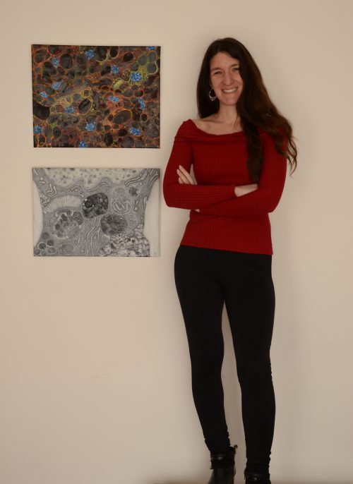
Our eleventh SciArt profile of the series features Ayelén Valko, a postdoc in Sebastian Schuck’s lab at Heidelberg University
Where are you originally from and what do you work on now?
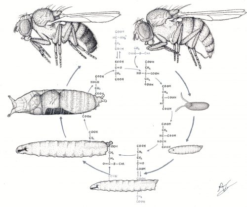
I am originally from Argentina, where I studied Biology at the University of Buenos Aires (UBA). During my PhD I explored the molecular mechanisms that trigger starvation-induced macro-autophagy in the fruit fly, Drosophila melanogaster. This is a physiological process by which cells digest their own cellular material in an attempt to compensate for nutrient deprivation. Later on, I became interested in studying a different kind of autophagic degradation, called micro-ER-phagy, by which the endoplasmic reticulum (ER) is eliminated during ER stress, and for which the underlying mechanisms are not completely understood. With this aim I joined Dr Sebastian Schuck’s laboratory at Heidelberg University one year ago, as a postdoctoral fellow, where I am using budding yeast as a model organism to tackle this issue.
Were you always going to be a scientist?
Definitely yes! According to my family, when I was 5 or 6 years old I used to spend hours in the garden observing insects and plants, and drawing them on notebooks. At around 10, my dad gave me a small microscope, an old and very simple device that belonged to him, which I used to look at water samples and fragments of leaves. I remember my excitement seeing for the first time this amazing tiny world through the lens of this enigmatic artefact. That was when I knew that I wanted to hold to that feeling and keep exploring that microscopic universe.
And what about art – have you always enjoyed it?
For me art has always been as important as science. It was also during my childhood that I became interested in depicting human faces, as I thought that by doing so I would be able to catch the essence of each of my family members on paper. Later on, as an adult, I started to see art as a useful way of conveying my appreciation of life and nature, and share with the general audience the beauty hidden inside a cell, which my work as a scientist allows me to grab. To do so, in parallel to my scientific training, I took several art courses at the National University of Art in Argentina. After having been trained in different art techniques, I specialized in scientific and naturalistic illustration. My works have been exhibited in several galleries and museums back in Argentina, among them the Quinquela Martín Museum and the Rómulo Raggio Foundation in Buenos Aires. I have also described two of these scientific paintings in a commentary article for the Autophagy journal.
What or who are your most important artistic influences?
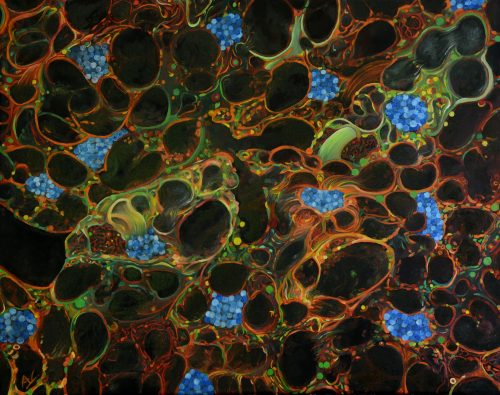
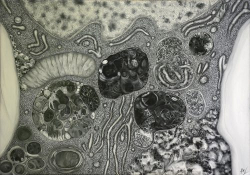
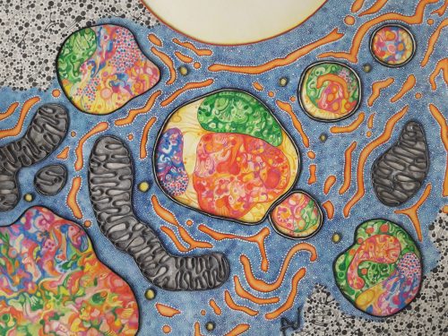
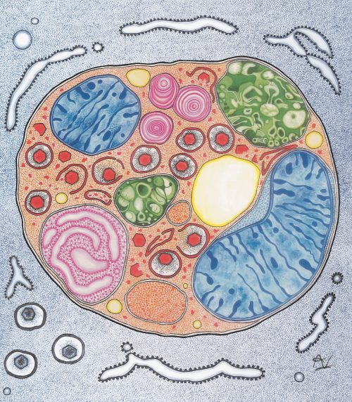
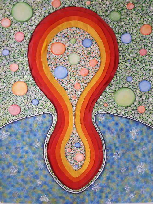
The main inspiration for my scientific artwork comes from my own photographs of cells and tissues that I need to take for my scientific work. The complexity of these subcellular worlds and their biological processes are constantly feeding my imagination. In fact, I am motivated not only by their mystery and beauty but also by the fact that this magnificent cellular universe remains veiled to the general public. Thus, my main aim is to create attractive paintings that can appeal to the general public, as an instrument of enjoyment and popularisation of science. But, obviously, not only cells inspire me. There are a large number of artists, that belong to very different artistic traditions, whose work has affected me deeply. I feel very moved by the incredible representation of hell and heaven by Hieronymus Bosch, the monochromatic landscapes depicting alien universes by H. R. Giger, Remedios Varo’s surrealist paintings with alchemic creatures, and many other exponents of the Surrealist movement such as Salvador Dali and Joan Miró, just to name a few.
“My main aim is to create attractive paintings that can appeal to the general public, as an instrument of enjoyment and popularisation of science.”
How do you make your art?
The challenge of making art based on scientific images lays in the need to represent the elements of study using artistic resources without completely losing the rigor of scientific observations. For me, this means that subcellular elements should be recognisable in the final artwork. However, my paintings are not in any way copies of photographs, but instead are the product of a creative process inspired by my observations and planned carefully. The first step of this process is sketching the original idea, ordering the elements of the composition harmoniously and attractively. It is only then that I decide the best technique for the work, as well as the kind of support in which I am going to make the artwork. I usually employ oil colours, acrylics, watercolours, Indian ink, collage, or a combination of them. As for the support, I usually employ canvas, a particular type of paper or cardboard.
Does your art influence your science at all, or are they separate worlds?
Art can influence science for sure! I think both activities are connected in deeply synergistic ways. In general, to understand a new idea or fact, normally we have the intuitive tendency to draw it. It is often considered that our understanding of a particular subject can increase by the act of trying to put what we are learning on paper, and for this, of course, science is no exception. To represent a scientific phenomenon, either artistically or schematically, one needs to develop a high level of comprehension of the fact. I have found that by drawing outlines of the experimental designs and the expected results, I usually can visualize better the biological questions that I am trying to answer. And this practice has often led me to new ideas for future paintings. Thus, my artistic and scientific lives nurture each other, giving me a unique point of view that ended up being essential for my scientific and artistic projects.
What are you thinking of working on next?
Apart from two cover images for different scientific journals on which I am currently working, and some other art projects, I am planning to study which are the elements or features that generate a stimulating and intriguing scientific artwork. My feeling is that the general audience is not as enthused by scientific art as it is by the rest of figurative art. To address this issue, I plan to systematically analyse people’s responses to a collection of slightly different cellular paintings, in order to identify which elements can maximise the identification of the viewer with the artwork. In line with this, I also want to explore interdisciplinary approaches to divulge science. I would like to work with professionals from other disciplines, such as psychologists, graphic designers, and physicians, to study different ways to convey to the general public scientific knowledge through art.

For more images check out Ayelén’s instagram page or contact her at ayevalko@gmail.com. Her personal artistic website will be online soon.
We’re looking for new people to feature in this series – whatever kind of art you do, from sculpture to embroidery to music to drawing, if you want to share it with the community just email thenode@biologists.com (nominations are also welcome!)
 (6 votes)