There is no doubt we are in the information age, and this is especially true in our world of biological research. We are ever more reliant on finding and accessing data, from genetic and genomic to epigenetic, transcriptomic and expression data. Searching and sorting through published data can be time consuming and difficult. Another challenge can be navigating resources. With the wealth of reagents, it can be difficult to figure out what is available and best suited for your system. This is especially true when our work involves an organism that is less widely used than rodents. Enter the individual model organism databases (MODs). These are total game changers for our research. By curating data and providing a “one stop shop” for resources specifically related to your model organism of choice we can easily find what we need to move our research forward. But these databases don’t run themselves. They require expertise to keep them current and functional and this expertise requires funding. MODs are currently supported by grants from the NIH and this support is absolutely essential to the continuation of this resource. But continued funding isn’t guaranteed. What can we as individuals due to help keep this funding going? We can let the NIH know just how vital MODs are to us and our research. The NIH is currently soliciting feedback about use of scientific data sources and tools, including MODs. If your MOD is important to you, let the NIH know. A great response from the community could be the difference in securing that next funding cycle. Below is a collection of voices from around the community sharing their stories of what their MOD means to them and their research. We hope that these testimonials inspire you to share your own stories. Please help our community and provide feedback to the NIH through the survey link by October 15. https://www.surveymonkey.com/r/5NGLNNJ
Heather Ray, PhD
Assistant Professor, Idaho State University Department of Biological Sciences
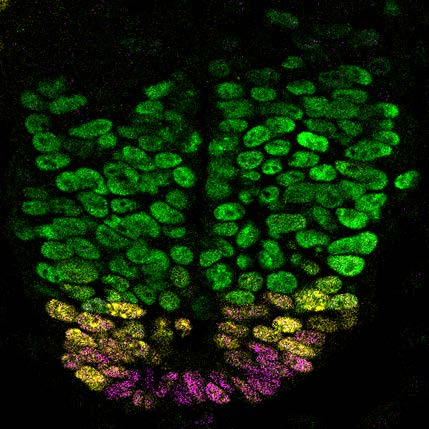
I began working with Xenopus laevis for the first time at the start of my postdoc. Besides beginning a new research project, I needed to get up to speed quickly on the specifics of working with a new organism. I spent many hours scouring Xenbase and it was all of the resources available in this database that helped me get my research up and running quickly. I continue to access Xenbase almost every day for a variety of purposes but most notably to access genomic and related gene expression data. The genetics of Xenopus laevis are complex as this species is a pseudo-tetraploid with differing alloalleles. With Xenbase, I can easily access the genetic sequences for each alloallele to identify the similarities and differences for primer design, gene targeting strategies, etc. I can also easily visualize gene expression data to compare expression patterns of each alloallele over developmental time.
I have recently started my independent lab and Xenbase is now an essential resource for training and education. It has great visuals to teach about embryonic cell fate, morphogenesis, and identifying developmental staging which I use in my undergraduate developmental biology course. It helps the students in my lab find the resources they need to conduct their research and aid in their independence. Without Xenbase as a resource, I would lose countless hours of valuable time. Instead of having a single resource with easily accessed species-specific information, I would have to search through an endless amount of primary literature and synthesize information that is reported in separate places. The time and effort that has gone into curating the interfaces on Xenbase should not have to be repeated a million times over by every individual investigator.
Joaquin Navajas Acedo, PhD
Postdoc, Schier Lab, Biozentrum, University of Basel
“Where is this gene expressed?”, “I wonder if there is an mCherry version of this transgene”, “Oh, lab X is looking for a Technician!”. I have been using Zebrafish as a model organism for my research for around 8 years now, and I don’t think I have spent more than a week without using any of ZFIN’s varied features.
Model organism databases like ZFIN are a central pillar of our work and must be protected at all cost. I cannot imagine doing my job without such a centralized and well curated database. This is thanks to the fantastic job that the folks at ZFIN do, but we should strive for giving them all the space and resources they need, and keep their funding. It would be amazing that some day ZFIN’s features expand to a point where it becomes an Alexandria’s Library where people come, interact and brainstorm together, present their work and leave with new ideas and expertise. A ZFIN Fish Virtual Forum, where this and other models can be integrated.
Money is a little price to pay to have such wonderful resources that ultimately benefit everyone.
[Remember that ZFIN is now citable. Please, do it generously! https://www.biorxiv.org/content/10.1101/2021.09.22.461443v1]
Allyson Angermeier
PhD candidate, University of Alabama Birmingham Department of Cell, Developmental and Integrative Biology
As a graduate student who entered the Xenopus research field with minimal knowledge of Xenopus, either conceptual or technical, the wealth of information about all things Xenopus on the Xenbase website has been (and continues to be) a cornerstone for my early career development. My lack of knowledge and skill was glaringly obvious during the first few months in my thesis lab and I desperately wanted to seek resources that could help me. I distinctly remember the moment when a senior lab mate showed me the Xenbase website. I was amazed, and quite frankly, I was relieved to have a seemingly overwhelming amount of information curated in an easily navigable fashion. I initially spent much of my time on Xenbase searching genes of interest, easily comparing their RNA (and now, protein) expression levels, and reading linked references to know more about these genes. I watched beautiful videos of Xenopus embryo development, learned how to time embryo development between Stages, and read numerous protocols designed and time-tested by others in the field. Over the past 4 years, I have continued to rely on Xenbase for pertinent scientific information as well as community updates and conference information. Xenbase has been magnificently and carefully designed to not only support those just beginning their research with Xenopus but continues providing excellent scientific support over a career.
Recently in the literature is the surge of large datasets generated via -omic experiments and analyses. These datasets can be extremely challenging to navigate and identify helpful or relevant information to one’s research. Xenbase compiles and presents these large datasets in a more accessible, meaningful way for researchers. One can easily obtain information to help design experiments, such as using RNA-seq datasets to identify highly expressed RNA isoforms for a gene, or finding the gene structures for L and S alloalleles of a gene to decide whether translational or splicing-blocking morpholinos would be best to achieve gene knockdown. Furthermore, Xenbase is also continuously improving to better serve our froggy community – they frequently add new features such as protein expression information that I previously mentioned and most recently, a new phenotype search function.
I am extremely grateful that Xenbase was funded by the NIH and established prior to my starting graduate school. I know that should I not have had the critical information that Xenbase organizes about Xenopus and associated techniques, it would have immensely slowed my progress as a Xenopus researcher. During graduate school, we are taught to be reliable, honest, independent researchers but scientific information does not need to be a constantly frustrating, convoluted process of diving into deep “rabbit holes”. Xenbase does a fantastic job of organizing data and placing it at a researcher’s finger tips. I truly hope that our community can rally behind Xenbase to keep it running for the generations of scientists to come after us, as I know how important it has been and continues to be for me.
Andrew Plygawko, PhD
Postdoctoral Fellow, Campbell lab, University of Sheffield, UK
My current project uses single-cell RNA sequencing data to identify genes required for Drosophila embryonic midgut development, and I don’t think it’s an exaggeration to say this would be almost impossible without FlyBase. Being able to browse the collective findings from a century’s research streamlines this analysis by distilling the immense amounts of information available into a single webpage that tells me about a gene’s structure, phenotypes, existing tools and more. I’m hopeful that the further identification of gene expression patterns using single-cell techniques will lead to a greater integration of these datasets into FlyBase in the future to build on existing knowledge. This would make it easier for researchers to identify orthologues which are expressed in homologous tissues in other species, accelerating the process through which flies can be established as a model system for understanding development and disease.
Ann Miller, PhD
Associate Professor, University of Michigan Department of Molecular, Cellular, and Developmental Biology
Xenbase is essential for my group to successfully carry out our research! Lab members use Xenbase at least 1x/week. When I polled my lab members about their most used features of Xenbase, they responded:
• We all use Xenbase for up-to-date information on genes of interest: getting sequences, finding alloalleles, looking for related papers using Xenopus, using RNAseq/proteomics data to get a sense of expression levels during relevant stages, looking at spatially localized expression levels at different developmental stages using in situ hybridization data.
• We all use Xenbase when looking up potential morpholinos for proteins of interest and antibodies that have been verified experimentally in Xenopus.
• We all use Xenbase to look up Xenopus protocols.
• We all use Xenbase to consult the Nieuwkoop and Faber Xenopus developmental staging series – and download images of different stages for presentations.
• We use Xenbase to access the lineage map information to do lineage-specific microinjections.
• In addition to the above, I have used Xenbase to access useful material for teaching, writing animal care protocols and grant proposals, and gain support for human disease modeling in Xenopus.
• One of my newer PhD students says: “Xenbase provides an excellent starting point for many of the processes, genes, and small molecule treatments that I’m interested in using for my projects. I also find the educational/background material (such as developmental videos) highly useful while building background for presentations”
• One of my senior lab members says: “I use this as a resource for my current research and also as a resource for undergraduates who join the lab and are not familiar with the model system. The images and movies of Xenopus development are excellent for helping new students learn the basic developmental biology. It is especially helpful that this information is gathered together in a single location so that researchers and students can quickly and easily find the information they need.”
• Another senior lab member says: “I often reference the data on mRNA expression during development as a way to determine whether a gene is of interest for my research. This type of data is not something that we could generate in our own lab, and it is helpful that the Xenopus community openly shares that information on Xenbase. Referencing gene expression data on Xenbase saves time and helps focus my efforts on genes that are most important for the developmental stage I am interested in.”
It would be a major blow to the community and to my group’s research if Xenbase did not exist. For example, it would be difficult to design primers/morpholinos/guide RNAs, time spent searching for background source material and supporting papers would be significantly longer, it would be harder to find trusted protocols and materials when trying out new experiments. Xenbase is a reliable resource that my group uses to quickly and easily find information about the Xenopus model system. It provides foundational knowledge for researchers who are new to working with Xenopus and catalogs up-to-date genomic information that is essential for our research. Finally, the staff members at Xenbase are knowledgeable, supportive, and flexible. They are always happy to explain new Xenbase features and very receptive to new ideas to make Xenbase more useful to the community.
Sumantra Chatterjee, PhD
Research Assistant Professor, NYU Grossman School of Medicine
I have been trained both as a developmental biologist as human geneticist working on deciphering the functional consequences of genetic mutations leading to congenital diseases like Hirschsprung disease (HSCR; congenital enteric aganglionosis). This research has necessitated the extensive use of mouse models as access to human fetal tissues is limited. I have extensively used MGI in the last 15 years to utilize its curated data of various mutant mouse models and their expression patterns to complement our transcriptomics studies of the developing mouse enteric nervous system (ENS).
The MGI data has been extremely useful to us to validate many of our finding without the need for generating multiple mouse lines. Without this it would have been extremely difficult to fully flesh out the downstream effect of many genes we observe disrupted in the ENS and build a comprehensive gene regulatory network, which is disrupted in HSCR. Now that we have started our forays in looking ta human fetal tissue, the MGIs vertebrate homology dataset has been very crucial in trying to integrate our mouse data with the human data in a logical comprehensive manner rather than a piecemeal gene name by name comparison. I cannot imagine the progress I have made in my career studying rare congenital diseases would have been possible without MGI. Finally, it would be good if we can start adding some of the multiple single cell genomics data that is been generated in various mouse tissues at different developmental stages. The cell state changes die to specific genetic mutations would be a great addition to the phenotypic panel.
John Wallingford, PhD
Professor and Mr. and Mrs. Robert P. Doherty, Jr. Regents Chair in Molecular Biology, University of Texas College of Natural Sciences
We use Xenbase every day. In fact, for our new proteomics analysis, we may sometimes query Xenbase hundreds of times per day. We literally could not continue the more cutting-edge omics approaches without this crucial resource.
Grégoire Michaux, PhD
Principle Investigator IGDR, France
As a C. elegans lab we use Wormbase on a daily basis, for two main reasons. First, examine various gene features (sequence, genetic localization, expression pattern, homology, phenotypes, relevant literature, etc). The other frequent use is the Blast tool allowing the fast identification of homologs and paralogs of genes from other organisms in the C. elegans genome. Without Wormbase it would take ages to find these data and I used it so frequently that I cannot imagine my work without it.
Ideally, I would love to see Wormbase converging with Flybase and other similar online resources to make it easier to explore other databases.
Ben Steventon, PhD
University Lecturer, University of Cambridge
Our lab uses ZFIN on a more-or-less continuous basis. We are constantly using it to look up gene expression patterns of interest- something that is becoming of increasing importance now that we are discovering new genes of interest while combing through transcriptomic datasets. How would we ever assign cell types to clouds of scRNAseq data without such an easy access to gene expression patterns? In addition to this, we use the database to look for available mutants and transgenic lines. A huge amount of effort has gone into building resources such as these, including continual engagement with the community’s needs. For these reasons MODs are essential resources for all and need to be maintained. An exciting direction for such databases would be to link in with single cell datasets, so that one could click on a dot and go straight to a gene expression and back. This would allow for a much more rapid exploration and validation of cell type assignment.
Richard Behringer, PhD
Professor, University of Texas MD Anderson Cancer Center Department of Genetics
Xenbase has some very useful information and tools for education. In the Marine Biological Laboratory Embryology course, the Frog Module exploits Xenbase for that information. We also use it here in our first-year graduate course during Developmental Biology week.
For examples, the Anatomy and Development section is very useful for embryo staging. In addition, the fate maps tool is really useful for students. The Movies section has numerous time-lapse movies of different periods of X. laevis and X. tropicalis development that lets students see the morphological changes that occur over time.
Dan Bergstralh, PhD
Assistant Professor, University of Rochester
I use Flybase multiple times a week, and I’ve been doing that for over a decade now. I love it. It’s an invaluable resource for our research and we use it many ways, including: to investigate and to track down alleles, to examine patterns of gene expression, and to help identify homologs in other species. I can’t imagine working without it!
 (No Ratings Yet)
 Loading...


