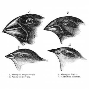
Charles Darwin’s famous finches from the Galapagos Islands
In this episode of Genetics Unzipped Kat Arney explores the myths and misconceptions behind two of the most iconic images in evolutionary biology: the much-parodied March of Progress – a series of human ancestors walking across the page, portraying the inexorable journey from monkey to man – and the famous finches of the Galapagos islands, which are supposedly the inspiration for Charles Darwin’s theory of natural selection.
Where did these infamous images come from, and do they really show what everyone seems to think they do?
If you enjoy the show, please do rate and review and spread the word. And you can always send feedback and suggestions for future episodes and guests to podcast@geneticsunzipped.com Follow us on Twitter – @geneticsunzip
The Lyons Lab at Scripps Institution of Oceanography (a department at U.C. San Diego) is recruiting a full-time Postdoctoral Scholar to support research projects funded by an NIH MIRA award. The Lyons Lab (www.lyonslab.org) focuses on cell type differentiation and morphogenesis with a particular interest in how these processes evolve. The postdoc will contribute to our lab’s goal to build the first comprehensive developmental gene regulatory networks (GRN) controlling early events in molluscan development (e.g. germ-layer segregation, organizer signaling, and gastrulation). GRN analysis (both experimental and synthetic) will be carried out in the marine slipper snail Crepidula. The postdoc can take advantage of a growing tool kit for functional genomics in Crepidula, as well as a new marine transgenics facility for maintenance of stable lines. In collaboration with Dr. Lyons the postdoc will be responsible for leading an independent project of mutual interest that aligns with the postdoc’s career goals.
The postdoc will join a motivated group of students, staff, and postdocs who are broadly interested in cell and developmental biology of marine organisms. Additionally, the Lyons Lab offers a broad range of other systems for comparative developmental studies among molluscs and echinoderms, and provides a highly interdisciplinary and collaborative environment within our group, and with other labs at the Scripps Institution of Oceanography (https://scripps.ucsd.edu/), with U.C. San Diego’s main campus (https://ucsd.edu/), and with other institutions in the greater San Diego Area.
Qualifications:
We are seeking talented applicants who have a Ph.D. degree (or are close to earning one), and training in molecular biology, cell biology developmental biology, bioinformatics, or a related field. Candidates with expertise in cis-regulatory element analysis, network inference, and bioinformatics are encouraged to apply.
Strong experience and interest in one or more of the following areas is preferred, but not required:
Genomics: Preparation of samples and analysis of data for RNA-seq, ATAC-seq, scRNA-seq, BAC libraries, etc.
CRISPR/Cas9: Design and delivery (microinjection, electroporation) of CRISPR/Cas9 components for induction of DNA double-strand breaks.
Transgenesis: Generation of transient and stable transgenes via transposons, CRISPR/Cas9 genome editing, etc.
To Apply:
Submit the following items, as a single PDF, to Dr. Lyons (d1lyons@ucsd.edu):
1) Cover letter explaining your interest in the position and qualifications
2) CV
3) Statement of research/career goals
4) Names and contact information for at least three references
Review of applications will begin immediately and continue until position is filled.
We are seeking EVODEVO POSTDOC candidates to apply any of the 3 currently open calls to join our lab in BARCELONA.
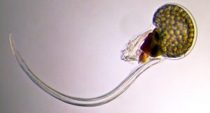
Our lab studies the chordate model Oikopleura dioica to better the impact of gene loss on the evolution of mechanisms of development and gene regulatory networks, with special interest in heart and muscle development. Click here for a tour “A day in our lab” posted in The Node
We have also engaged a new EcoEvoDevo line investigating if the developmental mechanisms of marine embryos are ready to respond to climate change, including biotoxins derived from algal blooms. Click here for a tour on this new EcoEvoDevo adventure.
Our approaches include single-cell RNAseq, Embryo microinjection, RNAi, Confocal-Microscopy, Bioinformatics and soon CRISPR
Currently there are 3 open calls.
-> CALL 1: Beatriu de Pinos. 3 year-postdoc + starting grant. Deadline: February 3rd 2020. Requirement: to have defended the PhD within the period January 1st 2012 – December 31st 2017; 2-years of postdoctoral experience; less than 12 months living in Spain the last 3 years.
-> CALL 2: Juan de la Cierva – “Training”. 2 year-postdoc. Deadline: January 22nd 2020. Requirement: to have defended the PhD within the period January 1st 2018 – December 31st 2019.
-> CALL 3: Juan de la Cierva – “Incorporation”. 2 year-postdoc. Deadline: January 21st 2020. Requirement: to have defended the PhD within the period January 1st 2015 – December 31st 2017.
CONTACT: Interested candidates, please send an email to Cristian Cañestro (canestro@ub.edu) ASAP, including a brief letter of interest, a brief CV, including list of publications with their impact factor and quartile, and technical skills (specially those related with our approaches) all together in ONE single pdf file.
A 2 year post-doctoral position is available starting early 2020 to study transcriptional control of neurogenesis in Xenopus and mouse. The research will be conducted in the laboratory of Developmental Genetics (http://gendev.ulb.ac.be/bellefroidlab/) of the Université Libre de Bruxelles (ULB) Neuroscience Institute (UNI) (https://uni.ulb.ac.be/) in Gosselies (30 km south of Brussels).
Balancing neural progenitor cell (NPC) self-renewal and neuronal differentiation is essential for generating cells in correct numbers and diverse types during neural development. As such, neurogenesis is tightly regulated by a complex array of transcription factors that work in concert to coordinate NPC maintenance, proliferation and differentiation. Our focus is on the Dmrt3 and Dmrt5 transcription factors that we have identified as critical regulators of cortex development and on Prdm12 that we found to be required for the development of pain-sensing neurons.
Our aim is to better understand how these transcriptional regulators function in vivo by identifying genome wide their direct targets and interacting partners. As most remains to be discovered about the mechanism of action of these transcription regulators, results of this project should uncover novel essential aspects of corticogenesis and nociceptor development and functioning.
We are looking for highly motivated candidates with an experience in mouse genetics, cell and molecular biology techniques (ChIP-seq, RNA-seq, AP-MS,…) and preferentially a background in neuroscience. Interested candidates should send a letter of motivation (before end of February) describing past research experiences and full CV to:
Eric Bellefroid (ebellefr@ulb.ac.be), together with the name and e-mail address of 2 references.
Selected recent related publications:
Desmaris et al. (2018). Dmrt5, Dmrt3 and Emx2 cooperatively block Gsx2 at the pallium-subpallium boundary to maintain cortical identity in dorsal telencephalic progenitors. J. of Neurosci. 38, 9105-9121.
Desiderio et al. (2019). Prdm12 directs nociceptive sensory neuron development by regulating the expression of the NGF receptor TrkA. Cell Reports 26, 3522-3536.
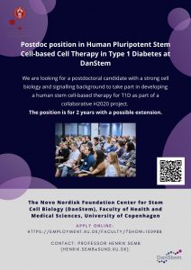
We are looking for a postdoctoral candidate with a strong cell biology and signalling background to take part in developing a human stem cell-based therapy for T1D as part of a collaborative H2020 project. The position is for 2 years with a possible extension.
Qualifications
The candidate is required to hold a PhD degree in pluripotent stem cell/developmental biology
Postdoctoral experience in the same areas is an advantage
We are looking for candidates with hands-on experience or an interest in expanding their knowledge in human pluripotent stem cell maintenance and differentiation, 3D culture of pluripotent stem cells, various cell and molecular biological methods, flow cytometry and confocal microscopy
Experience in differentiation towards pancreatic lineages is a plus
Finally, we are looking for applicants with a good track record of peer reviewed scientific publications and team work
Employment conditions
The position is for 2 years with a possible extension. The employment is planned to start 1 April or upon agreement.
For further information, please contact Professor Henrik Semb (henrik.semb@sund.ku.dk).
In keeping with a time-honoured tradition, we recently flooded Twitter with 12 beautiful developmental biology GIFs. They came from papers published this year and feature all kinds of systems and visual styles – here they are for posterity! Let us know your favourite in the comments
Whole-embryo developmental imaging of zebrafish spinal cord neurogenesis
Genetics Unzipped is delighted to host a short series of podcasts recorded at the 2019 Galton Institute symposium – New Light on Old Britons – which took place at the Royal Society in London at the end of October. Reporter Georgia Mills talks to some of the leading researchers uncovering the hidden stories of the people of the British Isles.
Who were these ancient Britons? Where did they come from and what were they like? What’s the real story behind the romantic myths about the Celts? And what can modern genetic and archaeological techniques tell us about their lives and loves?
Professor Ian Barnes and Dr Selina Brace, ancient DNA researchers at the Natural History Museum in London, discuss how their work on ancient DNA is shedding light on the British population from the Mesolithic to the Bronze Age.
Professor Sir Walter Bodmer FRS from the Weatherall Institute, Oxford, explains what we know so far about genetic structure and origins of populations of the British Isles.
The Celts are one of the most famous – and misunderstood – people who lived in ancient Britain. Professor Sir Barry Cunliffe CBE, FBA from the University of Oxford explores the myths and the reality.
The Latin American Society for Developmental Biology was founded almost two decades ago and has become a vital hub for researchers across the region. Its biennial meetings bring the community together and also attract leading researchers based elsewhere. At the end of October this year the meeting was held in Buenos Aires, the grand old capital of Argentina which sits on the widest river in the world and is the second most populous city on the continent (if, for the sake of a statistic, you count its greater metropolitan area). I got to go on behalf of the Node, Development and The Company of Biologists – these kinds of conferences are a great opportunity for us to keep in touch with our communities across the world, to listen and to let people know how we can help them. It was of course a great privilege to go, especially since it was my first time in South America.
We were welcomed to the conference by Pablo Wappner, one of the local organisers and LASDB President (Juan Riesgo-Escovar would take up the reins from Pablo once the meeting finished). He emphasised how the meeting represented excellent science on a low budget, and thanked the many invited speakers who had covered part or all of their travel expenses to get to Buenos Aires. Funding came up a lot in conversations throughout the conference – I had expected as much given recent (and not so recent) news stories, and also this Development interview I did with Brazilian researchers Maurício Rocha-Martins and Mariana Silveira. I don’t really want to dwell on it too much here but it’s worth recognising how many hurdles scientists face in the region, and how much great science gets done in spite of these hurdles.
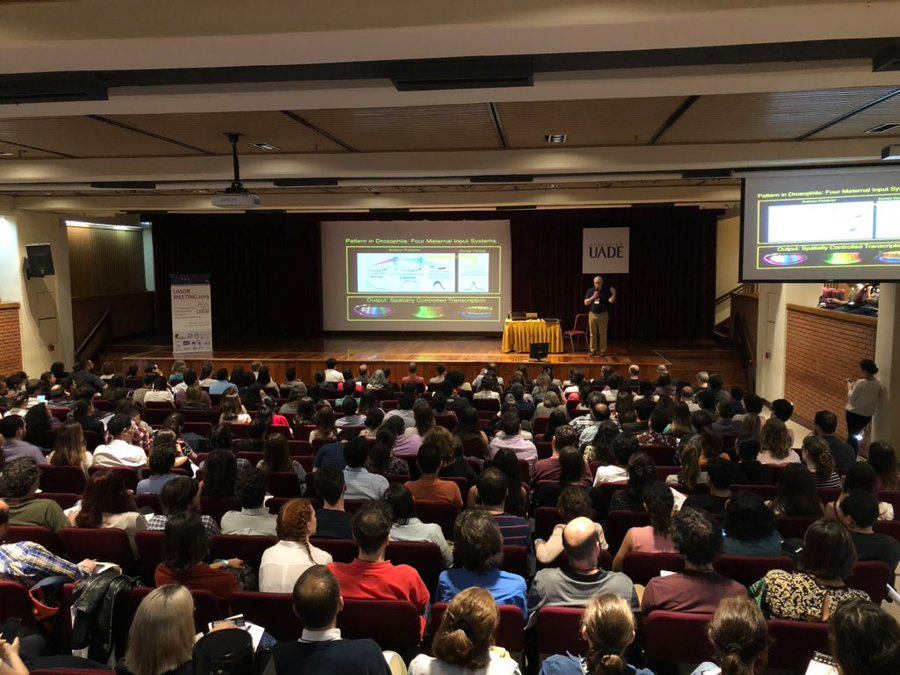
Here I’ll just share some of my talk highlights, depending on how legible my notes were and not at all exhaustively. Nobel Laureate Eric Wieschaus (Princeton University, US) kicked things off. Forty years of work on the four maternal systems that pattern the early fly embryo were now culminating with the aid of some extremely clever (and eye-watering) genetics. He was now in the position to subtract and add each system in turn to assess their interdependence. I noted down something he said in response to an audience question about whether Bicoid behaved like other morphogens – “All life is specific, specific solutions to specific problems”. The point beingI think that we need not be too preoccupied with how general a given mechanism is.
Eric Wieschaus opening the conference (photo courtesy of LASDB Facebook page)
Eugenia del Pino (Universidad Católica del Ecuador, Quito) was awarded the LASDB Prize, which recognises an ‘outstanding researcher who has made a major contribution to developmental biology, and has shown an active involvement in Latin American science.’ Before her talk we watched video messages from her previous students, now based all over the world, who thanked her for her support and guidance – it seems she really is at the heart of Ecuadorian developmental biology. She told us that when she started her lab in Quito, she was quite isolated, but she used this isolation as an advantage, in that she had minimal distractions. She gave a career perspective on the different ways frogs make eggs and undergo gastrulation, in particular focusing on marsupial frogs, which carry their eggs on their back. This research started when she found one such species (Gastrotheca riobambae) in the garden of her university, and developed it into a developmental model. Her talk was a testament to how biodiversity can be harnessed to understand basic problems in development and evolution.
Miguel Concha (Universidad de Chile, Santiago) presented his lab’s work on collective migration in the zebrafish embryo. Combining imaging, biomechemics and modelling, he described how mechanical interactions between epithelial cells (the enveloping layer) and mesenchymal cells (progenitors of the laterality organ) drives tissue morphogenesis. He was also spoke a little about the political situation in Chile, and the societal inequalities that had been driving their current protests. I’d see saw students watching live feeds of the protests on their phones (between talks of course!). Pablo Strobl-Mazzulla (InTeCh, Chascomús, Argentina) discussed the role of folate in development. Several congenital defects in humans are associated with folate deficiencies, notably failure of neural tube closure, and Strobl-Mazulla wanted to know why some tissues seem particularly sensitive to folate deficiency. After knocking down the two ways folate can get into the cell (a receptor and a channel), his lab observed reduced methylation of genes required for neural crest fate.
Igor Schneider (Federal University of Pará, Brazil) posed the question of whether the famous regeneration capability of salamanders really does stand out so much among vertebrates. His lab found that lungfishes (which are a sister group to tetrapods) can regenerate their fins pretty well and in a manner similar to salamanders, morphologically and molecularly. He further showed data on regeneration in paddle fish, birchir and gar, and argued that broader phylogenetic sampling will help uncover the shared and core mechanisms of regeneration. We heard a plea from Guillermo Oliver (Northwestern University, US) for developmental biology to stop overlooking the lymph vasculature, which has been increasingly implicated in various aspects of human health and is particularly interesting in terms of the plasticity of cell fates. He described his lab’s work on obesity – mice heterozygous for Prox (which controls lymphatic fate) get fat, and this has to do with leaky lymphatics in the small intestine. Many other fascinating discoveries await in this tissue hiding in plain sight.
How can a single signalling pathway can instruct diverse cell fates at different points during development? Trudi Schupbach (Princeton University, US) addressed this question using ERK signalling in Drosophila egg patterning as an example. ERK’s effects differed depending on combinatorial signalling with other pathways, the levels and duration of the signal, and feedback mechanisms. Irene Miguel Aliaga (Imperial College, London) emphasised the concept of ‘continued development’ in adult organs which can be resized and remodelled depending on environmental cues or physiological requirements. She described her lab’s work on cross-organ sexually dimorphic communication – testes talking to guts to change metabolism to suit the testes (if i have that right!).
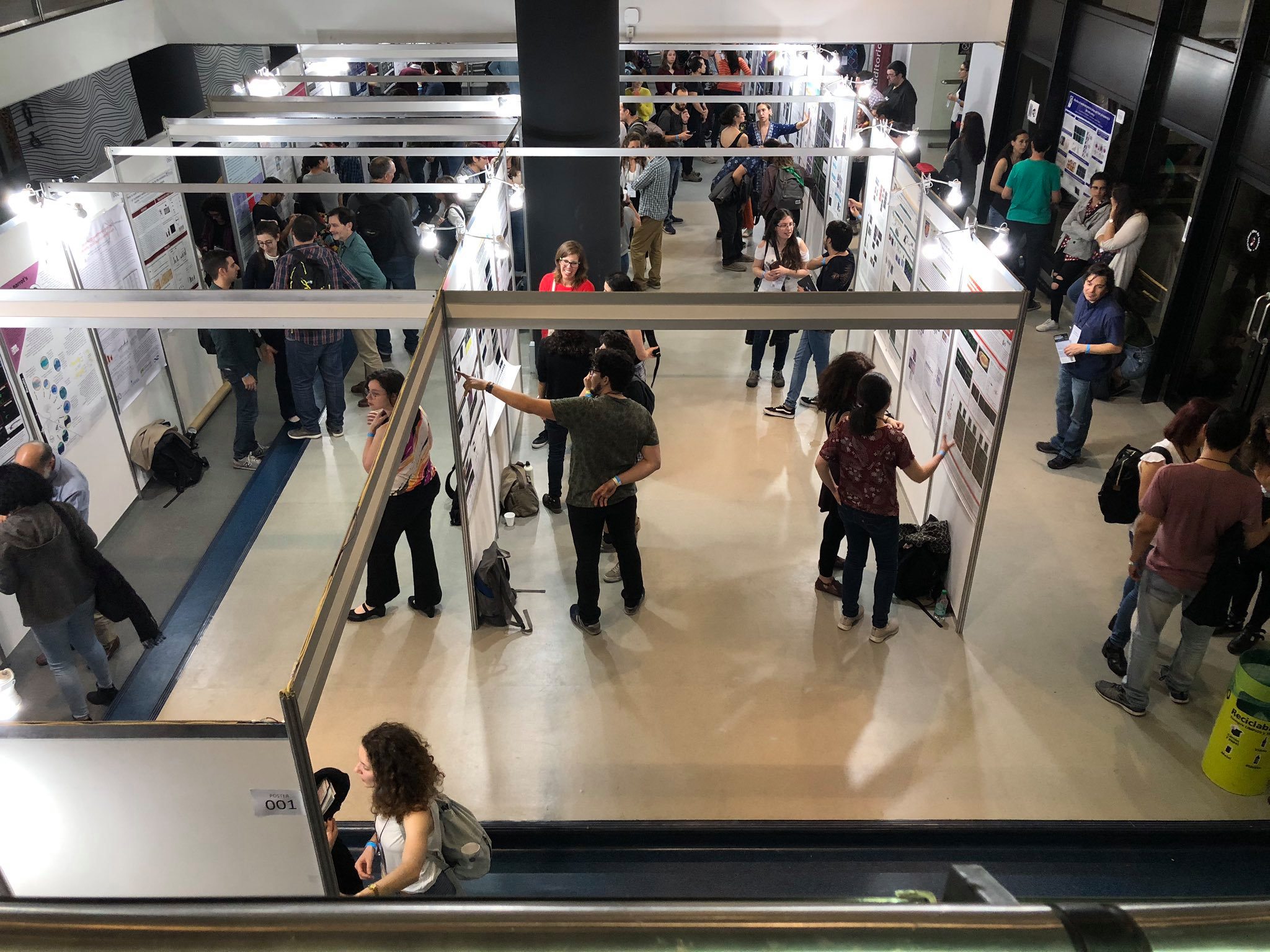
Poster session in full swing (photo courtesy of LASDB Facebook page)
At the meeting, I got to interview Roberto Mayor for Development (and also Trudi Schupbach – look out for the interviews in January), and he told me that one of the things that made the region special in terms of science were the students, who were always amongst the best in the world. And in the poster session I could see what he meant – there were so many great posters from PhD students, Master’s students and even undergraduates. We also heard a very impressive talk from Sandra Edwards, a PhD student from Santiago in Chile, who explored the role of calcium in the cytoskeleton of Drosophila immune cells with the aid of some gorgeous movies.
Another notable session in the conference was The Gordon Research Conference Power Hour. This was chaired by Marianne Bronner of Caltech and Nancy Gray, President and Chief Executive Officer of the GRC. Gray told us that the GRC are looking to increase Latin American representation at their conferences, and introduced their Power Hours, time set aside at GRCs dedicated to ‘discuss[ing] challenges women face in science and issues of diversity and inclusion’. And we had a Power Hour of our own – the audience was split into groups and asked to discuss a question or two. My group were asked What skills do you need to become a successful scientist?, and we had a lively debate on the relative importance of communication, luck and strategy (thanks to my group for putting up with the one person who spoke no Spanish). Other groups tackled sexism, microagression, working patterns and bullying, and at the end a speaker from each group shared the discussions. Nancy Gray then gave her card to all the speakers – this would give them free registration to a GRC of their choice! It was amazingly generous, potentially life-changing act.
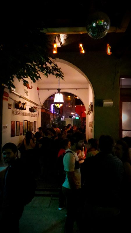
A rather gloomy photo of the final dinner/drinks, note disco ball
The conference dinner on the final night was held in a brew pub – for something like three US dollars we got a night of unlimited beer and pizza (I struggled throughout to work out how much things cost with the exchange rate, though as a local told me: ‘whatever it is, it’s much much cheaper than England!’). It was a fun informal occasion that suited the tone of the whole meeting. I also had half a day either side of the conference to explore Buenos Aires, an amazingly energetic place, with endlessly interesting architecture, crazy buses, crazier taxis, quality food and art, and even some wildlife (in the Reserva Ecológica Costanera Sur not far from the centre – bird fans take note). The next LASDB meeting will be in Chile in 2021.
Buenos Aires skylines.
Congratulations to all the poster prize winners!
1st Prize: Nicolas Cumplido, FONDAP Center for Genome Regulation, Santiago, Chile.
2nd Prize: Nicolas Vazquez. LBD-INTECH, Chascomús, Buenos Aires, Argentina
3rd Prize: Pablo Guzmán Palma. Pontifical Catholic University of Chile, Santiago, Chile
4th prize: Sofia Polcowñuk . Laboratorio de genética del comportamiento, Fundación Instituto Leloir (FIL-IIBBA-CONICET), Buenos Aires, Argentina
Centro Andaluz de Biología del Desarrollo (CABD), Seville.
Juan R. Martinez-Morales Lab (jrmarmor@upo.es) is recruiting competitive
postdoctoral researchers to participate in an interdisciplinary project on comparative
tissue morphogenesis
Research project/ Research Group description:
The main focus of our laboratory is to study cellular and molecular mechanisms
involved in the morphogenesis of the vertebrate eye. Using the teleost models
zebrafish and medaka, we investigate the machinery driving the folding of the retinal
neuroepithelium.. Although it is generally accepted that optic cup formation follows a
tissue-intrinsic program in vertebrates, several mechanisms have been postulated
(basal constriction, rim involution) and some important differences in cell behaviour
have been reported among species. Many important questions still remain open. Which
is the relative contribution of each mechanism to the folding of the optic cup? Do they
act in a cooperative manner? Do they have the same regulatory weight in different
species? To answer these questions, we aim to integrate genetic information, imaging
analysis of cell shape changes, and tensional forces distribution into coherent
computational models able to predict the key morphogenetic rules that shape the entire
organ. We plan to extend our observations to mammalian tissue, by examining cell
shape changes and tensions in 3D retinal organoids developed in vitro. A key aspect of
the project is to understand how classical signalling events, which have proved to be
essential for the proper patterning of the organ, act in coordination with tensional
forces. This highly interdisciplinary approach, combining genetics, imaging, biophysics
and computational modelling should yield information relevant not only to understand
optic cup formation, but also to deduce general self-organization principles of the living
matter.
Relevant Publications:
Letelier et al. (2018a) Nat Genetics, Letelier et al (2018b) PNAS, Nicolás-Pérez et
al (2016). eLife; Gago-Rodrigues et al (2015). Nat Comm ; Tena et al (2014) Genome Research. 24(7):;
Bogdanovic et al (2012). Developmental Cell. 23 (4).
For a full list: https://www.ncbi.nlm.nih.gov/pubmed/?term=Martinez-morales+JR
Job position description:
Given the interdisciplinary character of the project above described, we are seeking
talented and highly motivated postdoctoral researchers with a background either in
Developmental Biology or in Biophysics. Previous experience with teleost models will
be well received. The candidates should have good communication skills, critical for
ensuring the success of the team effort. We offer full support to apply for external
funding in upcoming postdoctoral call “Juan de la Cierva” (deadline-21st January 2020).
 (No Ratings Yet)
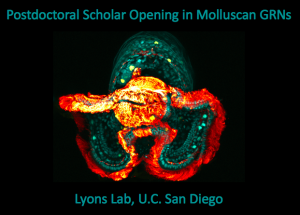
 (1 votes)
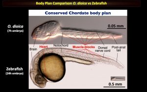


 Genetics Unzipped is delighted to host a short series of podcasts recorded at the 2019 Galton Institute symposium – New Light on Old Britons – which took place at the Royal Society in London at the end of October. Reporter Georgia Mills talks to some of the leading researchers uncovering the hidden stories of the people of the British Isles.