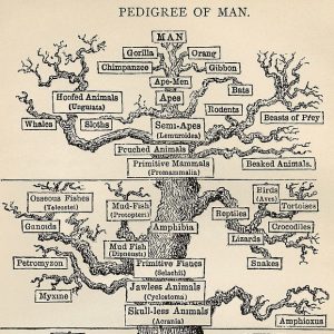
In this episode from our centenary series exploring 100 ideas in genetics, we’re uprooting the tree of life – asking whether we should believe our eyes or our sequencing machines when it comes to deciding what makes a species.
Plus, the greatest comebacks of all time – we look at the science of de-extinction and find out whether Jurassic Park could ever become a reality.
If you enjoy the show, please do rate and review and spread the word. And you can always send feedback and suggestions for future episodes and guests to podcast@geneticsunzipped.com Follow us on Twitter – @geneticsunzip
Two weeks ago, we had the opportunity to attend the Company of Biologists Workshop, “Understanding Birth Defects in the Genomic Age”. This workshop brought together a diverse collection of basic developmental biologists, human geneticists and clinicians to discuss the current challenges and opportunities in the field of birth defects research. We can almost guarantee you that none of the groups of attendees would normally overlap at any other meeting. And yet, what quickly became apparent was that there was so much in common between everyone in attendance.
The setting for the meeting was stunning. Set at Wiston House in the English countryside not too far from Brighton, history oozed from every corner of the building. From the delicious catering, to the beautiful (but rainy) countryside walk, to the very interesting talk on the history of the house, this venue provided an epic background for what would be an extremely stimulating three and a half days.
The meeting was unique right from the start. It was definitely the smallest meeting we had ever attended. There were only about 33 people total, and we each had the opportunity to introduce ourselves to the group on the first day. We were asked to give a few slides introducing ourselves as well as a “problem” (a topic that we would like help with) and “solution” (something that we were good at/could use to help others). Right away this encouraged discussion and it became quickly apparent how each of our skillsets would be able to help not just each others’ research, but contribute a unique perspective to the group.
Participants at the Workshop
In addition to traditional scientific talks, we had the opportunity to sit down and talk amongst the group about some of the issues which we would like to address. Collectively, birth defects are the number one cause of infant mortality in the USA, but it is sometimes challenging to justify studying individual rare developmental defects. Many of our discussions centred around how to increase public awareness of our research and to emphasize the huge impact of return of results to families – even if a genetic diagnosis doesn’t lead to a treatment, patients and families can be hugely comforted to know more about the underlying cause. John Wallingford gave a stirring talk about the disturbing history of society’s (including the medical profession’s) poor treatment of individuals with congenital anomalies. There was unanimous agreement amongst the group that we needed a new term for birth defects that was both more sensitive and more inclusive. There was much discussion, but the group was not able to come to a consensus as to a better term moving forward. One of our favourite suggestions was that perhaps we needed to create a new word entirely!
One of the goals of the workshop was to generate actual concrete steps to address the above issues. Working groups were established to draft a white paper, review articles, social media campaigns, and other methods to increase the awareness of this very important field of research. One of they key action items became bringing together a similar group of people again to build on the ideas started at this workshop. The Company of Biologists is already planning a meeting on developmental disorders for 2021; hopefully this will provide another opportunity for a meeting of minds to continue to forward the cause.
As two early career researchers funded by the Company of Biologists to attend the workshop, we gained great insight into the diverse approaches being used to study birth defects research. In addition, it provided a unique opportunity to discuss and learn about larger scope projects such as impacting public awareness and advocating for increased funding for a particular area of research.
At the conclusion of the meeting everyone left inspired and ready to act. We are looking forward to 2021!
Find out more about The Company of Biologists’ Workshops, including our 2020 schedule and details of how to apply for funded places, here:
Welcome to our monthly trawl for developmental biology (and related) preprints.
This month we found preprints detailing extensive mouse and fly knockout resources, exploring bacterial influences on development, and investigating mechanics in vivo and in silico. They were hosted on bioRxivandarXiv. Let us know if we missed anything. Use these links to get to the section you want:
Apcdd1 is a dual BMP/Wnt inhibitor in the developing nervous system and skin
Alin Vonica, Neha Bhat, Keith Phan, Jinbai Guo, Lăcrimioara Iancu, Jessica A. Weber, Amir Karger, John W. Cain, Etienne C. E. Wang, Gina M. DeStefano, Anne H. O’Donnell-Luria, Angela M. Christiano, Bruce Riley, Samantha J. Butler, Victor Luria
Syndecan-3 enhances anabolic bone formation through WNT signalling
Francesca Manuela Johnson de Sousa Brito, Andrew Butcher, Addolorata Pisconti, Blandine Poulet, Amanda Prior, Gemma Charlesworth, Catherine Sperinck, Michele Scotto di Mase, George Bou-Gharios, Robert Jurgen van ’t Hof, Anna Daroszewska
A TBX5 dosage-sensitive gene regulatory network for human congenital heart disease
Irfan S. Kathiriya, Kavitha S. Rao, Giovanni Iacono, W. Patrick Devine, Swetansu K. Hota, Michael H. Lai, Bayardo I. Garay, Reuben Thomas, Andrew P. Blair, Henry Z. Gong, Lauren K. Wasson, Piyush Goyal, Tatyana Sukonnik, Gunes A. Akgun, Laure D. Bernard, Brynn N. Akerberg, Fei Gu, Kai Li, William T. Pu, Joshua M. Stuart, Christine E. Seidman, J. G. Seidman, Holger Heyn, Benoit G. Bruneau
A novel microRNA-based strategy to expand the differentiation potency of stem cells
María Salazar-Roa, Marianna Trakala, Mónica Álvarez-Fernández, Fátima Valdés-Mora, Cuiqing Zhong, Jaime Muñoz, Yang Yu, Timothy J. Peters, Osvaldo Graña, Rosa Serrano, Elisabet Zapatero-Solana, María Abad, María José Bueno, Marta Gómez de Cedrón, José Fernández-Piqueras, Manuel Serrano, María A. Blasco, Da-Zhi Wang, Susan J. Clark, Juan Carlos Izpisua-Belmonte, Sagrario Ortega, Marcos Malumbres
PI 3-kinase delta enhances axonal PIP3 to support axon regeneration in the adult CNS
Amanda C Barber, Rachel S Evans, Bart Nieuwenhuis, Craig S Pearson, Joachim Fuchs, Amy R MacQueen, Susan van Erp, Barabara Haenzi, Lianne A Hulshof, Andrew Osborne, Raquel Conceicao, Sarita S Deshpande, Joshua Cave, Charles ffrench-Constant, Patrice D Smith, Klaus Okkenhaug, Britta J Eickholt, Keith R Martin, James W Fawcett, Richard Eva
A cell surface arabinogalactan-peptide influences root hair cell fate
Cecilia Borassi, Javier Gloazzo Dorosz, Martiniano M. Ricardi, Laercio Pol Fachin, Mariana Carignani Sardoy, Eliana Marzol, Silvina Mangano, Diana Rosa Rodríguez Garcia, Javier Martínez Pacheco, Yossmayer del Carmen Rondón Guerrero, Silvia M. Velasquez, Bianca Villavicencio, Marina Ciancia, Georg Seifert, Hugo Verli, José M. Estevez
A simple method for spray-on gene editing in planta
Cara Doyle, Katie Higginbottom, Thomas A. Swift, Mark Winfield, Christopher Bellas, David Benito-Alifonso, Taryn Fletcher, M. Carmen Galan, Keith Edwards, Heather M. Whitney
Lgr5+ telocytes are a signaling hub at the intestinal villus tip
Keren Bahar Halpern, Hassan Massalha, Rachel K. Zwick, Andreas E. Moor, David Castillo-Azofeifa, Milena Rozenberg, Lydia Farack, Adi Egozi, Dan R. Miller, Inna Averbukh, Yotam Harnik, Noa Weinberg-Corem, Frederic J. de Sauvage, Ido Amit, Ophir D. Klein, Michal Shoshkes-Carmel, Shalev Itzkovitz
Novel function of TRIP6, in brain ciliogenesis
Shalmali Shukla, Pavel Urbanek, Lucien Frappart, Ronny Hänold, Sigrun Nagel, Shamci Monajembashi, Paulius Grigaravicius, Woo Kee Min, Alicia Tapias, Olivier Kassel, Heike Heuer, Zhao-Qi Wang, Aspasia Ploubidou, Peter Herrlich
Large-scale transgenic Drosophila resource collections for loss- and gain-of-function studies
Jonathan Zirin, Yanhui Hu, Luping Liu, Donghui Yang-Zhou, Ryan Colbeth, Dong Yan, Ben Ewen-Campen, Rong Tao, Eric Vogt, Sara VanNest, Cooper Cavers, Christians Villalta, Aram Comjean, Jin Sun, Xia Wang, Yu Jia, Ruibao Zhu, Pin Peng, Jinchao Yu, Da Shen, Yuhao Qiu, Limmond Ayisi, Henna Ragoowansi, Ethan Fenton, Senait Efrem, Annette Parks, Kuniaki Saito, Shu Kondo, Liz Perkins, Stephanie E. Mohr, Jianquan Ni, Norbert Perrimon
A resource of targeted mutant mouse lines for 5,061 genes
Marie-Christine Birling, Atsushi Yoshiki, David J Adams, Shinya Ayabe, Arthur L Beaudet, Joanna Bottomley, Allan Bradley, Steve DM Brown, Antje Bürger, Wendy Bushell, Francesco Chiani, Hsian-Jean Genie Chin, Skevoulla Christou, Gemma F Codner, Francesco J DeMayo, Mary E Dickinson, Brendan Doe, Leah Rae Donahue, Martin D Fray, Alessia Gambadoro, Xiang Gao, Marina Gertsenstein, Alba Gomez-Segura, Leslie O Goodwin, Jason D Heaney, Yann Hérault, Martin Hrabe de Angelis, Si-Tse Jiang, Monica J Justice, Petr Kasparek, Ruairidh E King, Ralf Kühn, Ho Lee, Young Jae Lee, Zhiwei Liu, K C Kent Lloyd, Isabel Lorenzo, Ann-Marie Mallon, Colin McKerlie, Terrence F Meehan, Stuart Newman, Lauryl MJ Nutter, Goo Taeg Oh, Guillaume Pavlovic, Ramiro Ramirez-Solis, Barry Rosen, Edward J Ryder, Luis A Santos, Joel Schick, John R Seavitt, Radislav Sedlacek, Claudia Seisenberger, Je Kyung Seong, William C Skarnes, Tania Sorg, Karen P Steel, Masaru Tamura, Glauco P Tocchini-Valentini, Chi-Kuang Leo Wang, Hannah Wardle-Jones, Marie Wattenhofer-Donzé, Sara Wells, Brandon J Willis, Joshua A Wood, Wolfgang Wurst, Ying Xu, IMPC Consortium, Lydia Teboul, Stephen A Murray
Minimal genome-wide human CRISPR-Cas9 library
Emanuel Gonçalves, Mark Thomas, Fiona M Behan, Gabriele Picco, Clare Pacini, Felicity Allen, David Parry-Smith, Francesco Iorio, Leopold Parts, Kosuke Yusa, Mathew J Garnett
A portable and cost-effective microfluidic system for massively parallel single-cell transcriptome profiling
Chuanyu Liu, Tao Wu, Fei Fan, Ya Liu, Liang Wu, Michael Junkin, Zhifeng Wang, Yeya Yu, Weimao Wang, Wenbo Wei, Yue Yuan, Mingyue Wang, Mengnan Cheng, Xiaoyu Wei, Jiangshan Xu, Quan Shi, Shiping Liu, Ao Chen, Ou Wang, Ming Ni, Wenwei Zhang, Zhouchun Shang, Yiwei Lai, Pengcheng Guo, Carl Ward, Giacomo Volpe, Lei Wang, Huan Zheng, Yang Liu, Brock A. Peters, Jody Beecher, Yongwei Zhang, Miguel A. Esteban, Yong Hou, Xun Xu, I-Jane Chen, Longqi Liu
bioRxiv: the preprint server for biology
Richard Sever, Ted Roeder, Samantha Hindle, Linda Sussman, Kevin-John Black, Janet Argentine, Wayne Manos, John R. Inglis
Plagiarism in Brazil: A perspective of 25,000 PhD holders across the sciences
Sonia MR Vasconcelos, Hatisaburo Masuda, Martha Sorenson, Francisco Prosdocimi, Marisa Palácios, Edson Watanabe, José Carlos Pinto, José Roberto Lapa e Silva, Adalberto Vieyra, André Pinto, Jesús Mena-Chalco, Mauricio Sant’Ana, Miguel Roig
Heart development in mammals is a beautifully complex process. Patterning, proliferation and differentiation are all coordinated with cell movements and tissue morphogenesis (for instance elongation, fusion, folding, looping). However, our knowledge of the molecular regulators of heart development currently outstrips what we know about the morphogenetic processes that create the functional structure. A new paper in Development seeks to understand tissue-level morphogenesis – and its molecular control – in the mouse secondary heart field. We caught up with first author Ding Li and his postdoctoral advisor Jianbo Wang, Associate Professor at the University of Alabama at Birmingham, to find out more about the paper.
Ding (L) and Jianbo (R)
Jianbo, can you give us your scientific biography and the questions your lab is trying to answer?
JW I have always been fascinated by developmental biology, and how limited sets of highly conserved genetic circuits can be used to create hugely diverse structures and organs in different organisms. I did my PhD with Terry Magnuson, investigating epigenetic regulation in embryonic and extra-embryonic development in the mouse. For my postdoctoral training, I studied mouse dishevelled genes during embryogenesis with Tony Wynshaw-Boris. Dishevelled is a multi-functional, modular protein that is crucial for both canonical and non-canonical Wnt/planar cell polarity (PCP) signalling in flies and frogs. To functionally dissect dishevelled genes in the mouse, I created domain-deletion and point mutations in Dvl2 to specifically block either pathway. It turns out that most of the defects in Dvl2 mutants, from neural tube closure to inner ear and heart development, arise from disruption of PCP signalling. After starting my own lab, I have continued to use the mouse as a model to explore the role of PCP-mediated morphogenesis in different contexts of mammalian development. In addition, we are trying to understand the impact of PCP gene variants on human biology and congenital defects. Finally, in collaboration with my colleague Chenbei Chang, we are also using Xenopus to decipher the mechanisms and logics of PCP during tissue morphogenesis.
Ding, how did you come to work in the Wang lab, and what drives your research today?
DL Back in 2012, I was searching for a postdoc training opportunity in America. Jianbo’s ad caught my attention because I was interested in cardiovascular biology and heart development research. I had a nice interview with Jianbo on the phone, came to his lab in June 2012, and started a seven-year-long journey. I recently started a job in a clinical genomics laboratory, overseeing sequencing of human patient samples. I still however pay attention to the latest developments and advances in the field of cardiovascular development, an interest planted deep inside me.
Why do you think knowledge of heart morphogenesis has lagged behind knowledge of the signalling and transcriptional networks involved in its patterning?
DL & JW This probably has multiple reasons. Historically, our fundamental understanding of how the heart forms is largely from studies of key transcription factors and signalling pathways, so naturally there are more labs and people working on them. This trend has been further fuelled by the rapid advance in genomic technology, which has made it feasible to delineate molecularly how signalling crosstalk acts upon epigenetic and transcriptional hierarchies for cardiac specification and differentiation. The knowledge from these studies holds tremendous potential for translational medicine, such as stem cell-based regenerative approaches for cardiac repair. These studies are exciting and significant. They have been, and will continue to be, one of the main driving forces of the field.
Studies of morphogenesis, on the other hand, require greater attention to biology at the cellular and tissue levels. They are time consuming and labour intensive to carry out, the data tend to be noisier, and the results are more likely to be regarded as ‘descriptive and correlative’ rather than ‘mechanistic and causal’. So there is probably less impetus for these studies. But sometimes detailed descriptions at the cellular and tissue levels are important because they inform us about things that molecular and genomic studies cannot, such as spatial organization and the temporal order of events underlying heart development.
Additionally, compared with other fields, studying heart morphogenesis is uniquely challenging because the rapid beating makes live imaging of the heart and its surrounding cardiac progenitor field quite difficult. Some new technologies, such as light-sheet microscopy and high-resolution episcopic microscopy, will help to overcome the technical hurdles for morphogenesis studies, whereas other technologies, such as single cell RNAseq and spatial genomics, may help to bridge the gap between our knowledge of heart patterning and morphogenesis.
Can you give us the key results of the paper in a paragraph?
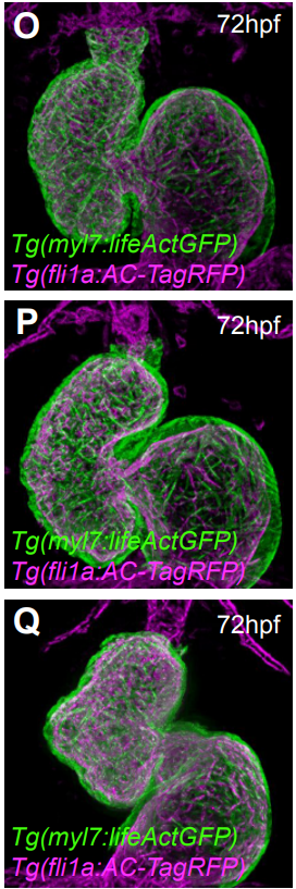
DL & JW Our studies first reveal that in the mouse, the secondary heart field population (SHF) in the splanchnic mesoderm (SpM-SHF) normally grows in a polarized fashion to preferentially elongate anteroposteriorly. Loss of Wnt5a, however, leads to isotropic expansion of the SpM-SHF. We provide evidence that Wnt5a may act through PCP effector and formin protein Daam1 to form horizontally oriented actomyosin cables in the medial SpM-SHF, thereby generating the mechanical force to constrict SHF widening and promote its lengthening. Genetic labelling and tracing reveal that the Wnt5a lineage is a unique SHF subpopulation specified as early as embryonic day (E)7.5, and undergoes bi-directional deployment towards both the arterial and venous poles to contribute specifically to the pulmonary trunk and atrial septum, respectively. In Wnt5a null mutants, the Wnt5a lineage fails to extend into the arterial and venous poles, causing both outflow tract and atrial septation defects, both of which can be rescued by an activated form of Daam1. Interestingly, the Wnt5a lineage in the SHF also contributes to the pulmonary mesenchyme and proper morphogenesis of the airway, suggesting the intriguing idea that during the evolution of tetrapods for terrestrial life, Wnt5a/PCP was recruited by the cardiopulmonary progenitors to orchestrate morphogenesis of both the pulmonary airway and cardiac septation necessary for pulmonary circulation.
3D reconstruction of a wild-type heart at E13.5.
Is Wnt5a acting locally or at a distance in the SHF?
DL & JW This is a question that we debated a lot in the lab. The conventional wisdom in the Wnt field seems to be that the Wnt ligand does not travel very far, probably less than ten cell diameters away from the source. Our anti-Wnt5a antibody staining also shows a very restricted distribution in the caudal SHF, largely overlapping with the domain of Wnt5a mRNA expression. But we need to be cautious about the sensitivity of antibody staining. The fact that the SHF deployment defect in Wnt5a null mutants is not restricted to the Wnt5a lineage per se raises the possibility that Wnt5a may signal at some distance. In the limb, we have very strong genetic evidence that Wnt5a produced at a defined location can signal hundreds of microns away. The action of Wnt5a could be tissue specific though, so to address this question, we will need to tag Wnt5a and visualize its distribution.
Does your data have any relevance to the idea of cardiopulmonary progenitors?
DL & JW The idea of ‘cardiopulmonary progenitors’ was initially proposed by Ed Morrisey’s group to explain co-development of the heart and lung during the evolutionary adaptation to terrestrial life. They had found that the posterior SHF contains multipotent progenitors contributing to both the venous pole of the heart and pulmonary mesoderm. Our Wnt5a lineage-tracing data support this idea, and further demonstrate that the cardiopulmonary progenitors may also contribute to pulmonary trunk at the arterial pole, in addition to the dorsal mesenchymal protrusion (DMP) at the venous pole. Formation of the pulmonary trunk and DMP-mediated septation of the atria are both necessary to fully separate pulmonary from systemic circulation.
Recent studies from Ivan Moskowitz’s group further demonstrated that expression of Tbx5 in cardiopulmonary progenitors initiates a signalling cascade to specify pulmonary fate in the adjacent endoderm. We found that deleting Wnt5a using SHF-specific Mef2cCre causes not only cardiac septation but also airway morphogenesis defects. Therefore, we speculate that in addition to adopting the Tbx5 transcription network for pulmonary fate specification, the cardiopulmonary progenitors may have also recruited Wnt5a/PCP as a genetic circuit to orchestrate morphogenesis of the heart and lung during their co-evolution in tetrapods.
When doing the research, did you have any particular result or eureka moment that has stuck with you?
DL I would say the most exciting moment during this project was when I finished the first 3D reconstruction of E10.5 wild-type and Wnt5a null mutant embryos. For the first time I was able to turn around and really see the whole heart in relation to the SHF on the computer display, not just a series of individual sections. That feeling was indescribable and the memory will definitely stay with me forever.
That feeling was indescribable and the memory will definitely stay with me forever
And what about the flipside: any moments of frustration or despair?
DL I have never had a project completely free of frustration, so of course this project was no exception. Here and there, we had big or small troubles with techniques, but somehow found ways to overcome and move on. The biggest frustration came from the clonal analysis. We tried to genetically label a few Wnt5a lineage cells by using our Wnt5aCreER mouse line and R26R-confetti (Brainbow) mouse line, and to test whether their deployment to the arterial and venous poles of the heart would change over time. However, the labelling efficiency of Wnt5aCreER with the confetti reporter was extremely low and we could not get any interpretable result after many tries. Finally, we gave up and had to submit the manuscript without this piece of data.
So what next for you after this paper?
DL After finishing the revision of this manuscript, I accepted a job offer from a clinical genomics laboratory, and started a career path in medical genetics and diagnosis. Although not dealing with embryos anymore, I do benefit a lot at work from my past life in research.
Where will this work take the Wang lab?
JW There are several things that we would like to pursue. First, we want to examine further the role of Wnt5a and PCP signalling in the co-morphogenesis of the heart and lungs. Second, we want to define further the action of Wnt5a by determining its signalling range, and whether it functions in a permissive or instructive fashion. Finally, we want to understand how the human WNT5A point mutations perturb its function during development and result in congenital birth defects.
Finally, let’s move outside the lab – what do you like to do in your spare time in Birmingham?
DL I like hiking. My favourite trail is on the top of the Red Mountain, where Birmingham’s famous Vulcan statue stands. Walking along the trail and overlooking downtown Birmingham is just really relaxing.
JW I was surprised to find how liveable Birmingham was when I first moved here from San Diego, and discovered a large number of fantastic restaurants of different cuisines. When my kids were younger, I hiked and mountain biked a lot with them in the nearby mountains, and practiced karate with them. Now I tend to do activities that are more relaxing, like yoga and golf. The Robert Trent Jones golf trail is a series of world-class golf courses built throughout the state of Alabama, and there are two of them within a 10 minute drive from my house.
The Department of Tissue Dynamics & Regeneration (@ Max Planck Institute for Biophysical Chemistry) is recruiting a new research technician!
The department of Dr. Jochen Rink studies the fascinating ability of planarian flatworms to regenerate complete animals from tiny tissue pieces. The department employs a broad range of techniques to understand the molecular mechanisms of regeneration, but also carries out field work to study the evolutionary mechanisms that determine why some worms regenerate, while others cannot.
The technician will work on specific research projects in the department, independently or as part of a team. In addition, he/she will also help with general lab management and organization.
This project will study alternative transcript expression, splicing and function of TCF/LEF genes in the Wnt signalling pathway. This is a close collaboration with the research group of David Ferrier at the Scottish Oceans Institute in St. Andrews who will study the evolution and genomics of TCF/LEF genes and will also involve Seb Shimeld and Nanopore in Oxford and Nori Satoh in Okinawa, Japan.
The Fang research group (https://www.northeastern.edu/fang/) seeks outstanding candidates to fill TWO post-doctoral positions in the Department of Electrical and Computer Engineering at Northeastern University in Boston, USA. The candidates will be working on research projects related to soft neural interfaces.
The post-doctoral candidates should meet the basic requirements of training/expertise as described below:
In vivo neural recording and biocompatibility studies. Candidates should have ample experience and capability to conduct in vivo neural engineering studies (surgical implant of electrodes in the brain, in vivo recording/stimulation, and histological analysis).
OR
Active matrix data acquisition system development. Candidate should have experience and capability to develop large-throughput data acquisition system for actively multiplexed electrode array to record extracellular neuronal signals.
These positions are available immediately until filled. Qualified candidates can send their (a) CV, (b) three representative publications, and (c) names and contact info of at least two references to h.fang@northeastern.edu.
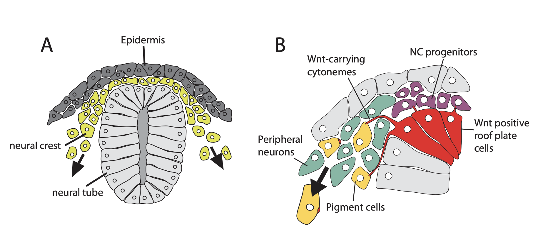
Dissecting the function of Wnt signalling in zebrafish neural crest development.
Project Description
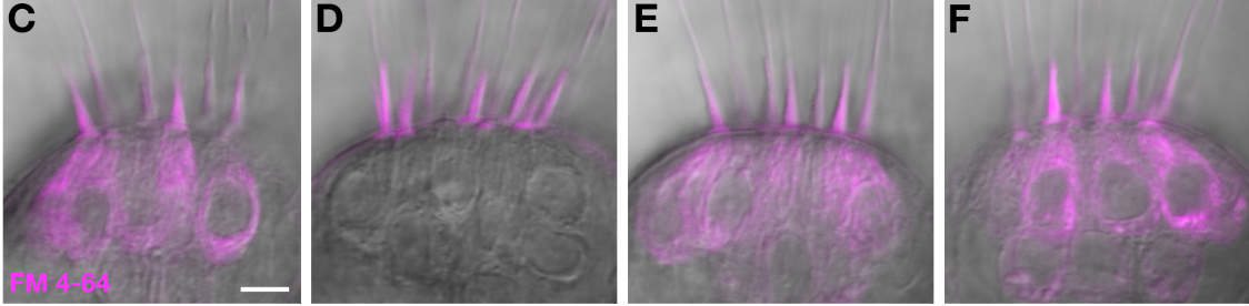
The neural crest (NC) is a stem-cell-like cell population, which is unique to vertebrates. NC cells can become peripheral neurons and glia, pigment cells and cartilage. NC cells migrate a long distance from their birthplace to the site where they function (Fig. 1A). Along the way, NC cells are exposed to signals promoting the variety of cell fates. We have shown that Wnt signals regulate the balance between peripheral neurons and pigment cells (Vibert et al., 2017 Pigm Cell Melanoma). However, how Wnt activates pigment cell fate, and leaving others to adopt another fate, is not understood. Recently, we could show that Wnt transport is directed by long cellular extensions called cytonemes. (Stanganello et al., 2015; Nat Comms). The producing cell loads Wnt on cytonemes to send them to the receiving cell. So, the producers can control how much and how far Wnt is transported, but also which cells are contacted. We hypothesise that Wnt cytonemes are the underlying cellular mechanism driving diversification of the NC lineage (Fig. 1B).
Under the supervision of leading cell biologists in Exeter and Bath, the student will test this hypothesis by studying Wnt transport in zebrafish. The student will generate transgenic zebrafish lines with fluorescently tagged Wnt proteins. The student will use these fish lines to monitor Wnt trafficking by advanced high- and super-resolution microscopy. Simultaneously signal activation in the receiving neural crest cells will be described by using real-time PCR, and live reporter systems. Finally, the student will interfere with Wnt cytonemes by using chemical inhibitors and CRISPR-based mutations in zebrafish to study their impact on NC differentiation.
The student selected for this project will develop invaluable skill sets in experimental genetics, cell biology while also making a significant contribution to the development of and high-resolution in vivo imaging techniques. This combined skill set will make the candidate a highly desirable recruitment prospect for future academic and industrial employers.
The Living Systems Institute in Exeter and the Department of Biology in Bath, with complementary expertise in biosciences and high-resolution imaging, will be an optimal environment to conduct these doctoral training studies. We offer unique training opportunities for the PhD student as it allows the student to address critical problems in life sciences with state-of-the-art equipment in an interdisciplinary environment.
A fully-funded four-year SWBio DTP studentship will cover:
• a stipend* at the standard UKRI rate; currently £15,009 per annum for 2019-2020
• research and training costs
• tuition fees (at the standard UKRI rate)
• additional funds to support fieldwork, conferences and a 3-month internship
Modern scientific innovation involves the integration of diverse disciplines and the bridging of pure and application-oriented research. This requires a new generation of bioscientists who are trained to think and experiment beyond traditional boundaries. The recently established Living Systems Institute (LSI), University of Exeter, provides this opportunity by housing world-leading Biologists, Physicists, Mathematicians, Computational Scientists and Engineers using cutting-edge technologies. Our mission is to understand life across all scales – from individual atoms to whole organisms – and to discover approaches for improving health and treating disease. We are pleased to announce the first call for our interdisciplinary PhD programme for the autumn 2020 intake. We wish to recruit the best and most imaginative students from across the full range of disciplines to join our thriving community of over 60 PhD students.
For the first time in 20 years, researchers from across the continent gathered for the European Developmental Biology Congress in Alicante, Spain (October 23-26 2019). Fortunately, we won’t have to wait another 20 years for the next one – it will be held next in the UK in 2023. But in addition to a fantastic program of talks and posters, friendly networking opportunities and the biggest pan of paella I’ve ever seen, the Congress also featured a pre-meeting workshop on preprints, organised by Teresa Rayon (postdoc at The Francis Crick Institute and preLighter).
Teresa put together a panel of researchers and publishers to talk about the value of preprints to the community and how they might change scientific publishing. While, unfortunately, the weather prevented panel member Sofia Araujo (IRB Barcelona) from making it on time to introduce the session, Teresa stepped in to kick off what we hope was a useful session for those attending. Here, I summarise some of the key points and take-home messages from the session, and also invite you to take part in the survey (click on the link!) that Sofia and Teresa put together for the workshop. You can see the results from the people that filled it in during the session below, but we’d be interested in gathering more feedback from the community on how you use preprints.
The first encouraging outcome from the discussion was to find that all publishers/journals represented in the panel (The Company of Biologists, PLOS, EMBO Press, Developmental Biology and Mechanisms of Development) are fully supportive of preprints. As Monica Lodi (Publisher at Elsevier) explained, Developmental Biology and Mechanisms of Development – two of the journals she looks after – are fully preprint-friendly: authors can share their preprint anywhere at any time without compromising potential publication in the journals (though it’s worth noting that – while most journals now consider papers that have been printed – some do have restrictions on posting of revised manuscripts). The PLOS, EMBO Press and Company of Biologists journals also all facilitate simultaneous submission of papers to the journal and bioRxiv. And the EMBO Press journals, represented here by Ieva Gailite (Editor, The EMBO Journal) have even extended their ‘scooping protection policy’ to the point at which a preprint is submitted (within reason – see here for more details). As raised in the discussion, many editors are now actively scouting for potential submissions of preprints by browsing through bioRxiv or social media, though the ever-growing volume of the preprint literature limits the extent to which this can efficiently be done at most journals.
Despite a long history in the physical sciences, preprints have only recently made an impact in life sciences publishing. I gave a brief overview of The Company of Biologists’ history with preprints. Over the past 5 years, we’ve had a complete turnaround in policy – from considering preprint deposition as prior publication through to integrating fully with bioRxiv, encouraging preprinting and launching preLights – more on which later). In 2018, well over 10% of papers submitted to Development were co-submitted to bioRxiv. Many publishers have a difficult relationship with the preprint literature: it has the potential to disrupt both our models of peer review and our financial business models. But we also recognise the value for our communities, and preprints open the door to interesting new ventures – like preLights and Review Commons (more on this later too). I also outlined how preprints might change our current publishing system – by separating out dissemination of research results from their validation through peer review, by providing opportunities for new publication models (community peer review, overlay journals, journal-agnostic peer review), and potentially by accelerating the transition to Open Access (as preprints are generally free to read).
Talking of Open Access, Ines Alvarez-Garcia (Senior Editor, PLOS Biology) provided an overview of the principles of Plan S, the Open Access initiative from a group of (mainly European) funding agencies, and what they could mean for researchers. You can find the full list of Plan S principles here, and plenty more information about their implementation online, but the bottom line is that Plan S wants all research funded by its signatories to be published in fully Open Access journals or platforms (or in journals with a clear plan to transition to Open Access) from 2021 onwards. This is challenging for many journals, but publishers are working on ways of ensuring that authors can still publish in the journals of their choice, regardless of funding source.
EMBO have long been proponents of increased transparency in publishing, and are now integrating with bioRxiv to display peer review reports of papers under consideration at EMBO Press journals on bioRxiv – part of a new initiative from bioRxiv called Transparent Review in Preprints (TRiP for short). Both EMBO Press and eLife are trialling TRiP, whereby authors can choose to post the referee reports from the journal (whether the paper was accepted or rejected) alongside the preprint. Finally, Ieva introduced Review Commons, a journal-independent peer review platform that aims to reduce the time authors spend re-submitting their paper to multiple journals and focus reviewers’ attention objectively on the science rather than the ‘fit’ for a particular journal, while reducing the total reviewing burden. Both PLOS and The Company of Biologists (along with a number of other journals) are partnering with EMBO and ASAPbio on Review Commons, which should launch later this year.
After these publisher perspectives, two researchers gave their thoughts on the rise of preprints. Jesus Victorino, a PhD student in Miguel Manzanares’ lab and- like Teresa – a preLighter, focussed on the time taken for papers to be published and the problems this can cause particularly for early career researchers – who may need their work in the public domain quickly to apply for the next position. He also pointed out that, as we all know, peer review is not perfect, and that reading the preprint literature can make you think more deeply about the validity of a particular piece of research. Finally, he explained what he gets out of being involved with preLights: improving his writing skills, interacting with the authors of preprints he writes about, and engaging with the preLights community.
The final perspective came from Fernando Casares, a PI at the CABD in Seville. Fernando’s support for preprints was summed up in 5 points:
Communication: a primary purpose of science is to communicate your results and preprints allow you to do this quickly, and to promote your work to your network and beyond.
Priority: once the preprint is online, this gives you a claim to the discovery – even if it then takes some time for the paper to be published.
Material for grant proposals or job applications: means the people assessing you can actually see the work you’ve done rather than just taking your word for it.
As a ‘psychological aid’: it can take a long time to go from submission of a paper to acceptance, and this can be pretty demoralising. At least if the preprint is out, there’s proof – for yourself as much as anyone else – that the work is done.
To allow publication of otherwise ‘unpublishable’ work: data that would be hard to get formally published or where it’s not worth the effort to do so. Fernando pointed out that there are some places where people are assessed based on the average citation rate of papers they’ve published – in these cases, you might be cynically better off not publishing the work you think is unlikely to cite well. This isn’t good for science or the community, so at least you could make preprints available for work like this.
Fernando’s final point brought up the issue of how research and researchers are assessed – something we returned to in the discussion session. Clearly, the obsession with where you publish and how well you’re cited is a problem in research assessment – but not one that any of us have real solutions for at this point. But there is a movement to promote better practices, and if you’ve not heard of DORA, or read some of their case studies, I’d encourage you to take a look.
The discussion that followed the brief presentations from each panellist ranged from the practicalities of Review Commons to the economics of quality publishing, and from submission policies through to whether we still need journals in the age of preprinting (short answer – yes! – if you ask me anyway…!). Huge thanks to Teresa for organising the session and to everyone who came along. And, as final evidence that preprinting is definitely taking off in our community, take a look at this curated preList of preprints discussed over the course of the EDBC meeting.
 (No Ratings Yet)
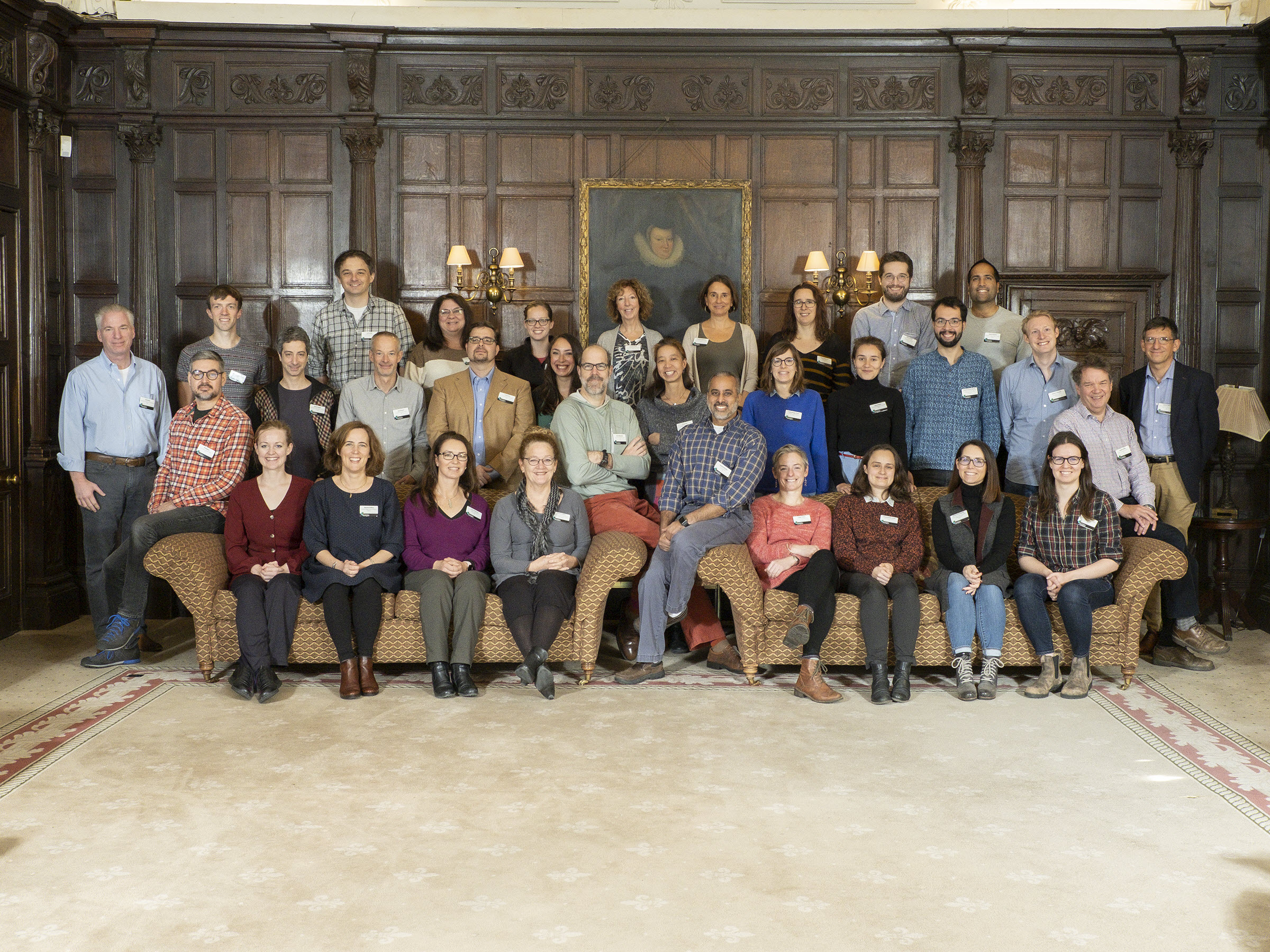
 (2 votes)


 (6 votes)