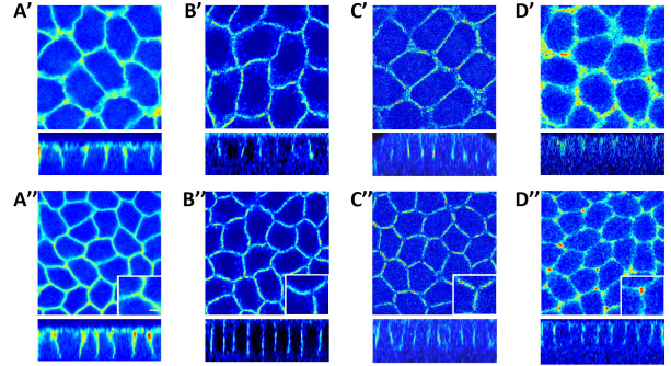
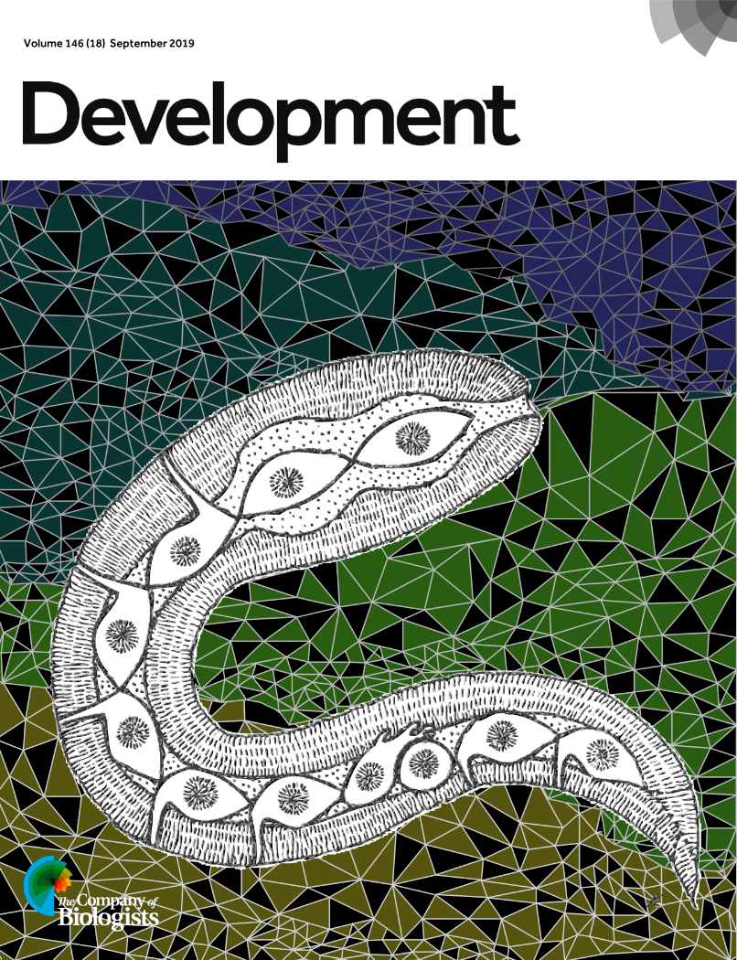
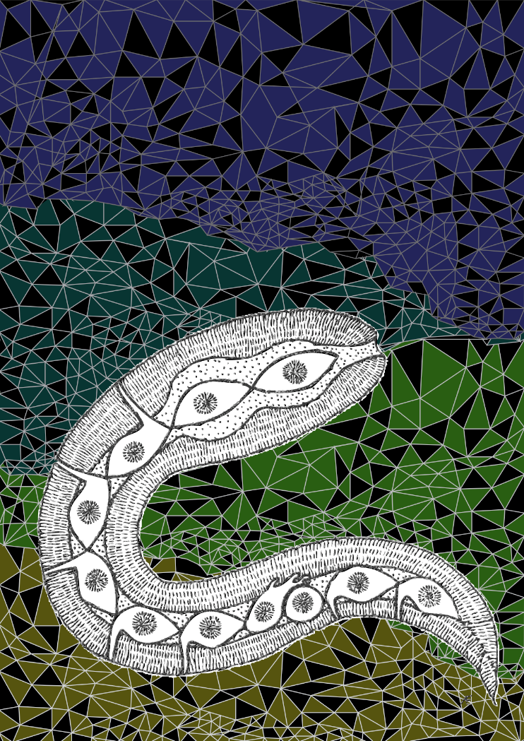
Development covers showcase the beauty of developmental biology. Embryos, tissues and cells are rendered in striking colour palettes and artistic arrangements. We mainly receive confocal image submissions but sometimes also EM and standard light microscopy. And sometimes, art – like our most recent cover, a schematic overview of C. elegans created by Annabel Ebbing, PhD student in Hendrik Korswagen’s labat the Hubrecht Institute in The Netherlands and first author of a new paper on neuroblast migration. We caught up with Annabel to find out how her beautiful cover came about and how she thinks about art and science.
Annabel’s cover
Can you tell us about your PhD project and your new Development paper?
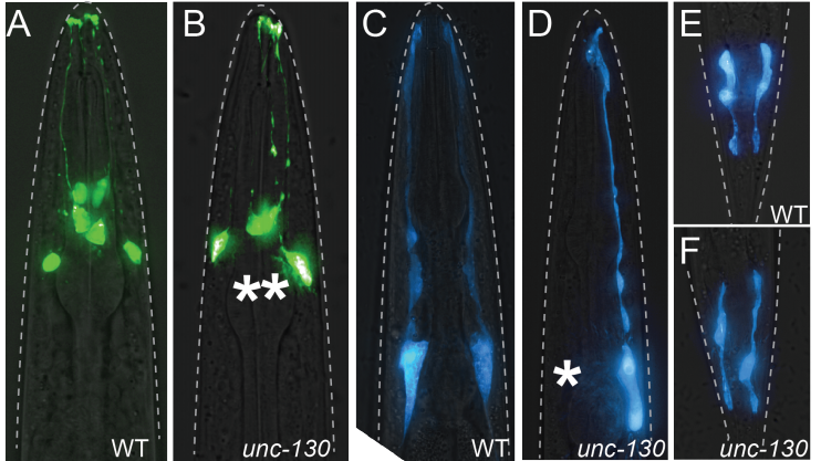
The project started a while back (in 2013), when I was still a Master’s intern student at the Korswagen lab. My direct supervisor was Teije (the shared first author on this paper) who helped me with the imaging and epistasis experiments to elucidate the role of the Fat-like cadherins CDH-4 and CDH-3 in Q neuroblast migration. In the meanwhile, he did similar experiments for UNC-40, DPY-19, and MIG-21. Finally, when I started my PhD I set out to combine the two projects. This was during the emergence of CRISPR as a standardized tool for C. elegans, which really helped the project get to the state where it is now. We managed to study the effects of tissue specific protein depletions and endogenous protein localization, which together really resolved some of the outstanding questions we had concerning the interplay between proteins and a possible molecular mechanism. All in all, there are several proteins involved in Wnt independent Q neuroblast migration; apart from their own roles they have overlapping functions as well, making it a complex and highly regulated mechanism controlling protrusion formation and directionality.
Have you always been interested in art as well as science?
Both art and science have always been fascinating subjects to me. Actually, I wanted to pursue a more artistic education at a certain point, but I decided to study biology in the end to feed my general curiosity. Pursuing a career in science did not make me leave the artistic side altogether though. I especially love thinking about scientific projects in a more abstract way. For me the process of visualization helps in my understanding of a certain subject.
Where did you get the idea for submitting cover art rather than something more standard?
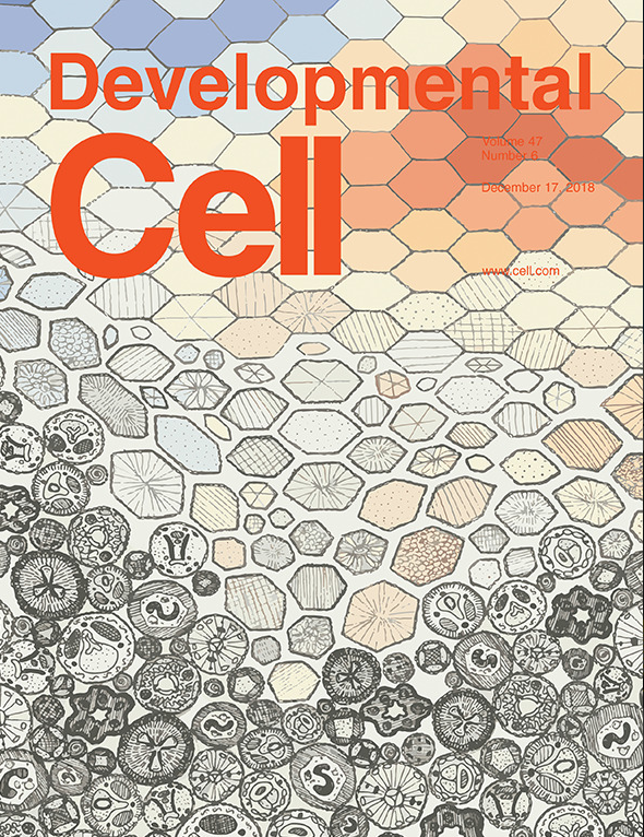
Well, last year we published a paper in Development Cell, for which I made the cover. So when we were asked to submit a cover proposal for Development, I decided to take a similar approach. I love drawing and painting and honestly thought the immunofluorescence images for this project were rather dull (sometimes you see these amazing structures of cells, organs, and even organisms and our imaging was just rather 2D). Let’s say, the choice to draw was quickly made.
Annabel’s Developmental Cell cover from last year
Can you tell us a little about the process of designing the piece?
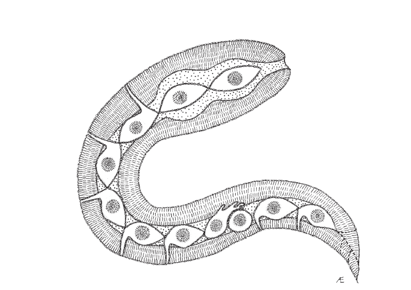
I started the piece by trying to make an abstract overview of a worm, including a migrating Q neuroblast, using simple lines and dots. Later on, I imported the file in illustrator and made the background. Most covers of Development (if not all) have a dark/black background, so I decided to use dark colors, which made a nice contrast with the black and white worm.
The abstract worm drawing at the heart of Annabel’s piece
How did Aboriginal art influence the work?
Aboriginal art is an artform of storytelling; using simple lines and dots the Aborigines manage to tell entire stories and legends. Since the mechanism underlying the process of Wnt independent Q neuroblast migration is rather complex, I thought there would be no better way to illustrate it than in such a narrative-rich manner.
Annabel’s final piece (minus the Development augmentations)
What’s next for you after this paper?
I am submitting my thesis as we speak! So I guess the main focus for the coming months is the defense. Art wise that means I am illustrating a cover for every chapter, which is a lot of fun! Moreover, together with colleagues we are preparing another manuscript at the moment, which will hopefully be accepted sometime this winter.
Some more worm art from Annabel
Some previous artistic Development covers
This picture pays homage to M. C. Escher’s tessellation studies and Alan Turing’s reaction-diffusion mechanism. Both Turing and Escher were interested in pattern generation and the changes of organic forms, two phenomena central to evolutionary developmental biology, as addressed here in the context of periodic patterning in the turtle shell. From Moustakas-Verho, et al. https://dev.biologists.org/content/141/15/3033?iss=15
Taking the iconic example of the Nautilus pompilius shell from Darcy Thompson’s On Growth and Form, the rules of logarithmic spiral growth were abstracted as the foundation to develop a computational model. By experimenting with its parameters, an array of new shapes was created, highlighting the important role that computational modelling has in advancing our understanding of complex physical form in the field of developmental biology. Image created by Jennifer Ma (Zandstra lab, University of Toronto, Canada) and Matthew Spremulli (Living Architecture Systems Group and University of Toronto, Canada). To find out more, visit http://thenode.biologists.com/behind-the-cover/interview/.
Recently, two iconic developmental biology models entered into the single cell genomics era: chick and zebrafish. In this image, line art was traced using real embryo images for reference and filled with individual dots to represent the reduction of the whole embryo to its smallest structural, functional and biological unit: the cell. This cover was chosen by Special Issue guest editors Allon Klein and Barbara Treutlein from entries to Development’s cover competition. By Martin Estermann (Monash University, Australia).
Applications open for the Graduate School of Life Science
The Graduate School Life Science Munich (LSM) offers an international doctoral programme to highly motivated and academically qualified next generation researchers at one of Europe’s top Universities. LSM members are internationally recognized for their innovative research approaches and technologies, they are aiming to answer essential questions relevant to basic and applied biological and biochemical research. Within their own research group or in collaboration with a specialized research group on campus, LSM doctorates are given the opportunity to learn and command a variety of techniques. Furthermore, the graduate programme holds various workshops and seminars that strengthen and prepare doctorates for a successful career as scientists.
With over 40 research groups from the Faculty of Biology and the Faculty of Chemistry and Pharmacy of Ludwig Maximilian University (LMU) München, the LSM is located in the Biocenter. Its prominent location within the HighTechCampus in Martinsried south of Munich, contributes to the enormous possibilities for support, interdisciplinarity and constant scientific input from the surrounding laboratories. Available research projects cover areas from Biochemistry, Bioinformatics, Cell and Developmental Biology, Epigenetics, Genetics, Human Biology and Bioimaging, Pharmacology, and Plant Biology.
All courses, lectures and seminars at the LSM are held in English. Thus, selected candidates have to be fluent in both written and spoken English.
Successful students of the graduate school will be awarded a doctoral degree (Dr. rer. nat.) by the LMU after 3 to 4 years. Candidates need to prove a strong qualitative background as well as interest and ability to conduct independent research.
The doctoral programme is open for students who hold either a master´s or diploma degree, as well as to exceptional candidates with a four years bachelor degree (with written thesis).
LSM calls for doctoral applications on a yearly basis, currently open from the 14th of October until the 29th of November 2019. Applicants are selected in a multi-step process through our online portal, thus ensuring openness and fairness throughout the application procedure. Every complete submission is evaluated by the LSM coordinator. Applications will be independently reviewed by several faculty members of the LSM Graduate School. Based on academic qualification, research experience, motivation, scientific background and the letters of recommendation, candidates will be selected to participate in the LSM Interview week. After thorough evaluation through the LSM committee board members, successful candidates will be invited to join the LSM Graduate School.
We are looking for a highly motivated and talented PhD candidate to investigate the role of enteric glial cells in the assembly and maintenance of the intrinsic neural circuits that regulate gastrointestinal function and homeostasis.
During the course of this PhD research project, which will take place at Maastricht University (The Netherlands), novel genetic and neurophysiological tools will be combined with cutting-edge live cell microscopy techniques to study the role of enteric glial cells in enteric neuron connectivity. You will make use of in vivo models to investigate how enteric glial cells adapt to safeguard gastrointestinal function and homeostasis. You will run a research project in an international multidisciplinary lab and collaborate with (inter)national research groups to complement the know-how within our research group with state-of-the-art external expertise. This PhD position is part of a NWO-funded Vidi project, entitled “Glia of the bowel, landscapers in the second brain” that was recently awarded to Dr. Werend Boesmans.
Additional information about the vacancy can be obtained from: dr. Werend Boesmans (w.boesmans[at]maastrichtuniversity.nl). Interested candidates should send a letter of motivation, a detailed CV and the contact details of two referents.
The Buckley lab at the department of Physiology, Development and
Neuroscience (PDN), University of Cambridge is recruiting a Research
Assistant or Research Associate to help further develop their
optogenetic technology. The lab uses optogenetic and live confocal
imaging approaches within the whole zebrafish neural tube and mammalian
embryo stem cell (mESC) culture to manipulate the polarity of single
cells. In combination with CRISPR-mediated functional knock down
experiments, we are directly testing the role of cell polarity in
building epithelial integrity during organ development and breaking in
it during normal developmental processes such as EMT and during abnormal
disease states.
The post is for 1 year in the first instance from early 2020 and funding
is available for potential contract extension.
An Engineer position in Molecular Biology is available starting January 2020 in the group of Thomas Lecuit at the Developmental Biology Institute of Marseille (IBDM, CNRS UMR7288), Marseille, France. Funding is provided by an ERC grant. The appointment will be made for 1 year, with a possible extension to up to 4 years.
We are seeking a highly-motivated candidate with strong expertise in Molecular Biology. Experience in CRISPR/CAS9 editing and/or Drosophila genetics is valued. The working language in the laboratory is English, so the candidate should have a good practice in English.
The Engineer will perform standard molecular biology techniques and CRISPR/Cas9 gene editing in vivo. We are seeking a highly motivated and flexible individual to join our team.
Essential Skills
Advanced knowledge of molecular biology and associated practical techniques. Experimental dexterity and attention to detail; quality driven.
Excellent organizational and planning skills.
A high level of interpersonal and communications skills.
Ability to follow instructions accurately and efficiently, correctly interpret scientific data and work under supervision.
Position Summary:
The MBL is seeking a candidate for the position of Postdoctoral Scientist in the laboratory of Dr. Blair Paul to investigate accelerated protein evolution in aquatic bacteria. The lab uses computational and molecular tools to study processes that diversify microbial genes and to understand the functional importance of hypervariable proteins. To this end, the prospective research will involve a synergistic combination of experimental biology and bioinformatics to investigate genome variation and protein evolution/optimization in microorganisms and their viruses. The ideal candidate will bring expertise in molecular biology and microbial genetics, which will be complemented with training in genome bioinformatics through this postdoc experience.
Additional information:
The position is for one year with anticipated extension to 2+ years, contingent on performance. For more information about MBL and living on Cape Cod, please visit: https://www.mbl.edu/hr/employment/our-community/.
Basic qualifications:
A Ph.D. in biology, microbiology, molecular biology, or a related field is required.
Preferred qualifications:
Experience in the following areas is desirable: microbiology (including isolation or cultivation from environmental populations), molecular techniques (especially bacterial or archaeal genetics), or protein biochemistry.
Development invites you to submit your latest research for our upcoming special issue: The Origins and Mechanisms of Developmental Disorders. This issue will be coordinated by two guest editors: Sally Dunwoodie and John Wallingford.
Developmental disorders are among the most lethal diseases of childhood, and this recent decade has witnessed significant advances in our understanding of their genetic and environmental basis. Studies in model systems, alongside clinical research and human genetics, have been foundational to this effort but major challenges remain. Congenital disorders are characterised by a high incidence of sporadic cases, a low recurrence risk, and variable penetrance and expressivity of identified genetic mutations even within families, indicating that gene-gene and gene-environment interactions are likely to be important. In model systems, the impact of various teratogens and environmental factors on embryogenesis demonstrate both the robustness and the fragility of specific developmental processes. In mammals, placental function and the pathophysiology of the mother are also disruptors of embryogenesis.
Development has a long history of publishing leading research in these areas and, for this special issue, we welcome submissions encompassing congenital disorders, teratogenic and environmental impacts on development, and links between development and paediatric tumours.
The issue will be published in mid-2020 (although note that, in our new ‘continuous publication‘ model, we will be able to publish your article as soon as it is accepted; you will not have to wait for the rest of the issue to be ready) and the deadline for submission of articles is 31 January 2020*. This special issue will be widely promoted online and at key global conferences – guaranteeing maximum exposure for your work. Please refer to our author guidelines for information on preparing your manuscript for Development, and submit via our online submission system. Please highlight in your cover letter that the submission is to be considered for this special issue. Prospective authors are welcome to send presubmission enquiries, or direct any queries, to dev.specialissue@biologists.com
We also welcome proposals for Review articles; if you’re interested in contributing a Review, please send a brief synopsis of your proposed article to dev.specialissue@biologists.com by 31 October 2019.
* Please note that not all articles accepted for publication will be included in the special issue; they may instead be published in earlier or later issues of the journal.
Why choose Development?
Submissions handled by expert academic Editors
Competitive decision speeds and rapid publication
Format-free submission
Strong commitment at first decision – over 95% of invited revisions accepted
Free to publish – no page or colour charges, no hidden fees
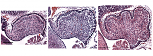
The Lawton Lab is seeking a talented and motivated graduate student to study the cell and tissue mechanics regulating cerebellar morphogenesis and brain folding.
Sagittal midline sections of mouse cerebellum at embryonic day E16.5, E17.5 and E18.5 show the initiation of folding.
The beautiful and robust folds of the human cerebral cortex and the cerebellum increase the synaptic volume and compartmentalize the neural circuits. We have previously shown that the murine cerebellum initiates folding, without a pre-pattern, through differential expansion – between an outer fluid-like layer and an inner solid mass – together with radial and circumferential tension. However, it is not known how the folding amount or pattern is set. Nor is it known how the measured tension is regulated or how the fluidity of the outer layer is maintained. The Lawton Lab uses developmental and mechanical assays, live-imaging, and quantitative analysis to address these issues and to discover the cellular and emergent tissue-level regulation of brain folding.
This project will provide training in classical developmental biology assays, ex vivo slice culture, histology, confocal microscopy, and quantitative analysis.
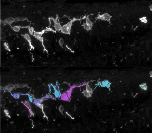
Granule cell precursors labeled with membrane GFP. Cell shapes are masked and quantified
Applying:
Position is to begin fall semester of 2020.
The ideal candidate will have a strong interest in developmental biology, and some experience with microscopy, image analysis, and Matlab. Those with a background in mathematics or physics are also strongly encouraged to apply.
Please send a statement of research interest and a CV with references to alawton@biology.msstate.edu
Apply your developmental biology / molecular biology / neurobiology skills to the problem of brain cancer.
The lab of Dr. Jennifer Chan seeks to recruit a motivated postdoctoral fellow to investigate cell fate decisions during the process of brain tumour development and progression. Research in the lab focuses on growth factor signalling and transcriptional regulation as determinants of neural precursor identity and fate. We use models that include patient-derived glioma cultures, xenografts, and engineered mouse models generated from in utero and postnatal electroporation to address our research questions.
The successful candidate will collaborate with investigators in bioinformatics to apply tools of genomics, epigenomics, and transcriptomics to further define key alterations during neoplastic transformation, and will collaborate with medicinal chemists to determine if identified alterations may be potential therapeutic targets. Early-stage postdocs (within 3 years of receiving PhD or equivalent) with experience in advanced immunohistochemistry, fluorescence microscopy, gene editing, and molecular biology; and/or experience in genomic approaches like RNA-seq, ChIP-seq, ATAC-seq will be preferentially considered.
The position is available immediately.
Other information
Located in Calgary, Alberta, Canada, the Chan Lab is part of the Charbonneau Cancer Institute at the University of Calgary’s Cumming School of Medicine. Within the Charbonneau Institute, we are part vibrant multidisciplinary research groups focused on childhood cancers and brain cancers. Calgary is a very livable and family-friendly city located less than an hour’s drive from the Canadian Rocky Mountains – a haven for outdoor enthusiasts.
Application details
Submit a brief letter of interest, your academic CV, and the names and contact information of at least three references.
Applications should be submitted as a single PDF file and sent as an email with the subject line “Post-Doctoral Fellowship, Glioma Biology” to: jawchan@ucalgary.ca
Welcome to our monthly trawl for developmental biology (and related) preprints.
In recent preprint news, CSHL, which runs bioRxiv, launched Transparent Review in Preprints (TRiP), a new project enabling journals and peer review services to post peer reviews of submitted manuscripts. In linked news EMBO Press and ASAPbio launched Review Commons, a platform that peer-reviews research manuscripts in the life sciences before submission to a journal, and enables authors to publicly post the reviews and their own response to them bioRxiv. Finally, PeerJ Preprints, regular source of preprints for this list, announced it would no longer be accepting new content. So long and thanks for all the preprints!
This month we found a typical wealth and breadth of preprints hosted on bioRxiv, PeerJ, andarXiv. Let us know if we missed anything, and use these links to get to the section you want:
Essential omega-3 fatty acids tune microglial phagocytosis of synaptic elements in the developing brain
C. Madore, Q. Leyrolle, L. Morel, J.C. Delpech, A.D. Greenhalgh, C. Lacabanne, C. Bosch-Bouju, J. Bourel, A. Thomazeau, K.E. Hopperton, S. Beccari, A. Sere, A. Aubert, V. De Smedt-Peyrusse, C. Lecours, K. Bisht, L. Fourgeaud, S. Gregoire, L. Bretillon, N. J. Grant, J. Badaut, P. Gressens, A. Sierra, O. Butovsky, M.E. Tremblay, R.P. Bazinet, C. Joffre, A. Nadjar, S. Layé
STAG2 cohesin is essential for heart morphogenesis
Magali De Koninck, Eleonora Lapi, Claudio Badia-Careaga, Itziar Cossio, Daniel Gimenez-Llorente, Miriam Rodriguez-Corsino, Elena Andrada, Andres Hidalgo, Miguel Manzanares, Francisco X Real, Ana Losada
The enteric nervous system of the human and mouse colon at a single-cell resolution
Eugene Drokhlyansky, Christopher S. Smillie, Nicholas Van Wittenberghe, Maria Ericsson, Gabriel K. Griffin, Danielle Dionne, Michael S. Cuoco, Max N. Goder-Reiser, Tatyana Sharova, Andrew J. Aguirre, Genevieve M. Boland, Daniel Graham, Orit Rozenblatt-Rosen, Ramnik J. Xavier, Aviv Regev
Systematic assessment of regulatory effects of human disease variants in pluripotent cells
Marc Jan Bonder, Craig Smail, Michael J. Gloudemans, Laure Frésard, David Jakubosky, Matteo D’Antonio, Xin Li, Nicole M. Ferraro, Ivan Carcamo-Orive, Bogdan Mirauta, Daniel D. Seaton, Na Cai, Danilo Horta, YoSon Park, HipSci Consortium, iPSCORE Consortium, GENESiPS Consortium, PhLiPS Consortium, Erin N. Smith, Kelly A. Frazer, Stephen B. Montgomery, Oliver Stegle
PI 3-kinase delta enhances axonal PIP3 to support axon regeneration in the adult CNS
Amanda C. Barber, Rachel S. Evans, Bart Nieuwenhuis, Craig S. Pearson, Joachim Fuchs, Amy R. MacQueen, Susan van Erp, Barbara Haenzi, Lianne A. Hulshof, Andrew Osborne, Raquel Conceicao, Sarita S. Deshpande, Joshua Cave, Charles ffrench-Constant, Patrice D. Smith, Klaus Okkenhaug, Britta J. Eickholt, Keith R. Martin, James W. Fawcett, Richard Eva
Evolution of the growth plate into a spatially separated structure allows bone growth on land
Meng Xie, Pavel Gol’din, Anna Nele Herdina, Jordi Estefa, Ekaterina V Medvedeva, Lei Li, Phillip T Newton, Svetlana Kotova, Boris Shavkuta, Aditya Saxena, Lauren T Shumate, Brian Metscher, Karl Großschmidt, Shigeki Nishimori, Anastasia Akovantseva, Irene Linares Arregui, Paul Tafforeau, Kaj Fried, Mattias Carlström, Andras Simon, Christian Gasser, Henry M Kronenberg, Murat Bastepe, Kimberly L. Cooper, Peter Timashev, Sophie Sanchez, Igor Adameyko, Anders Eriksson, Andrei S Chagin
Profiling cellular diversity in sponges informs animal cell type and nervous system evolution
Jacob M. Musser, Klaske J. Schippers, Michael Nickel, Giulia Mizzon, Andrea B. Kohn, Constantin Pape, Jörg U. Hammel, Florian Wolf, Cong Liang, Ana Hernández-Plaza, Kaia Achim, Nicole L. Schieber, Warren R. Francis, Sergio Vargas R., Svenja Kling, Maike Renkert, Roberto Feuda, Imre Gaspar, Pawel Burkhardt, Peer Bork, Martin Beck, Anna Kreshuk, Gert Wörheide, Jaime Huerta-Cepas, Yannick Schwab, Leonid L. Moroz, Detlev Arendt
Universality of clone dynamics during tissue development
Steffen Rulands, Fabienne Lescroart, Samira Chabab, Christopher J. Hindley, Nicole Prior, Magdalena K. Sznurkowska, Meritxell Huch, Anna Philpott, Cedric Blanpain, Benjamin D. Simons
Negligible-Cost and Weekend-Free Chemically Defined Human iPSC Culture
Hui-Hsuan Kuo, Xiaozhi Gao, Jean-Marc DeKeyser, K. Ashley Fetterman, Emily A. Pinheiro, Carly J. Weddle, Hananeh Fonoudi, Michael V. Orman, Marisol Romero-Tejeda, Mariam Jouni, Malorie Blancard, Tarek Magdy, Conrad L. Epting, Alfred L. George Jr., Paul W. Burridge
Cell type specific novel lincRNAs and circRNAs in the BLUEPRINT haematopoietic transcriptomes atlas
Luigi Grassi, Osagie G. Izuogu, Natasha A.N. Jorge, Denis Seyres, Mariona Bustamante, Frances Burden, Samantha Farrow, Neda Farahi, Fergal J. Martin, Adam Frankish, Jonathan M. Mudge, Myrto Kostadima, Romina Petersen, John J. Lambourne, Sophia Rowlston, Enca Martin-Rendon, Laura Clarke, Kate Downes, Xavier Estivill, Paul Flicek, Joost H.A. Martens, Marie-Laure Yaspo, Hendrik G. Stunnenberg, Willem H. Ouwehand, Fabio Passetti, Ernest Turro, Mattia Frontini
Anthony Etuk, Felix Shaw, Alejandra Gonzalez-Beltran, David Johnson, Marie-Angélique Laporte, Philippe Rocca-Serra, Elizabeth Arnaud, Medha Devare, Paul J Kersey, Susanna-Assunta Sansone, Robert P Davey


 (8 votes)
 (No Ratings Yet)
 (1 votes)