December in preprints
Posted by the Node, on 3 January 2019
Welcome to our monthly trawl for developmental biology (and related) preprints.
December’s haul includes a succession of preprints on Drosophila patterning (embryos, wings, brains and intestines), single cell investigations into the neural crest, hair cells, spinal cord and retina, a comparison of primate brain organoids, and plant development covered from root to shoot.
The preprints were hosted on bioRxiv, PeerJ, and arXiv. Let us know if we missed anything, and use these links to get to the section you want:
Developmental biology
| Stem cells, regeneration & disease modelling
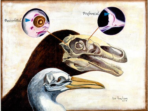
Evo-devo & evo
Cell biology
Modelling
Tools & resources
Research practice & education
Why not…
Developmental biology
| Patterning & signalling
Dynamics of Spaetzle morphogen shuttling in the Drosophila embryo shapes pattern
Neta Rahimi, Inna Averbukh, Shari Carmon, Eyal D Schejter, Naama Barkai, Ben-Zion Shilo
Speeding up anterior-posterior patterning of insects by differential initialization of the gap gene cascade
Heike Rudolf, Christine Zellner, Ezzat El-Sherif
Precise spatial scaling in the early fly embryo
Victoria Antonetti, William Bialek, Thomas Gregor, Gentian Muhaxheri, Mariela Petkova, Martin Scheeler
Evidence of functional long-range Wnt/Wg in the developing Drosophila wing epithelium
Varun Chaudhary, Michael Boutros

Rab converter DMon1 constitutes a novel node in the brain-gonad axis essential for female germline maturation
Neena Dhiman, Girish Deshpande, Girish S Ratnaparkhi, Anuradha Ratnaparkhi
Makorin1 controls embryonic patterning by alleviating Bruno-mediated repression of oskar translation
Annabelle Dold, Hong Han, Niankun Liu, Andrea Hildebrandt, Mirko Brüggemann, Cornelia Rücklé, Anke Busch, Petra Beli, Kathi Zarnack, Julian König, Jean-Yves Roignant, Paul Lasko
Serial synapse formation through filopodial competition for synaptic seeding factors
Mehmet Neset Ozel, Abhishek Kulkarni, Amr Hasan, Josephine Brummer, Marian Moldenhauer, Ilsa-Maria Daumann, Heike Wolfenberg, Vincent Dercksen, Ferdi Ridvan Kiral, Martin Weiser, Steffen Prohaska, Max von Kleist, Peter Robin Hiesinger
The Tenets of Teneurin: Conserved Mechanisms Regulate Diverse Developmental Processes in the Drosophila Nervous System
Alison T DePew, Michael A Aimino, Timothy J Mosca
A refutation to ‘A new A-P compartment boundary and organizer in holometabolous insect wings.’
Peter A. Lawrence, Jose Casal, Jose F. de Celis, Gines Morata
The Notch and EGFR signaling regulate caspase inhibitor Diap1 to match supply with intestinal demand
Tobias Reiff, Zeus A Antonello, Esther Ballesta-Illan, Laura Mira, Salvador Sala, Maria Navarro, Luis M Martinez, Maria Dominguez
Enhancer priming enables fast and sustained transcriptional responses to Notch signaling
Julia Falo-Sanjuan, Nicholas C Lammers, Hernan G Garcia, Sarah Bray
Dynamics of Notch-dependent transcriptional bursting in its native context
ChangHwan Lee, Heaji Shin, Judith Kimble
The Caenorhabditis elegans HAM-1 protein modifies G protein signaling and membrane extension to reverse the polarity of asymmetric cell division
Jerome Teuliere, Gian Garriga
Symmetry breaking in the embryonic skin triggers a directional and sequential front of competence during plumage patterning
Richard Bailleul, Carole Desmarquet-Trin Dinh, Magdalena Hidalgo, Camille Curantz, Jonathan Touboul, Marie Manceau
Scale invariance of BMP signaling gradients in zebrafish
Yan Huang, David Umulis
Embryo geometry drives formation of robust signaling gradients through receptor localization
Zhechun Zhang, Steven Zwick, Ethan Loew, Joshua S Grimley, Sharad Ramanathan
Arkadia degrades SNON to activate level-specific NODAL responses
Jonathon M Carthy, Marilia Ioannou, Vasso Episkopou
Wnt/β-catenin signaling is required for the development of multiple nephron segments
Patrick Deacon, Charles W Concodora, Eunah Chung, Joo-Seop Park

Blood vessels guide Schwann cell migration in the adult demyelinated CNS through Eph/ephrin signaling
Beatriz Garcia-Diaz, Corinne Bachelin, Fanny Coulpier, Gaspard Gerschenfeld, Cyrille Deboux, Violetta Zujovic, Patrick Charnay, Piotr Topilko, Anne Baron-Van Evercooren
CDX4 regulates the progression of neural maturation in the spinal cord
Piyush Joshi, Andrew J. Darr, Isaac Skromne
Posterior axis formation requires Dlx5/Dlx6 expression at the neural plate border
Nicolas Narboux-Neme, Marc Ekker, Giovanni Levi, Eglantine Heude
Loss of YAP/TAZ impaired the proliferation and differentiation ability of neural progenitor cells
Shanshan Kong, Xinwei Cao
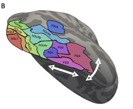
Human visual cortex is organized along two genetically opposed hierarchical gradients with unique developmental and evolutionary origins
Jesse Gomez, Zonglei Zhen, Kevin Weiner
Longitudinal dissection in brain organoids at single cell resolution uncovers the developmental role of GSK3 in human corticogenesis
Alejandro Lopez Tobon, Carlo Emanuele Villa, Cristina Cheroni, Sebastiano Trattaro, Nicolo Caporale, Paola Conforti, Raffaele Iennaco, Maria Lachgar, Marco Tullio Rigoli, Berta Marco de la Cruz, Pietro Lo Riso, Erika Tenderini, Flavia Troglio, Marco de Simone, Isabel Liste, Stefano Piccolo, Giuseppe Macino, Massimigliano Pagani, Elena Cattaneo, Giuseppe Testa
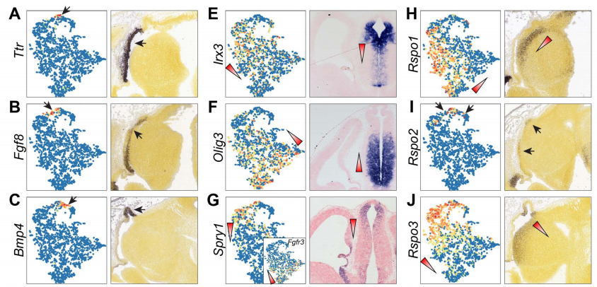
Defining developmental diversification of diencephalon neurons through single-cell gene expression profiling
Qiuxia Guo, James Y.H. Li
Prediction and control of symmetry breaking in embryoid bodies by environment and signal integration
Naor Sagy, Shaked Slovin, Maya Allalouf, Maayan Pour, Gaya Savyon, Jonathan Boxman, Iftach Nachman
Dissecting the dynamics of signaling events in the BMP, WNT, and NODAL cascade during self-organized fate patterning in human gastruloids.
Sapna Chhabra, Lizhong Liu, Ryan Goh, Aryeh Warmflash
RLIM enhances BMP signalling mediated fetal lung development in mice
Molka Kammoun, Elke Maas, Nathan Criem, Joost Gribnau, An Zwijsen, Joris Robert Vermeesch

Mitofusin 1 is required for the oocyte-granulosa cell communication that regulates oogenesis
Thiago S Machado, Karen F. Carvalho, Bruna M. Garcia, Amanda F. Zangirolamo, Carolina H. Macabelli, Fabricia H. C. Sugiyama, Mateus P. Grejo, Jose Djaci Augusto Neto, Fernanda K. S. Ribeiro, Fabiana D. Sarapiao, Flavio V. Meirelles, Francisco E. G. Guimaraes, Lena Pernas, Marcelo M. Seneda, Marcos R. Chiaratti
Fetal and trophoblast PI3Kp110α have distinct roles in regulating resource supply to the growing fetus
Jorge Lopez-Tello, Vicente Perez-Garcia, Jaspreet Khaira, Laura C Kusinski, Wendy N Cooper, Adam Andrani, Imogen Grant, Edurne Fernandez de Liger, Myriam Hemberger, Ionel Sandovici, Miguel Constancia, Amanda N Sferruzzi-Perri
Novel cytokine interactions identified during perturbed hematopoiesis
Madison Ski Krieger, Joshua M Moreau, Haiyu Zhang, May Chien, James L Zehnder, Martin A. Nowak, Morgan Craig
Fibroblast growth factor receptors function redundantly during zebrafish embryonic development
Dena M Leerberg, Rachel E Hopton, Bruce W Draper
Differential physiological role of BIN1 isoforms in skeletal muscle development, function and regeneration
Ivana Prokic, Belinda Simone Cowling, Candice Kutchukian, Christine Kretz, Hichem Tasfaout, Josiane Hergueux, Olivia Wendling, Arnaud Ferry, Anne Toussaint, Christos Gavriilidis, Vasugi Nattarayan, Catherine Koch, Jeanne Lainné, Roy Combe, Laurent Tiret, Vincent Jacquemond, Fanny Pilot-Storck, Jocelyn Laporte
| Morphogenesis & mechanics
Shaping the zebrafish myotome by differential friction and active stress
Sham Tlili, Jianmin Yin, Jean-Francois Rupprecht, Gauthier Weissbart, Jacques Prost, Timothy E Saunders
Cell size heterogeneity early in development is required for collective cell migration during gastrulation in zebrafish
Triveni Menon, Rahul Kumar, Sreelaja Nair

Imaging mechanical properties of sub-micron ECM in live zebrafish using Brillouin microscopy
Carlo Bevilacqua, Héctor Sánchez Iranzo, Dmitry Richter, Alba Diz-Muñoz, Robert Prevedel
Geometry of epithelial cells provides a robust method for image based inference of stress within tissues
Nicholas Noll, Sebastian J. Streichan, Boris I. Shraiman
Netrin/UNC-6 triggers actin assembly and non-muscle myosin activity to drive dendrite retraction in the self-avoidance response.
Lakshmi Sundararajan, Cody Smith, Joseph Watson, Bryan Millis, Matthew Tyska, David Miller
Measurement of junctional tension in epithelial cells at the onset of primitive streak formation in the chick embryo via non-destructive optical manipulation
Valentina Ferro, Manli Chuai, David McGloin, Cornelis Weijer
Tonotopy of the mammalian cochlea is associated with stiffness and tension gradients of the hair cell’s tip-link complex.
Mélanie Tobin, Vincent Michel, Nicolas Michalski, Pascal Martin
Liquid-crystal organization of liver tissue
Hernan Morales-Navarrete, Hidenori Nonaka, Andre Scholich, Fabian Segovia-Miranda, Walter de Back, Kirstin Meyer, Roman L Bogorad, Victor Koteliansky, Lutz Brusch, Yannis Kalaidzidis, Frank Julicher, Benjamin M. Friedrich, Marino Zerial
Confinement-induced transition between wave-like collective cell migration modes
Vanni Petrolli, Magali Le Goff, Monika Tadrous, Kirsten Martens, Cédric Allier, Ondrej Mandula, Lionel Hervé, Silke Henkes, Rastko Sknepnek, Thomas Boudou, Giovanni Cappello, Martial Balland
Sustained oscillations of epithelial cell sheets
Gregoire Peyret, Romain Mueller, Joseph d’Alessandro, Simon Begnaud, Philippe Marcq, Rene-Marc Mege, Julia Yeomans, Amin Doostmohammadi, Benoit Ladoux
Extracellular Matrix acts as pressure detector in biological tissues
Monika E Dolega, Benjamin Brunel, Magali Le Goff, Magdalena Greda, Claude Verdier, Jean-Francois Joanny, Pierre Recho, Giovanni Cappello
Force inference predicts local and tissue-scale stress patterns in epithelia
Weiyuan Kong, Olivier Loison, Pruthvi Chavadimane Shivakumar, Claudio Collinet, Pierre-François Lenne, Raphaël Clément
YAP/TAZ-TEAD Activity Links Mechanical Cues To Cell Progenitor Behavior During Hindbrain Segmentation
Adria Voltes, Covadonga F Hevia, Chaitanya Dingare, Simone Calzolari, Javier Terriente, Caren Norden, Virginie Lecaudey, Cristina Pujades
The Caspase-3 homolog DrICE regulates endocytic trafficking during Drosophila tracheal morphogenesis
Saoirse McSharry, Greg J Beitel
Radial F-actin Organization During Early Neuronal Development
Durga Praveen Meka, Robin Scharrenberg, Bing Zhao, Theresa Koenig, Irina Schaefer, Birgit Schwanke, Oliver Kobler, Sergei Klykov, Melanie Richter, Dennis Eggert, Sabine Windhorst, Carlos G. Dotti, Michael R. Kreutz, Marina Mikhaylova, Froylan Calderon de Anda
| Genes & genomes

Reconstruction of the global neural crest gene regulatory network in vivo
Ruth M Williams, Ivan Candido-Ferreira, Emmanouela Repapi, Daria Gavriouchkina, Upeka Senanayake, Jelena Telenius, Stephen Taylor, Jim Hughes, Tatjana Sauka-Spengler
Lineage tracing on transcriptional landscapes links state to fate during differentiation
Caleb Weinreb, Alejo E Rodriguez-Fraticelli, Fernando D Camargo, Allon M Klein
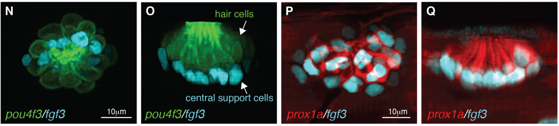
Single cell RNA-Seq reveals distinct stem cell populations that drive sensory hair cell regeneration in response to loss of Fgf and Notch signaling
Mark E. Lush, Daniel C. Diaz, Nina Koenecke, Sungmin Baek, Helena Boldt, Madeleine K. St. Peter, Tatiana Gaitan-Escudero, Andres Romero-Carvajal, Elisabeth Busch-Nentwich, Anoja Perera, Kate Hall, Allison Peak, Jeffrey S. Haug, Tatjana Piotrowski
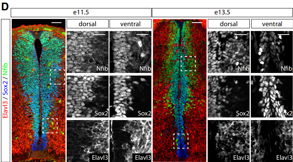
Single cell transcriptomics reveals spatial and temporal dynamics of gene expression in the developing mouse spinal cord
Julien Delile, Teresa Rayon, Manuela Melchionda, Ameila Edwards, James Briscoe, Andreas Sagner
Transcriptional logic of cell fate specification and axon guidance in early born retinal neurons revealed by single-cell mRNA profiling
Quentin Lo Giudice, Marion Leleu, Pierre J. Fabre
An integrated genome-wide multi-omics analysis of gene expression dynamics in the preimplantation mouse embryo
Steffen Israel, Mathias Ernst, Olympia Psathaki, Hannes C. A. Drexler, Ellen Casser, Yutaka Suzuki, Wojciech Makalowski, Michele Boiani, Georg Fuellen, Leila Taher
Genetic approaches in mice demonstrate that neuro-mesodermal progenitors express T/Brachyury but not Sox2
Dorothee Mugele, Dale Moulding, Dawn Savery, Matteo Mole, Nicholas Greene, Juan Pedro Martinez-Barbera, Andrew Copp
The Spatio-Temporal Control of Zygotic Genome Activation
George Gentsch, Nick D. L. Owens, James C. Smith
Pleomorphic Adenoma Gene 1 Is Needed For Timely Zygotic Genome Activation and Early Embryo Development
Elo Madissoon, Anastasios Damdimopoulos, Shintaro Katayama, Kaarel Krjutskov, Elisabet Einarsdottir, Katariina Mamia, Bert De Groef, Outi Hovatta, Juha Kere, Pauliina Damdimopoulou
Ezh2-dependent epigenetic reprogramming controls a developmental switch between modes of gastric neuromuscular regulation
Sabriya Syed, Yujiro Hayashi, Jeong-Heon Lee, Huihuang Yan, Andrea Lorincz, Peter R Strege, Gabriella B Gajdos, Srdjan Milosavljevic, Jinfu Nie, Juri J Rumessen, Simon J Gibbons, Viktor J Horvath, Michael R Bardsley, Doug D Redelman, Sabine Klein, Dieter Saur, Gianrico Farrugia, Zhiguo Zhang, Raul Urrutia, Tamas Ordog
Functional evaluation of transposable elements as transcriptional enhancers in mouse embryonic and trophoblast stem cells
Christopher D Todd, Ozgen Deniz, Miguel R Branco
PTBP2-dependent alternative splicing regulates protein transport and mitochondria morphology in post-meiotic germ cells.
Molly M Hannigan, Hisashi Fujioka, Adina Brett-Morris, Jason A Mears, Donny D Licatalosi
Linked-read sequencing of gametes allows efficient genome-wide analysis of meiotic recombination
Hequan Sun, Beth A Rowan, Pádraic J Flood, Ronny Brandt, Janina Fuss, Angela M Hancock, Richard W Michelmore, Bruno Huettel, Korbinian Schneeberger
CBX2 is required during male sex determination to repress female fate at bivalent loci
Sara Alexandra Garcia-Moreno, Yi-Tzu Lin, Christopher Futtner, Isabella Salamone, Danielle Maatouk, Blanche Capel
Exploring the role of Polycomb recruitment in Xist-mediated silencing of the X chromosome in ES cells
Aurelie Bousard, Ana Claudia Raposo, Jan Jakub Zylicz, Christel Picard, Vanessa Borges Pires, Yanyan Qi, Laurene Syx, Howard Y. Chang, Edith Heard, Simao Teixeira da Rocha
3D Chromatin Architecture Remodeling during Human Cardiomyocyte Differentiation Reveals A Novel Role of HERV-H In Demarcating Chromatin Domains
Yanxiao Zhang, Ting Li, Sebastian Preissl, Jonathan Grinstein, Elie Farah, Eugin Destici, Ah Young Lee, Sora Chee, Yunjiang Qiu, Kaiyue Ma, Zhen Ye, Quan Zhu, Hui Huang, Rong Hu, Rongxin Fang, Sylvia Evans, Neil Chi, Bing Ren
Human sperm chromatin forms spatially restricted nucleosome domains consistent with programmed nucleosome positioning
Wei-Hong Huang, Mei-Zi Zhang, Xiao-Min Cao, Feng-Qin Xu, Xiao-Wei Liang, Long-Long Fu, Fang-Zhen Sun, Xiu-Ying Huang
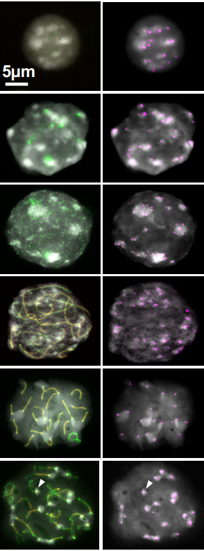
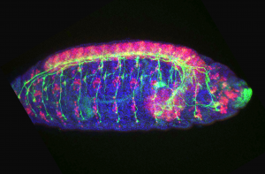
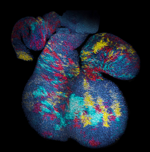
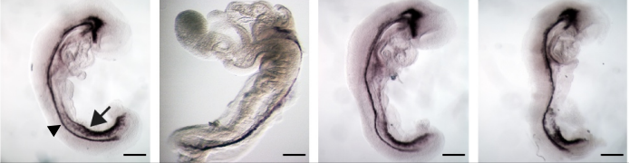
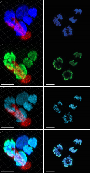
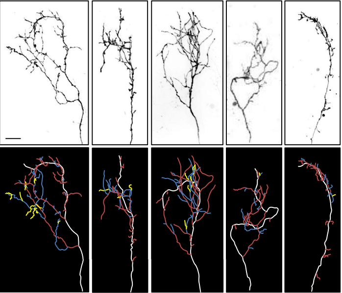
different stages of meiotic prophase I, from Chuong, et al.’s preprint
Heterochromatin Interactions Maintain Homologous Centromere Associations in Mouse Spermatocyte Meiosis
Hoa H Chuong, Craig Eyster, Chih-Ying Lee, Roberto Pezza, Dean Dawson
Time-resolved succession of epigenetic regulation during early mammalian development
Hebing Chen, Hao Li, Shuai Jiang, Xin Huang, Wanying Li, Ruijiang Li, Zhuo Zhang, Hao Hong, Chenghui Zhao, Xiaochen Bo
The WT1-BASP1 complex is required to maintain the differentiated state of taste receptor cells
Yankun Gao, Debarghya Dutta Banik, Stefan Roberts, Kathryn Medler
LEDGF and HDGF2 relieve the nucleosome-induced barrier to transcription
Gary LeRoy, Ozgur Oksuz, Nicolas Descostes, Yuki Aoi, Rais Ganai, Havva Ortabozkoyun, Jia-Ray Yu, Chul-Hwan Lee, James Stafford, Ali Shilatifard, Danny Reinberg
PLZF limits enhancer activity during hematopoietic progenitor aging
Mathilde Poplineau, Julien Vernerey, Nadine Platet, Lia Nguyen, Leonard Herault, Michela Esposito, Andrew Saurin, Christel Guilouf, Atsushi Iwama, Estelle Duprez
Gene-centric functional dissection of human genetic variation uncovers regulators of hematopoiesis
Satish K Nandakumar, Sean K McFarland, Laura Marlene Mateyka, Caleb A Lareau, Jacob C Ulirsch, Leif S Ludwig, Gaurav Agarwal, Jesse M Engreitz, Bartlomiej Przychodzen, Marie McConkey, Glenn S Cowley, John G Doench, Jaroslaw Maciejewski, Benjamin L Ebert, David E Root, Vijay G. Sankaran
Mutations in the zebrafish hmgcs1 gene reveal a novel function for isoprenoids during red blood cell development.
Jose A Hernandez, Victoria L Castro, Nayel Reyes-Nava, Laura P Montes, Anita M Quintana
Basal Role for Nrf2-transgne on transcriptional (mRNA/miRNA) regulation in the mouse myocardium
Arun Jyothidasan, Gobinath Shanmugam, John Zhang, Brain Dally, David Crossman, Rajasekaran Namakkal Soorappan
Dynamics Of Cardiomyocyte Transcriptome And Chromatin Landscape Demarcates Key Events Of Heart Development
Michal Pawlak, Katarzyna Z Kedzierska, Maciej Migdal, Karim Abu Nahia, Jordan A Ramilowski, Lukasz Bugajski, Kosuke Hashimoto, Aleksandra Marconi, Katarzyna Piwocka, Piero Carninci, Cecilia L Winata
miRNAs, target genes expression and morphological analysis on the heart in gestational protein-restricted offspring
José A.R. Gontijo, Heloisa Balan Assalin, Patrícia Aline Boer
Dynamics of microRNA expression during mouse prenatal development
Rabi Murad, Sorena Rahmanian, Alessandra Breschi, Weihua Zeng, Brian A Williams, Mark Mackiewicz, Brian Roberts, Sarah Meadows, Dianne Moore, Carrie Davis, Diane Trout, Chris Zaleski, Alexander Dobin, Lei-Hoon Sei, Jorg Drenkow, Alex Scavelli, Thomas R Gingeras, Barbara Wold, Richard M. Myers, Roderic Guigo, Ali Mortazavi
The landscape of DNA methylation associated with the transcriptomic network in laying hens and broilers gets insight into embryonic muscle development in chicken
Zihao Liu, Xiaoxu Shen, Shunshun Han, Yan Wang, Qing Zhu, Can Cui, Haorong He, Jing Zhao, Yuqi Chen, Yao Zhang, Lin Ye, Zhichao Zhang, Diyan Liu, Xiaoling Zhao, Huadong Yin
Sang Hwan Kim, Ji Hye Lee, Jong Taek Yoon
Neuroblast-specific chromatin landscapes allow integration of spatial and temporal cues to generate neuronal diversity in Drosophila
Chris Q Doe, Sonia Sen, Sachin Chanchani, Tony Southall
Ribosomal DNA and the rDNA-binding protein Indra mediate non-random sister chromatid segregation in Drosophila male germline stem cells
George Watase, Yukiko Yamashita
Discovery of Alstrom syndrome gene as a regulator of centrosome duplication in asymmetrically dividing stem cells in Drosophila.
Cuie Chen, Yukiko Yamashita
Separate Polycomb Response Elements control chromatin state and activation of the vestigial gene
Kami Ahmad, Amy E Spens
The coordination of terminal differentiation and cell cycle exit is mediated through the regulation of chromatin accessibility
Yiqin Ma, Daniel J McKay, Laura Buttitta
A gene expression atlas of embryonic neurogenesis in Drosophila reveals complex spatiotemporal regulation of lncRNAs.
Alexandra L McCorkindale, Philipp Wahle, Sascha Werner, Irwin Jungreis, Peter Menzel, Chinmay J Shukla, Ruben Lopes Pereira Abreu, Rafael Irizarry, Irmtraud Meyer, Manolis Kellis, Robert P Zinzen
Age-dependent changes in transcription factor FOXO targeting in Drosophila melanogaster
Allison Birnbaum, Xiaofen Wu, Marc Tatar, Nan Liu, Hua Bai
Female genetic contributions to sperm competition in Drosophila melanogaster
Dawn S. Chen, Sofie Y.N. Delbare, Simone L. White, Jessica L. Sitnik, Martik Chatterjee, Elizabeth L. DoBell, Orli D. Weiss, Andrew G. Clark, Mariana F. Wolfner
A Regulatory Loop between the Retinoid-Related Orphan Nuclear Receptor NHR-23 and let-7 family microRNAs Modulates the C. elegans Molting Cycle
Ruhi Patel, Alison R Frand
LIN-15B promotes enrichment of H3K9me2 on the promoters of a subset of germline genes that are repressed in somatic cells in C. elegans
Andreas Rechtsteiner, Meghan E. Costello, Thea A. Egelhofer, Jacob M. Garrigues, Susan Strome, Lisa Petrella
| Stem cells, regeneration & disease modelling
Geometry alone influences stem cell differentiation in a precision 3D printed stem cell niche
Elisabetta Prina, Laura Sidney, Maximilian Tromayer, Jonathan Moore, Robert Liska, Marina Bertolin, Stefano Ferrari, Andrew Hopkinson, Harminder Dua, Jing Yang, Ricky Wildman, Felicity RAJ Rose
Lgr5+ stem/progenitor cells reside at the apex of the embryonic hepatoblast pool
Nicole Prior, Christopher J Hindley, Fabian Rost, Elena Melendez Esteban, Winnie W. Y. Lau, Berthold Gottgens, Steffen Rulands, Benjamin D Simons, Meritxell Huch
Homeostatic and tumourigenic activity of SOX2+ pituitary stem cells is controlled by the LATS/YAP/TAZ cascade
Emily J Lodge, Alice Santambrogio, John P Russell, Paraskevi Xekouki, Thomas Jacques, Randy Johnson, Selvam Thavaraj, Stefan R Bornstein, Cynthia Lilian Andoniadou
Differential cell fates of muscle stem cells are accompanied by symmetric segregation of canonical H3 histones in vivo
Brendan Evano, Gilles Le Carrou, Genevieve Almouzni, Shahragim Tajbakhsh

Bioprinted pluripotent stem cell-derived kidney organoids provide opportunities for high content screening.
J. William Higgins, Alison Chambon, Kristina Bishard, Anke Hartung, Derek Arndt, Jamie Brugnano, Pei Xuan Er, Kynan T Lawlor, Jessica M Vanslambrouck, Sean Wilson, Alexander N Combes, Sara E Howden, Ker Sin Tan, Santhosh V Kumar, Lorna J Hale, Benjamin Shepherd, Stephen Pentoney, Sharon C Presnell, Alice E Chen, Melissa H Little
Cell division history determines hematopoietic stem cell potency
Fumio Arai, Patrick S Stumpf, Yoshiko M Ikushima, Kentaro Hosokawa, Aline Roch, Matthias P Lutolf, Toshio Suda, Ben D MacArthur
Maintenance of active chromatin states by Hmgn1 and Hmgn2 is required for stem cell identity
Sylvia Garza-Manero, Abdulmajeed A. A. Sindi, Gokula Mohan, Ohoud Rehbini, Valentine H. M. Jeantet, Mariarca Bailo, Faeezah Abdul Latif, Maureen West, Ross Gurden, Lauren Finlayson, Silvija Svambaryte, Adam G. West, Katherine West
MicroRNA-deficient embryonic stem cells acquire a functional Interferon response
Jeroen Witteveldt, Lisanne Iris Knol, Sara Macias
Comparative RNAi Screens in Isogenic Human Stem Cells Reveal SMARCA4 as a Differential Regulator
Ceren Güneş, Maciej Paszkowski-Rogacz, Susann Rahmig, Shahryar Khattak, Martin Wermke, Andreas Dahl, Martin Bornhäuser, Claudia Waskow, Frank Buchholz
An acute immune response underlies the benefit of cardiac adult stem cell therapy
Ronald Vagnozzi, Marjorie Maillet, Michelle Sargent, Hadi Khalil, Anne Katrine Johansen, Jennifer Schwanekamp, Allen J York, Vincent Huang, Matthias Nahrendorf, Sakthivel Sadayappan, Jeffery D Molkentin
Ageing affects DNA methylation drift and transcriptional cell-to-cell variability in muscle stem cells
Irene Hernando-Herraez, Brendan Evano, Thomas Stubbs, Pierre-Henri Commere, Stephen Clark, Simon Andrews, Shahragim Tajbakhsh, Wolf Reik
Loss of muscle stem cells in aged mice is replenished by muscle-secreted niche factor G-CSF
Hu Li, Qian Chen, Changyin Li, Ran Zhong, Yixia Zhao, Dahai Zhu, Yong Zhang
Environmental Optimization Enables Maintenance of Quiescent Hematopoietic Stem Cells Ex Vivo
Hiroshi Kobayashi, Takayuki Morikawa, Ayumi Okinaga, Fumie Hamano, Tomomi Hashidate-Yoshida, Shintaro Watanuki, Daisuke Hishikawa, Hideo Shindou, Fumio Arai, Yasuaki Kabe, Makoto Suematsu, Takao Shimizu, Keiyo Takubo
Mechanobiological Conditioning of Mesenchymal Stem Cells Enhances Therapeutic Angiogenesis by Inducing a Hybrid Pericyte-Endothelial Phenotype
Jason Lee, Kayla Henderson, Miguel Armenta-Ochoa, Austin Veith, Pablo Maceda, Eun Yoon, Lara Samarneh, Mitchell Wong, Andrew Dunn, Aaron Baker
Hierarchical stem cell topography splits growth and homeostatic functions in the fish gill
Julian Stolper, Elizabeth Mayela Ambrosio, Diana-Patricia Danciu, David Elliott, Kiyoshi Naruse, Anna Marciniak-Czochra, Lazaro Centanin
Region-specific regulation of stem cell-driven regeneration in tapeworms
Tania Rozario, Edward B Quinn, Jianbin Wang, Richard A Davis, Phillip A Newmark
General characterization of regeneration in Aeolosoma viride
Chiao-Ping Chen, Sheridan Ke-Wing Fok, Yu-Wen Hsieh, Cheng-Yi Chen, Fei-Man Hsu, Jiun-Hong Chen
UNC-16/JIP3 inhibits the function of the regeneration promoting isoform of DLK-1
Sucheta S Kulkarni, Seema Sheoran, Kunihiro Matsumoto, Naoki Hisamoto, Sandhya P Koushika
A metabolic switch from OXPHOS to glycolysis is essential for cardiomyocyte proliferation in the regenerating heart
Hessel Honkoop, Dennis de Bakker, Alla Aharonov, Fabian Kruse, Avraham Shakked, Phong Nguyen, Cecilia de Heus, Laurence Garric, Mauro Muraro, Adam Shoffner, Federico Tessadori, Joshua Peterson, Wendy Noort, George Posthuma, Dominic Grun, Willem van der Laarse, Judith Klumperman, Richard Jaspers, Kenneth Poss, Alexander van Oudenaarden, Eldad Tzahor, Jeroen Bakkers
Tissue repair in the mouse liver following acute carbon tetrachloride depends on injury-induced Wnt/β-catenin signaling
Ludan Zhao, Yinhua Jin, Katie Donahue, Margaret Tsui, Matt Fish, Catriona Logan, Bruce Wang, Roel Nusse
In vivo epigenetic editing of sema6a promoter reverses impaired transcallosal connectivity caused by C11orf46/ARL14EP neurodevelopmental risk gene
Cyril J. Peter, Atsushi Saito, Yuto Hasegawa, Yuya Tanaka, Gabriel Perez, Emily Alway, Sergio Espesio-gil, Tariq Fayyad, Chana Ratner, Aslihan Dincer, Achla Gupta, Lakshmi Devi, John G. Pappas, François M. Lalonde, John A. Butman, Joan C. Han, Schahram Akbarian, Atsushi Kamiya
Inhibition of Notch signaling rescues cardiovascular development in Kabuki Syndrome
Maria de los Angeles Serrano, Bradley L. Demarest, Tarlynn Tone-Pah-Hote, Martin Tristani, H. Joseph Yost
Transcriptional suppression from KMT2D loss disrupts cell cycle and hypoxic responses in neurodevelopmental models of Kabuki syndrome
Giovanni A Carosso, Leandros Boukas, Jonathan J Augustin, Ha Nam Nguyen, Briana L Winer, Gabrielle H Cannon, Johanna D Robertson, Li Zhang, Kasper D Hansen, Loyal A Goff, Hans T Bjornsson
Tracking dynamic changes in Alzheimer’s disease brain proteome reveals ageing-independent damage in Drosophila
Harry M Scholes, Adam Cryar, Fiona Kerr, David Sutherland, Lee A Gethings, Johannes P C Vissers, Jonathan G Lees, Christine A Orengo, Linda Partridge, Konstantinos Thalassinos
Microglial activation in an amyotrophic lateral sclerosis-like model caused by Ranbp2 loss and nucleocytoplasmic transport impairment in retinal ganglion neurons
Kyoung-in Cho, Dosuk Yoon, Minzhong Yu, Neal S Peachey, Paulo A Ferreira
Circuit dysfunction in SOD1-ALS model first detected in sensory feedback prior to motor neuron degeneration is alleviated by BMP signaling
Aaron Held, Paxton Major, Asli Sahin, Robert Reenan, Diane Lipscombe, Kristi Wharton
Zebrafish larvae as a model system for systematic characterization of drugs and genes in dyslipidemia and atherosclerosis
Manoj K Bandaru, Anastasia Emmanouilidou, Petter Ranefall, Benedikt von der Heyde, Eugenia Mazzaferro, Tiffany Klingstroem, Mauro Masiero, Olga Dethlefsen, Johan Ledin, Anders Larsson, Hannah L Brooke, Carolina Wahlby, Erik Ingelsson, Marcel den Hoed
Inner hair cell and neuron degeneration contribute to hearing loss in a DFNA2-like mouse model
Camila Carignano, Esteban P Barila, Ezequiel I Rias, Leonardo Dionisio, Eugenio Aztiria, Guillermo Spitzmaul
Developmental Dieldrin Exposure Alters DNA Methylation at Genes Related to Dopaminergic Neuron Development and Parkinson’s Disease in Mouse Midbrain
Joseph Kochmanski, Sarah E. VanOeveren, Alison I. Bernstein
Gene augmentation and read-through rescue channelopathy in an iPSC-RPE model of congenital blindness
Pawan K Shahi, Dalton Hermans, Divya Sinha, Simran Brar, Hannah Moulton, Sabrina Stulo, Katarzyna D Borys, Elizabeth Capowski, De-Ann M Pillers, David M Gamm, Bikash R Pattnaik
| Plant development
Control of stem-cell niche establishment in Arabidopsis flowers by REVOLUTA and the LEAFY-RAX1 module
Gregoire Denay, Gabrielle Tichtinsky, Marie Le Masson, Hicham Chahtane, Sylvie Huguet, Irene Lopez-Vidriero, Christian Wenzl, Jose-Manuel Franco-Zorrilla, Ruediger Simon, Jan U. Lohmann, Francois Parcy
Genetic and physical interactions between the organellar mechanosensitive ion channel homologs MSL1, MSL2, and MSL3 reveal a role for inter-organellar communication in plant development
Josephine Lee, Margaret Wilson, Ryan Richardson, Elizabeth Haswell
Excess light priming in Arabidopsis thaliana with altered DNA methylomes
Diep R Ganguly, Bethany AB Stone, Steven R Eichten, Barry J Pogson
Light remote control of alternative splicing in roots through TOR kinase
Stefan Riegler, Lucas Servi, Armin Fuchs, Micaela A. Godoy Herz, Maria Guillermina Kubaczka, Peter Venhuizen, Alois Schweighofer, Craig Simpson, John W.S. Brown, Christian Meyer, Maria Kalyna, Andrea Barta, Ezequiel Petrillo
Pheophorbide a, a chlorophyll catabolite may regulate jasmonate signalling during dark-induced senescence in Arabidopsis
Sylvain Aubry, Niklaus Fankhauser, Serguei Ovinnikov, Krzysztof Zienkiewicz, Ivo Feussner, Stefan Hortensteiner
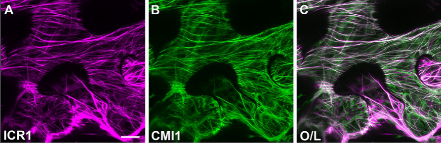
The Ca2+ sensor protein CMI1 fine tunes root development, auxin distribution and responses
Ora Hazak, Elad Mamon, Meirav Lavy, Hasana Sternberg, Smrutisanjita Behera, Ina Schmitz-Thom, Daria Bloch, Olga Dementiev, Itay Gutman, Tomer Danziger, Netanel Schwarz, Anas Abuzeineh, Keithanne Mockaitis, Mark Estelle, Joel Hirsch, Jörg Kudla, Shaul Yalovsky
Origin of gibberellin-dependent transcriptional regulation by molecular exploitation of a transactivation domain in DELLA proteins
Jorge Hernandez-Garcia, Asier Briones-Moreno, Renaud Dumas, Miguel A Blazquez
Anchorene is an endogenous diapocarotenoid required for anchor root formation in Arabidopsis
Kunpeng Jia, Alexandra J. Dickinson, Jianing Mi, Guoxin Cui, Najeh M. Kharbatia, Xiujie Guo, Erli Sugiono, Manuel Aranda, Magnus Rueping, Philip N. Benfey, Salim Al-Babili
An interaction map of transcription factors controlling gynoecium development in Arabidopsis
Humberto Herrera-Ubaldo, Sergio E. Campos, Valentin Luna Garcia, Victor M. Zuniga-Mayo, Gerardo Armas-Caballero, Alexander DeLuna, Nayelli Marsch-Martinez, Stefan de Folter
Arabidopsis TRM5 encodes a nuclear-localised bifunctional tRNA guanine and inosine-N1-methyltransferase that is important for growth.
Qianqian Guo, PeiQin Ng, Shanshan Shi, Diwen Fan, Jun Li, Hua Wang, Trung Do, Rakesh David, Parul Mittal, Ralph Bock, Ming Zhao, Wenbin Zhou, Iain R Searle
Functional characterization of Arabidopsis ARGONAUTE 3 in reproductive tissue
Pauline E Jullien, Stefan Grob, Antonin Marchais, Nathan Pumplin, Clement Chevalier, Caroline Otto, Gregory Schott, Olivier Voinnet
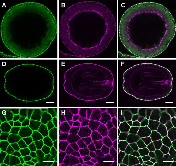
Arabidopsis Myosins XI Are Involved in Exocytosis of Cellulose Synthase Complexes
Weiwei Zhang, Chao Cai, Christopher J Staiger

βVPE is involved in tapetal degradation and pollen development by activating proprotease maturation in Arabidopsis thaliana
Ziyi Cheng, Bin Yin, Jiaxue Zhang, Yadi Liu, Bing Wang, Hui Li, Hai Lu
A novel role for Cyclic Nucleotide-Gated Ion Channel 2 (DND1) in auxin signaling
Sonhita Chakraborty, Masatsugu Toyota, Wolfgang Moeder, Kimberley Chin, Alex Fortuna, Marc Champigny, Steffen Vanneste, Simon Gilroy, Tom Beeckman, Keiko Yoshioka
A regulatory module controlling stress-induced cell cycle arrest in Arabidopsis
Naoki Takahashi, Nobuo Ogita, Tomonobu Takahashi, Shoji Taniguchi, Maho Tanaka, Motoaki Seki, Masaaki Umeda
Effects of FLOWERING LOCUS T on FD during the transition to flowering at the shoot apical meristem of Arabidopsis thaliana
Silvio Collani, Manuela Neumann, Levi Yant, Markus Schmid
Dicer-like 5 deficiency confers temperature-sensitive male sterility in maize
Chong Teng, Han Zhang, Reza Hammond, Kun Huang, Blake Meyers, Virginia Walbot

The dynamic association of SPO11-1 with conformational changes of meiotic axial elements in maize
Arnaud Ronceret, Inna Golubovskaya, Jia-Chi Ku, Ding Hua Lee, Ljudmilla Timofejeva, Ana Karen Gomez Angoa, Yu-Hsin Kao, Karl Kremling, Rosalind Williams-Carrier, Robert Meeley, Alice Barkan, W. Zacheus Cande, Chung-Ju Rachel Wang
A wheat/rye polymorphism affects seminal root length and is associated with drought and waterlogging tolerance
Tyson Howell, Jorge I. Moriconi, Xueqiang Zhao, Joshua Hegarty, Tzion Fahima, Guillermo Santa-Maria, Jorge Dubcovsky
Isolation and characterisation of mutants with altered seminal root numbers in hexaploid wheat
Oluwaseyi Shorinola, Ryan Kaye, Guy Golan, Zvi Peleg, Stefan Kepinski, Cristobal Uauy
Clonal seeds in hybrid rice using CRISPR/Cas9
Chun Wang, Qing Liu, Yi Shen, Yufeng Hua, Junjie Wang, Jianrong Lin, Mingguo Wu, Tingting Sun, Zhukuan Cheng, Raphael Mercier, Kejian Wang
Barley yield formation under abiotic stress depends on the interplay between flowering time genes and environmental cues
Mathias Wiegmann, Andreas Maurer, Anh Pham, Timothy March, Ayed Al-Abdallat, William Thomas, Hazel Bull, Mohammed Shahid, Jason Eglinton, Michael Baum, Andrew Flavell, Mark Tester, Klaus Pillen
Evo-devo & evo
Establishing Cerebral Organoids as Models of Human-Specific Brain Evolution
Alex A Pollen, Aparna Bhaduri, Madeline G Andrews, Tomasz J Nowakowski, Olivia S Meyerson, Mohammed A Mostajo-Radji, Elizabeth Di Lullo, Beatriz Alvarado, Melanie Bedolli, Max L Dougherty, Ian T Fiddes, Zev N Kronenberg, Joe Shuga, Anne A Leyrat, Jay A West, Marina Bershteyn, Craig B Lowe, Bryan J Pavolvic, Sofie R Salama, David Haussler, Evan Eichler, Arnold A Kriegstein
Establishment of the mayfly Cloeon dipterum as a new model system to investigate insect evolution
Isabel Almudi, Carlos Martin-Blanco, Isabel Maria Garcia-Fernandez, Adrian Lopez-Catalina, Kristofer Davie, Stein Aerts, Fernando Casares
Deep evolutionary origin of limb and fin regeneration
Sylvain Darnet, Aline Cutrim Dragalzew, Danielson Baia Amaral, Andrew W Thompson, Amanda N Cass, Jamily Lorena, Josane F Sousa, Carinne M Costa, Marcos P Sousa, Nadia B Froebisch, Patricia N Schneider, Marcus C Davis, Ingo Braasch, Igor Schneider
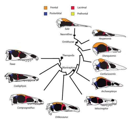
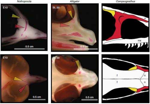
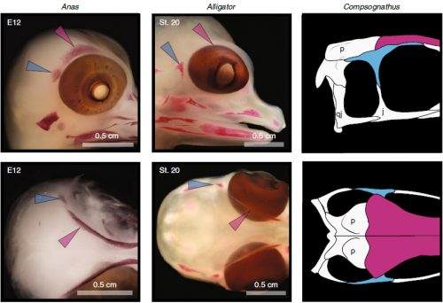
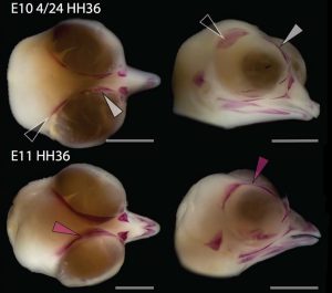
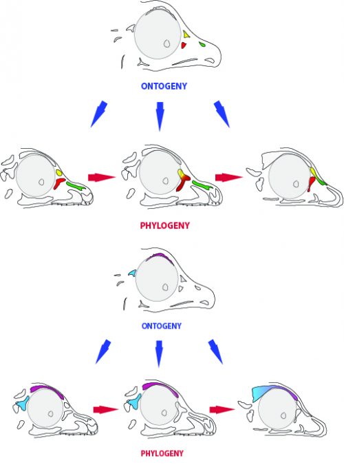
Evidence against tetrapod-wide digit identities and for a limited frame shift in bird wings
Thomas A Stewart, Cong Liang, Justin Cotney, James P Noonan, Thomas Sanger, Gunter Wagner
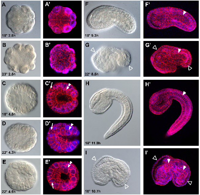
High temperature limits on developmental canalization in the ascidian Ciona intestinalis
Steven Q Irvine, Katherine B McNulty, Evelyn M Siler, Rose E Jacobson
Cadherin switch marks germ layer formation in the diploblastic sea anemone Nematostella vectensis
Ekaterina Pukhlyakova, Anastasia Kirillova, Yulia Kraus, Ulrich Technau
The genetic basis of hindwing eyespot number variation in Bicyclus anynana butterflies
Angel G Rivera-Colón, Erica Westerman, Steven van Belleghem, Antonia Monteiro, Riccardo Papa
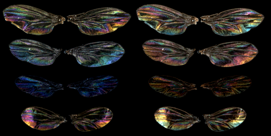
Sexual selection drives the evolution of wing interference patterns.
MF Hawkes, Eoin Duffy, Richa Joag, Alison Skeats, Jacek Radwan, Nina Wedell, Manmohan Sharma, DJ Hosken, Jollyon Troscianko
Silent crickets reveal the genomic footprint of recent adaptive trait loss
Sonia Pascoal, Judith E. Risse, Xiao Zhang, Mark Blaxter, Timothee Cezard, Richard J. Challis, Karim Gharbi, John Hunt, Sujai Kumar, Emma Langan, Xuan Liu, Jack G. Rayner, Michael G. Ritchie, Basten L. Snoek, Urmi Trivedi, Nathan Bailey

Anatomical diversification of a skeletal novelty in bat feet
Kathryn E Stanchak, Jessica H Arbour, Sharlene E Santana
Opsin gene evolution in amphibious and terrestrial combtooth blennies (Blenniidae)
Fabio Cortesi, Karen M Cheney, Georgina M Cooke, Terry Ord
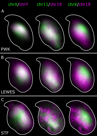
Automated nuclear cartography reveals conserved sperm chromosome territory localization across 2 million years of mouse evolution
Benjamin Matthew Skinner, Joanne Bacon, Claudia Cattoni Rathje, Erica Lee Larson, Emily Emiko Konishi Kopania, Jeffrey Martin Good, Nabeel Ahmed Affara, Peter James Ivor Ellis
Genome wide screen reveals a specific interaction between autosome and X that is essential for hybrid male sterility
Zhongying Zhao, Yu Bi, Xiaoliang Ren, Runsheng Li, Qiutao Ding, Dongying Xie
Contingency in the convergent evolution of a regulatory network: Dosage compensation in Drosophila
Doris Bachtrog, Chris Ellison
Recurrent gene amplification on Drosophila Y chromosomes suggests cryptic sex chromosome drive is common on young sex chromosomes
Doris Bachtrog, Chris Ellison
Parallel patterns of development between independent cases of hybrid seed inviability in Mimulus
Jenn M. Coughlan, John H. Willis
Adaptation to developmental diet influences the response to selection on age at reproduction in the fruit fly
Tina May, Joost van den Heuvel, Agnieszka Doroszuk, Katja Hoedjes, Thomas Flatt, Bas Zwaan
Evolution of sperm competition: Natural variation and genetic determinants of Caenorhabditis elegans sperm size
Clotilde Gimond, Anne Vielle, Nuno Silva Soares, Stefan Zdraljevic, Patrick McGrath, Erik Andersen, Christian Braendle
Sex chromosome evolution via two genes
Alex Harkess, Kun Huang, Ron van der Hulst, Bart Tissen, Jeffrey L Caplan, Aakash Koppula, Mona Batish, Blake C Meyers, Jim Leebens-Mack

A bird’s white-eye shot: looking down on a new avian sex chromosome evolution
Thibault Leroy, Yoann Anselmetti, Marie-Ka Tilak, Severine Berard, Laura Csukonyi, Maeva Gabrielli, Celine Scornavacca, Borja Mila, Christophe Thebaud, Benoit Nabholz
Programmed DNA elimination of germline development genes in songbirds
Cormac M. Kinsella, Francisco J. Ruiz-Ruano, Anne-Marie Dion-Côté, Alexander J. Charles, Toni I. Gossmann, Josefa Cabrero, Dennis Kappei, Nicola Hemmings, Mirre J. P. Simons, Juan P. M. Camacho, Wolfgang Forstmeier, Alexander Suh
The loci of behavioral evolution: Fas2 and tilB underlie differences in pupation site choice behavior between Drosophila melanogaster and D. simulans
Alison Pischedda, Michael P Shahandeh, Thomas L Turner
Recombination in a natural population of the bdelloid rotifer Adineta vaga
Olga A. Vakhrusheva, Elena A. Mnatsakanova, Yan R. Galimov, Tatiana V. Neretina, Evgeny S. Gerasimov, Svetlana G. Ozerova, Arthur O. Zalevsky, Irina A. Yushenova, Irina R. Arkhipova, Aleksey A. Penin, Maria D. Logacheva, Georgii A. Bazykin, Alexey S. Kondrashov
Genomic features of asexual animals
Kamil S. Jaron, Jens Bast, T. Rhyker Ranallo-Benavidez, Marc Robinson-Rechavi, Tanja Schwander
The quagga mussel genome and the evolution of freshwater tolerance
Andrew D Calcino, Andre Luiz de Oliveira, Oleg Simakov, Thomas Schwaha, Elisabeth Zieger, Tim Wollesen, Andreas Wanninger
The draft genome of an octocoral, Dendronephthya gigantea
Yeonsu Jeon, Seung Gu Park, Nayun Lee, Jessica A. Weber, Hui-Su Kim, Sung-Jin Hwang, Seonock Woo, Hak-min Kim, Youngjune Bhak, Sungwon Jeon, Nayoung Lee, Yejin Jo, Asta Blazyte, Taewoo Ryu, Yun Sung Cho, Hyunho Kim, Jung-Hyun Lee, Hyung-Soon Yim, Jong Bhak, Seungshic Yum
Cell biology
Cytoplasmic self-organization established by internal lipid membranes in the interplay with either actin or microtubules
Sindy Tang, Malte Renz, Tom Shemesh, Meghan Driscoll, Jennifer Lippincott-Schwartz
Myosin driven Actin Filament Sliding is Responsible for Endoplasmic Reticulum and Golgi Movement
Joseph F McKenna, Stephen E D Webb, Verena Kriechbaumer, Chris Hawes
Transcription factor TAp73 and microRNA-449 complement each other to support multiciliogenesis
Merit Wildung, Tilman Uli Esser, Katie Baker Grausam, Cornelia Wiedwald, Larisa Volceanov-Hahn, Dietmar Riedel, Sabine Beuermann, Li Li, Jessica Lynn Simcox Zylla, Ann-Kathrin Guenther, Magdalena Wienken, Evrim Ercetin, Zhiyuan Han, Felix Bremmer, Orr Shomroni, Stefan Andreas, Haotian Zhao, Muriel Lizé
On-site ribosome remodeling by locally synthesized ribosomal proteins in axons
Toshiaki Shigeoka, Max Koppers, Hovy Ho-Wai Wong, Julie Qiaojin Lin, Asha Dwivedy, Janaina de Freitas Nascimento, Roberta Cagnetta, Francesca van Tartwijk, Florian Strohl, Jean-Michel Cioni, Mark Carrington, Clemens F. Kaminski, William A. Harris, Hosung Jung, Christine E. Holt
Actomyosin-II facilitates long-range retrograde transport of large cargoes by controlling axonal radial contractility
Tong Wang, Wei Li, Sally Martin, Andreas Papadopulos, Golnoosh Shamsollahi, Vanessa Lanoue, Pranesh Padmanabhan, He Huang, Xiaojun Yu, Victor Anggono, Frederic Meunier

R51Q SNX10 induces osteopetrosis by promoting uncontrolled fusion of monocytes to form giant, non-functional osteoclasts
Maayan Barnea, Merle Stein, Sabina Winograd-Katz, Moran Shalev, Esther Arman, Ori Brenner, Fadi Thalji, Moien Kanaan, Hila Elinav, Polina Stepensky, Benjamin Geiger, Jan Tuckermann, Ari Elson
Dynamics of centriole amplification in centrosome-depleted brain multiciliated progenitors
Olivier MERCEY, Adel Al Jord, Philippe Rostaing, Alexia Mahuzier, Aurelien Fortoul, Amelie-Rose Boudjema, Marion Faucourt, Nathalie Spassky, Alice Meunier
Regulation of Cilia Abundance in Multiciliated Cells
Rashmi Nanjundappa, Dong Kong, Kyuhwan Shim, Tim Stearns, Steven Brody, Jadranka Loncarek, Moe Mahjoub
SGK regulates pH increase and cyclin B-Cdk1 activation to resume meiosis in starfish ovarian oocytes
Enako Hosoda, Daisaku Hiraoka, Noritaka Hirohashi, Saki Omi, Takeo Kishimoto, Kazuyoshi Chiba
Aberrant chromatin resolution in G2/M leads to chromosome instability
Lora Boteva, Ryu-Suke Nozawa, Catherine Naughton, Kumiko Samejima, William Earnshaw, Nick Gilbert
Rescue of DNA damage in cells after constricted migration reveals bimodal mechano-regulation of cell cycle
Yuntao Xia, Charlotte R Pfeifer, Kuangzheng Zhu, Jerome Irianto, Dazhen Liu, Kalia Pannell, Emily J Chen, Lawrence J Dooling, Roger A Greenberg, Dennis E Discher
Mitochondrial cristae biogenesis coordinates with ETC complex IV assembly during Drosophila maturation
Yi-fan Jiang, Hsiang-ling Lin, Li-jie Wang, Tian Hsu, Chiyu Fu
Rapid Whole Cell Imaging Reveals An APPL1-Dynein Nexus That Regulates Stimulated EGFR Trafficking
Harrison York, Amandeep Kaur, Abhishek Patil, Aditi Bhowmik, Ullhas K Moorthi, Geoffrey J Hyde, Hetvi Gandhi, Katharina Gaus, Senthil Arumugam
WNT vampirization by glioblastoma leads to tumor growth and neurodegeneration
Marta Portela Esteban, Varun Venkataramani, Natasha Fahey-Lozano, Esther Seco, Maria Losada-Perez, Frank Winkler, Sergio Casas-Tinto
Modelling

Elongated cells drive morphogenesis in a surface-wrapped finite element model of germband retraction
W. Tyler McCleery, Jim H Veldhuis, G. Wayne Brodland, Monica E Bennett, M. Shane Hutson
Theory of mechano-chemical patterning in biphasic biological tissues
Pierre Recho, Adrien Hallou, Edouard Hannezo
An individual-based mechanical model of cell movement in heterogeneous tissues and its coarse-grained approximation
Ryan Murphy, Pascal Buenzli, ruth E Baker, Matthew J Simpson
Cross-talk between Hippo and Wnt signalling pathways in intestinal crypts: insights from an agent-based model
Daniel Ward, Alexander G. Fletcher, Martin Homer, Lucia Marucci
Cell-based model of the generation and maintenance of the shape and structure of the multi-layered shoot apical meristem of Arabidopsis thaliana
Mikahl Banwarth-Kuhn, Ali Nematbakhsh, Kevin W. Rodriguez, Stephen Snipes, Carolyn G. Rasmussen, G. Venugopala Reddy, Mark Alber
Spatiotemporal Integration in Plant Tropisms
Yasmine Meroz, Renaud Bastien, L Mahadevan
Modifying Reaction Diffusion: A Numerical Model for Turing Morphogenesis and Ben Jacob Patterns
Kai Trepka
Modulation of tissue growth heterogeneity by responses to mechanical stress
Antoine Fruleux, Arezki Boudaoud
Tools & resources
Single-copy Knock-In Loci for Defined Gene Expression in C. elegans
Carlos G Silva-Garcia, Caroline Heintz, Sneha Dutta, Nicole M Clark, Anne Lanjuin, William B Mair
Endogenous CRISPR arrays for scalable whole organism lineage tracing
James Cotterell, James Sharpe
Strong gene activation with genome-wide specificity using a new orthogonal CRISPR/Cas9-based Programmable Transcriptional Activator.
Sara Selma, Joan Bernabe-Orts, Marta Vazquez-Vilar, Borja Diego, Maria Ajenjo, Victor Garcia-Carpintero, Antonio Granell, Diego Orzaez
Direct capture of CRISPR guides enables scalable, multiplexed, and multi-omic Perturb-seq
Joseph M Replogle, Albert Xu, Thomas M Norman, Elliott J Meer, Jessica M Terry, Daniel Riordan, Niranjan Srinivas, Tarjei S Mikkelsen, Jonathan S Weissman, Britt Adamson
A benchmark of computational CRISPR-Cas9 guide design methods
Jake Bradford, Dimitri Perrin
Towards best-practice approaches for CRISPR/Cas9 gene engineering
Claude Van Campenhout, Pauline Cabochette, Anne-Clemence Veillard, Miklos Laczik, Agnieszka Zelisko-Schmidt, Celine Sabatel, Maxime Dhainaut, Benoit Vanhollebeke, Cyril Gueydan, Veronique Kruys
Main constraints for RNAi induced by expressed long dsRNA in mouse cells
Tomas Demeter, Michaela Vaskovicova, Radek Malik, Filip Horvat, Josef Pasulka, Eliska Svobodova, Matyas Flemr, Petr Svoboda
Distinguishing cells from empty droplets in droplet-based single-cell RNA sequencing data
Aaron Lun, Samantha Riesenfeld, Tallulah Andrews, The Phuong Dao, Tomas Gomes, participants in the 1st Human Cell Atlas Jamboree, John Marioni
Snapshot: clustering and visualizing epigenetic history during cell differentiation
Guanjue Xiang, Belinda Giardine, Lin An, Chen Sun, Cheryl Keller, Elisabeth Heuston, David Bodine, Ross Hardison, Yu Zhang
An approach for accelerated isolation of genetically manipulated cell clones with reduced clonal variability
Natania Casden, Oded Behar
Cell type purification by single-cell transcriptome-trained sorting
Chloe S Baron, Aditya Barve, Mauro J Muraro, Gitanjali Dharmadhikari, Reinier van der Linden, Anna Lyubimova, Eelco JP de Koning, Alexander van Oudenaarden
SingleCellNet: a computational tool to classify single cell RNA-Seq data across platforms and across species
Yuqi Tan, Patrick Cahan
scAlign: a tool for alignment, integration and rare cell identification from scRNA-seq data
Nelson Johansen, Gerald Quon
Simultaneous profiling of chromatin accessibility and methylation on human cell lines with nanopore sequencing
Isac Lee, Roham Razaghi, Timothy Gilpatrick, Norah Sadowski, Fritz Sedlazeck, Winston Timp
Fragmentation Through Polymerization (FTP): A New Method to Fragment DNA for Next-Generation Sequencing
Konstantin B. Ignatov, Konstantin A. Blagodatskikh, Dmitry S. Shcherbo, Tatiana Kramarova, Yulia A. Monakhova, Vladimir M. Kramarov
High throughput genotyping of structural variations in a complex plant genome using an original Affymetrix® Axiom® array
Clément Mabire, Jorge Duarte, Aude Darracq, Ali Pirani, Hélène Rimbert, Delphine Madur, Valérie Combes, Clémentine Vitte, Sébastien Praud, Nathalie Riviere, Johann Joets, Jean-Philippe Pichon, Stéphane D Nicolas
Single-cell multi-omic profiling of chromatin conformation and DNA methylome
Dong-Sung Lee, Chongyuan Luo, Jingtian Zhou, Sahaana Chandran, Angeline Rivkin, Anna Bartlett, Joseph R Nery, Conor Fitzpatrick, Carolyn O’Connor, Jesse R Dixon, Joseph R. Ecker
ASCOT identifies key regulators of neuronal subtype-specific splicing
Jonathan P. Ling, Christopher Wilks, Rone Charles, Devlina Ghosh, Lizhi Jiang, Clayton P. Santiago, Bo Pang, Anand Venkataraman, Brian S. Clark, Abhinav Nellore, Ben Langmead, Seth Blackshaw
frenchFISH: Poisson models for quantifying DNA copy-number from fluorescence in situ hybridisation of tissue sections
Geoff Macintyre, Anna M Piskorz, Edith Ross, David B Morse, Ke Yuan, Darren Ennis, Jeremy A Pike, Teodora Goranova, Iain McNeish, James D Brenton, Florian Markowetz
Multiplexed detection of RNA using MERFISH and branched DNA amplification
Chenglong Xia, Hazen P Babcock, Jeffrey R Moffitt, Xiaowei Zhuang
DeepCell 2.0: Automated cloud deployment of deep learning models for large-scale cellular image analysis
Dylan Bannon, Erick Moen, Enrico Borba, Andrew Ho, Isabella Camplisson, Brian Chang, Eric Osterman, William Graf, David Van Valen
Transgenic Mice and Pluripotent Stem Cells Express EGFP under the Control of miR-302 Promoter
Karim Rahimi, Sara Parsa, Mehrnoush Nikzaban, Seyed Javad Mowla, Fardin Fathi
A fluorescent reporter enables instantaneous measurement of cell cycle speed in live cells
Anna E Eastman, Xinyue Chen, Xiao Hu, Amaleah A Hartman, Aria M Pearlman Morales, Cindy Yang, Jun Lu, Hao Yuan Kueh, Shangqin Guo
Surrogate R-spondin agonists for tissue-specific potentiation of Wnt signaling
Vincent C Luca, Yi Miao, Xingnan Li, Michael J Hollander, Calvin J Kuo, K. Christopher Garcia
Fast Objective Coupled Planar Illumination Microscopy
Cody J Greer, Timothy E Holy
Cytokit: A single-cell analysis toolkit for high dimensional fluorescent microscopy imaging
Eric Czech, Bulent Arman Aksoy, Pinar Aksoy, Jeffrey Hammerbacher
The Allen Cell Structure Segmenter: a new open source toolkit for segmenting 3D intracellular structures in fluorescence microscopy images
Jianxu Chen, Liya Ding, Matheus P. Viana, Melissa C. Hendershott, Ruian Yang, Irina A. Mueller, Susanne M. Rafelski
FishNET: An automated relational database for zebrafish colony management.
Abiud Cantu Gutierrez, Manuel Cantu Gutierrez, Alexander M. Rhyner, Oscar Ruiz, George T. Eisenhoffer, Joshua D Wythe
Adult zebrafish euthanasia: efficacy of anaesthesia overdose versus rapid cooling
Jorge M Ferreira, I Anna S Olsson, Ana M Valentim
Dissection of intestines from larval zebrafish for molecular analysis
Bilge San, Marco Aben, Gert Flik, Leonie Kamminga
Science Family skills: An Alexa Assistant Tailored for Laboratory Routine
Tiago Lubiana Alves, Andre A.N.A. Goncalves, Helder I Nakaya
Research practice & education
Talent Identification at the limits of Peer Review: an analysis of the EMBO Postdoctoral Fellowships Selection Process
Bernd Klaus, David del Alamo
Non-academic employability of life science PhDs: the importance of training beyond the bench
Sohyoung Her, Mathieu Jacob, Sharon Wang, Songyi Xu, David Sealey
Roles matter: Graduate student perceptions of active learning in the STEM courses they take and those they teach
Everett W. Wischusen, Lorelei Patrick, Leigh Anne Howell
A data-driven approach to reduce gender disparity in invited speaker programs at scientific meetings
Ann-Maree Vallence, Mark R Hinder, Hakeui Fujiyama
On the value of preprints: an early career researcher perspective
Sarvenaz Sarabipour, Humberto J Debat, Edward Emmott, Steven Burgess, Benjamin Schwessinger, Zach Hensel
The Case For and Against Double-blind Reviews
Amelia R Cox, Robert Montgomerie
Academic publishing empires need to go
Joona Lehtomäki, Johanna Eklund, Tuuli Toivonen
Open access policies of leading medical journals: a cross-sectional study
Tim S Ellison, Tim Koder, Laura Schmidt, Amy Williams, Christopher Winchester
Introduction to Genomic Analysis Workshop: A catalyst for engaging life-science researchers in high throughput analysis
Phillip Andrew Richmond, Wyeth W Wasserman
Using bioinformatics training to boost research capacities in resource-limited regions
Serghei Mangul, Lana Martin, Ben Langmead, Javier Sanchez Galan, Ian Toma, Pavel Pevzner, Eleazar Eskin
A comment on computational biology and connecting the dots.
Christopher J Lortie
Why not…
Where does time go when you blink?
Shany Grossman, Chen Guata, Slav Pesin, Rafael Malach, Ayelet N Landau
Stone Age “chewing gum” yields 5,700 year-old human genome and oral microbiome
Theis ZT Jensen, Jonas Niemann, Katrine Hoejholt Iversen, Anna K Fotakis, Shyam Gopalakrishnan, Mikkel HS Sinding, Martin R Ellegaard, Morten E Allentoft, Liam T Lanigan, Alberto J Taurozzi, Sofie Holtsmark Nielsen, Michael W Dee, Martin N Mortensen, Mads C Christensen, Soeren A Soerensen, Matthew J Collins, Tom Gilbert, Martin Sikora, Simon Rasmussen, Hannes Schroeder
 (No Ratings Yet)
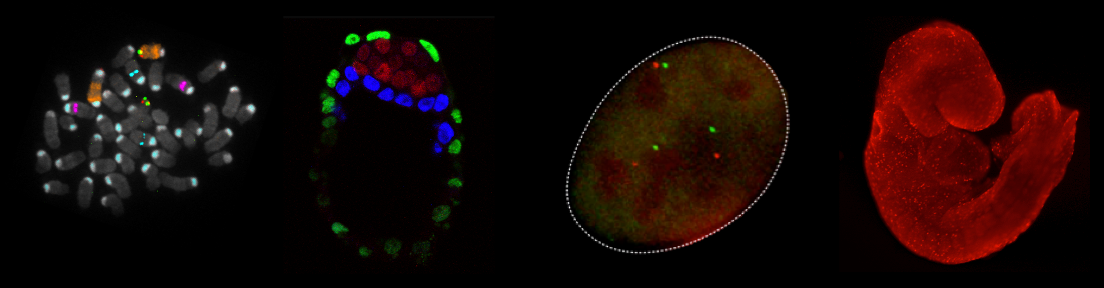


 (4 votes)