Neuronal migration is critical for mammalian brain development. In many migrating neurons, the nucleus translocates from the trailing to the leading edge of the cell in a manner dependent on the actin and microtubule cytoskeletons, but how these cytoskeletons interact and their relative contribution to the forces that move the nucleus has remained unclear. This week we feature a paper published in the latest issue of Development that uses live imaging to understand this process, revealing some fascinating cellular behaviours. We caught up with first author You Wu and his supervisor Mineko Kengaku, Professor at the Institute for Integrated Cell-Material Sciences at Kyoto University, to find out more about the story.
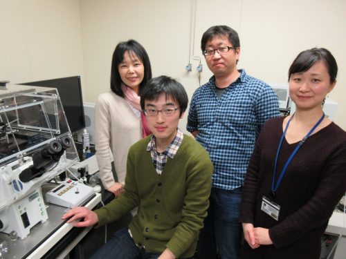
From left to right: Mineko Kengaku, You Wu, Hiroki Umeshima and Junko Kurisu.
Mineko, can you give us your scientific biography and the questions your lab is trying to answer?
MK We study how neurons are built up into the beautiful architecture of the brain cortex. A major effort of my lab has been devoted to live-imaging of growing neurons for a mechanistic understanding of brain formation, using the clear and simple cerebellar cortex as a model. We especially focus on the mechanisms and dynamics of cytoskeletal organization during migration of granule cells and dendrite arborization of Purkinje neurons, the two major events during the formation of the three-layered cortex of the cerebellum. I did my PhD study on the molecular mechanism of the primary axis formation in the Xenopus CNS, under supervision of Dr Harumasa Okamoto at the University of Tokyo. I wanted to further study the molecular signals regulating morphogenesis, and joined the laboratory of Cliff Tabin at Harvard Medical School as a post-doc. His lab had just discovered Sonic hedgehog as a bona fide morphogen in the chick embryo. I was extremely lucky to be present in the lab at moments when long-standing questions were clarified, such as the mechanisms of left-right asymmetry, bone formation, fore- and hind limb differentiation, etc. As a consequence of this rapid progress, many fundamental questions in morphogenetic signals had been clarified by the time I started my own lab at the RIKEN Brain Science Institute. The major interests in developmental biology then shifted toward stem cells and disease mechanisms. But I was still fascinated with animal morphology, and therefore decided to look into the beautiful shapes of neurons in the well-organized cortex of the brain.
And You, how did you come to be involved with this project?
YW I was attracted by the beautiful morphology of neurons and the structure of the cortex, and joined the Kengaku Lab as a Master’s student. I first worked on hippocampal neurons to seek how they achieve their unique dendritic shape by live-imaging. The dynamics of development really excited me. Around the time I completed my M.S. degree, I saw a movie of nuclear rotation in a migrating neuron, and was simply interested in the phenomenon.
What was known about the role of the cytoskeleton in CGC nuclear migration before your work?
MK & YW Unlike mesenchymal cell migration, neurons typically form a long leading process and move the nucleus into the leading process in a saltatory manner independently of the constant leading process extension. Since the discovery of an evolutionary conserved cytoplasmic dynein complex protein LIS1 as a responsible gene for smooth brain syndrome (type 1 lissencephaly), accumulating evidence has supported the model that the nucleus is pulled forward by dynein motor activity along the microtubules extended from the leading process. Subsequent studies have also implicated the contractile force of actomyosin in nuclear movement, although competing hypotheses for the site of actomyosin force generation have been proposed using different neuron models.
CGC migration has provided an in vitro model for cell biology study of neuronal migration ever since the first time-lapse observations in 1980s. Recent studies from the groups of Xiao-bing Yuan and David Solecki have provided strong evidence that actomyosin localized in the proximal leading process exerts the driving force for nuclear migration in CGCs. However, the precise interplay and relative contribution of actomyosin and microtubules during nuclear migration have remained to be elucidated.
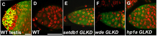
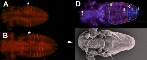
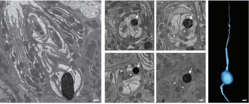
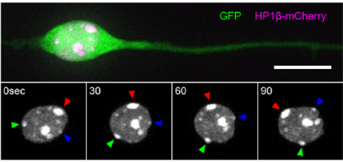
Heterochromatin spots reveal a migrating nucleus, from Figure 1, Wu, et al. 2018
Can you give us the key results of the paper in a paragraph?
MK & YW For better understanding of the complicated interplay between microtubule- and actin-dependent mechanisms driving nuclear migration in CGCs, we performed a high spatiotemporal resolution imaging using spinning-disk confocal microscopy. We found that the nucleus displayed unexpectedly dynamic motion during migration, including fast rotation in the soma. By quantitative 3D image analysis combined with molecular and pharmacological manipulations, we demonstrated that the nuclear rotation is driven by the same strain energy inducing nuclear translocation. Furthermore, among the two major cytoskeletal forces driving nuclear migration, rotation is solely dependent on microtubule systems but not actomyosin, while nuclear translocation requires both actomyosin and microtubules. We thus used nuclear rotation as a readout of microtubule-dependent tensile stress during nuclear migration, which led to the second major finding that both dynein and kinesin-1 are involved in the force transmission. Our results suggest that microtubules and associated bidirectional motors exert force to small points on the nuclear envelope via interaction with nesprins, and steer rotation or translocation depending on the position of the force points.
Nuclear dynamics of a migrating and a post-migratory CGC in vitro. Movie 1 from Wu, et al. 2018
I have to say that the videos in your paper are remarkably beautiful! Can you tell me what it was like to first see nuclear rotation in migrating CGCs?
MK Our co-author Hiroki Umeshima is highly skilled in live imaging, and he set up most of the imaging systems used in the present study. His first movie of nuclear rotation caught my eye, but I did not begin the present study for more than a year, as I was unsure if the rotation is a biologically significant event or an idling motion caused by excessive force to the nucleus in culture. But, the more movies I watched, the more confident I became that the rotation was driven by active forces on small points of the nucleus during nuclear migration. Around that time, You had developed his skill in image analysis, so I asked him if he was interested in analyzing this mysterious phenomenon, and he said ‘Yes’.
YW It was really interesting. I never imagined the nucleus would rotate so fast and frequently during migration. The movies also showed the dynamic deformation of the nucleus. I reckoned these motions could be indicating force transmission, and started to find a way to analyze the dynamics.
How do you think the opposing microtubule motors dynein and kinesin work together to drive unidirectional nuclear motion?
MK & YW We observed frequent switches of rotation direction both in vivo and in vitro (Movie 4 and 5), indicating that the nuclear surface readily moves in both anterograde and retrograde directions by dynamic interplay of the bidirectional motors on bipolar microtubules. Given that the polarity of perinuclear microtubules is mixed but strongly biased, dynein probably acts as the predominant motor and moves the nucleus along the retrograde microtubules (with minus-end toward the leading process) as previously indicated, while kinesin drives back-step movement or rotation. Involvement of bidirectional motors in nuclear migration along uniformly oriented microtubules has been found in long hypodermal precursor cells in nematode embryos (Bone et al., Development 2016; Fridolfsson and Starr, Dev. Biol. 2010). Both hypodermal cells and CGCs are elongated and move nuclei through narrow interstitial and tissue spaces, where dynamic bi-directional movement by opposing motors might adjust the precise positioning of the nucleus and help it squeeze through constrictions.
Nuclear dynamics of migrating CGCs in an organotypic slice. Movie 4 from Wu, et al. 2018.
When doing the research, did you have any particular result or eureka moment that has stuck with you?
I was excited to observe the dynamics of the nucleus in the organotypic slice culture. Until then, I was focused on the in vitro culture system, where the rotation and deformation occurs sporadically. I found the nucleus in 3D tissue much more dynamic, and there was almost no moment when the nucleus stayed still. It was interesting to see that forces seemed to be always applied to the nucleus, even if it was not translocating
And what about the flipside: any moments of frustration or despair?
While it was an exciting moment to see the nuclear motion in tissue, it was very challenging to analyse it quantitatively. It would be interesting and informative to characterize its properties in tissue precisely, but it was difficult to acquire enough resolution in live-imaging.
What next for you, You?
It is always fun for me to observe dynamic things. I am still interested in neural development and also in brain function, which is achieved by such well-organized structure, and am thinking about doing postdoc in related fields.
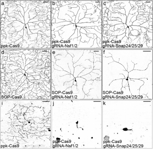
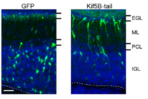
Kinesin inhibition disrupts CGC migration in vivo, from Figure 7, Wu, et al. 2018.
Where will this work take the Kengaku lab?
We want to further elucidate the actual crosstalk of microtubule motors and nuclear envelope during nuclear motion. New imaging techniques with higher spatiotemporal resolution might be required to pursue the next problem. Also, the dynamic deformation and rotation shown in the present study suggest that the nucleus is very soft and flexible in newborn neurons. We are interested in how the mechanical properties of the nucleus is determined, and if they contribute to neuronal migration.
Finally, let’s move outside the lab – what do you like to do in your spare time?
MK I must confess that I do not have so much spare time for special activities after research and family. I am a food fan and enjoy cooking every day. I sometimes eat-out with my family and friends on weekends. Luckily Kyoto is a great place to explore nice restaurants of various styles, from traditional Japanese Kaiseki to casual French bistro.
YW I like to sing songs, sometimes in a bit loud voice if nobody is by.
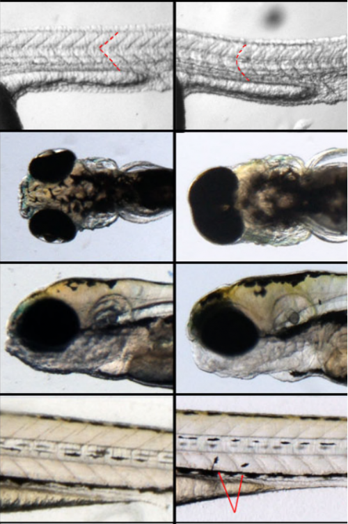
Here we discuss the curious case of female-restricted epilepsy, an unusual disorder caused by mutations in the Protocadherin 19 (PCDH19) gene. How changes in this cell adhesion gene cause seizures and intellectual disability in girls (but not boys) has been a mystery since this condition was first described over 20 years ago. By pursuing several lines of enquiry including in vitro cell sorting assays, CRISPR/Cas9 mouse models and patient MRIs, we have finally “cracked the case”, although intriguing questions remain about the neuronal pathology that underpins this unique condition.
PCDH19-GCE-no other disorder is quite like it
The most striking characteristic of PCDH19-GCE is its unique X-linked inheritance pattern which was first described by Ryan et al in 1997. Typically, X-linked disease-causing mutations affect males as they do not have a second wild type (WT) copy of the gene to compensate. As the name suggests, Protocadherin 19 Girls Clustering Epilepsy (PCDH19-GCE) is caused by mutation in the X-linked gene PCDH19 but this disorder does not follow the “typical” X-linked recessive disease inheritance pattern. Instead, heterozygous females (who have one WT copy and one mutated copy of the PCDH19) are affected while hemizygous males are not. A mysterious condition indeed!
It was not until 2008 that PCDH19 mutations were identified as causing the disease, and multiple hypotheses were put forward to explain its unusual inheritance pattern. It was initially proposed that PCDH19 mutations may be dominant negative, but the identification of whole gene deletions and examples of nonsense mediated decay provided strong evidence opposing this hypothesis (Dibbens et al., 2008). It was also suggested that males may be able to compensate for the loss of PCDH19 through the Y-linked PCDH11Y gene. However, this has also been ruled out due to the discovery of several mosaic males with early somatic mutations in PCDH19 that phenocopy affected girls (Depienne et al., 2009; Terracciano et al., 2016). The third and final hypothesis was based on two key facts. The first is that PCDH19 is located on the X-chromosome and is subject to X-inactivation. This mechanism ensures that females randomly “silence” one copy of the X chromosome in every cell to match the expression levels in males (who are XY). Thus, females with PCDH19 mutations have a mixture of WT and mutant neurons. The second was PCDH19’s known role as a homotypic cell adhesion molecule. Together these pieces of information led Dibbens et al , 2008 and Depienne et al 2009 to suggest that mosaic expression of PCDH19 in female brains leads to abnormal neuronal connections between PCDH19 WT and PCDH19 mutant cells affecting neural network formation and ultimately giving rise to seizures and intellectual disability. However, it was unclear how mosaicism could lead to PCDH19-GCE at the molecular, cellular and network level. Furthermore, this model did not explain why the complete absence of PCDH19 (in males) does not cause epilepsy.
The smoking gun: cell sorting assays provide evidence for a PCDH adhesion code
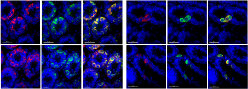
We attempted to unravel this mystery by combining complementary in vitro and in vivo experimental approaches. A valuable clue came from recent papers showing that clustered PCDHs (which are structurally similar to the non-clustered PCDH family to which PCDH19 belongs) act in combination to form complexes at the cell surface with highly specific homotypic binding affinities (Thu et al., 2014). This peaked our interest as it was unclear from the “cellular interference model” how the loss of a single cell adhesion molecule could disrupt normal interactions given that neurons typically express many PCDH family members. Using a co-immunoprecipitation and cell aggregation assays we demonstrated that PCDH19 can form promiscuous cis and highly specific trans interactions both of which are required to generate specific combinatorial binding affinities. We then mixed together two populations of cells expressing different combinations of PCDHs and observed segregation between the two populations when they differed by just a single PCDH (Figure 1). This confirmed that the “PCDH adhesion code” was acutely sensitive to the absence of PCDH19.
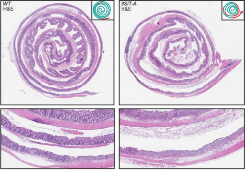
Figure 1 – Adhesion specificity is determined by single differences in PCDH expression. Two populations of cells expressing the same PCDHs show extensive mixing (left) compared to that of two populations with a single difference in PCDH expression, which display striking segregation (right).
The next step was to assess how disruption of PCDH19-dependent adhesion would impact the developing brain, where a complex array of cell adhesion molecules and binding partners are expressed. To investigate this, we turned to our previously characterised Pcdh19 Knockout (KO) mouse model (Pederick et al., 2016). Like humans, Pcdh19 is located on the X chromosome in mice and therefore heterozygous mice have mosaic expression of Pcdh19 due to X-inactivation. Interestingly, electrocorticogram analysis revealed significantly elevated activity in heterozygous mice when compared to WT controls. Importantly, this phenotype was not present in mice completely lacking Pcdh19, which matched the unique X-linked inheritance of PCDH19 epilepsy.
CRISPR/Cas9 joins the investigation
While the electrical phenotype of the mouse was consistent with humans, we couldn’t investigate the cellular impact of mosaic PCDH19 expression in the brain because we were lacking a specific PCDH19 antibody with which to identify PCDH19+ cells. We therefore turned to the CRISPR/Cas9 genome editing system and generated a mouse with an epitope-tagged (HA-FLAG) version of PCDH19, allowing us to identify PCDH19 WT cells with commercially available antibodies. To detect “PCDH19-expressing” Pcdh19 null cells we used the previously mentioned PCDH19 KO mouse which has a LacZ reporter. Now we were set to answer the key question- how does mosaic expression of Pcdh19 in heterozygous female brains affect the behaviour of the WT PCDH19 and null PCDH19 cell populations?
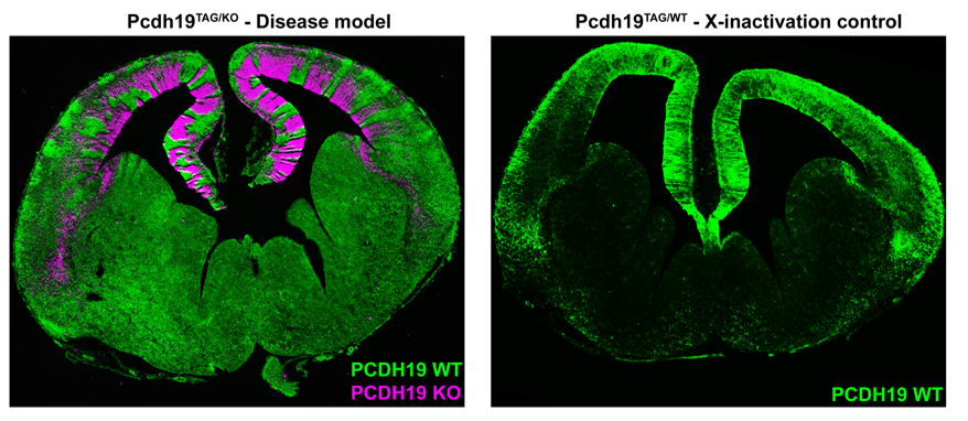
Simultaneous labelling of WT PCDH19 and null PCDH19 cells in PCDH19 heterozygous mice revealed a striking pattern of alternating PCDH19 +ve and PCDH19 -ve regions (Figure 2). This pattern was particularly obvious in the developing cortex where it resembled “tiger stripes”. Before getting too excited, we needed to address the possibility that the apparent segregation simply arose from X-inactivation and subsequent clonal expansion of PCDH19 +ve and PCDH19 -ve cells. We therefore generated “WT” mice with one tagged copy of PCDH19 and one (untagged) WT PCDH19 allele. This critical control was only possible because we had generated the tagged allele (i.e. even if we had a PCDH19 Ab we could not have done this experiment). Critically, there was no sign of the “tiger stripes” in this control- instead we detected small patches of tagged and untagged PCDH19 cells along the ventricle with subtle variations in the cortical plate (Figure 2). So, evidence for active cell segregation in heterozygous females was beginning to stack up. Interestingly, the abnormal segregation pattern of WT PCDH19 and null PCDH19 cells was different in each mouse, presumably caused by the random nature of X-inactivation. Since the symptoms of PCDH19 epilepsy are highly variable (even in identical twins (Higurashi et al., 2012)) it is possible that different segregation patterns contribute to the severity of the disorder. Although not performed in this study it would be interesting to correlate the X-inactivation patterns seen in individual mice with their ECoG recordings to identify any patterns that lead to higher electrical brain activity.
Figure 2 – Mosaic expression of PCDH19 leads to abnormal cell sorting in the developing cortex. Segregation of Pcdh19 +ve and Pcdh19 -ve cells results in distinct “tiger stripes” (left). Normal X-inactivation of Pcdh19 does not result in “tiger stripes” (right).
In our opinion the most interesting feature of PCDH19 epilepsy is not that mosaicism leads to disease but that individuals who completely lack PCDH19 do not have the disease. As mentioned above, homozygous null mice did not display increased electrical brain activity. We hypothesised that this phenotypic “rescue” was due to the uniform loss of Pchd19 and the consequent restoration of normal cell sorting during cortical development. To test this, we developed a strategy to rapidly assess in vivo cell sorting in Pcdh19 null embryos. We deleted the functional PCDH19TAG allele in heterozygous (PCDH19TAG/PCDH19LACZ) zygotes using CRISPR/Cas9, transferred the embryos to pseudopregnant females for further development, and then used X-Gal staining to track the location of the PCDH19 null cells. While negative (i.e. heterozygous) controls showed the expected segregation phenotype, no significant segregation occurred after deletion of the (WT) PCDH19TAG allele. These findings provided further evidence that abnormal cell sorting is caused by the differential adhesion affinities between WT PCDH19 and null PCDH19 cells. Furthermore, the absence of abnormal cell sorting in animals completely lacking PCDH19 provides a clear cellular phenotype that explains the unique inheritance pattern of PCDH19 epilepsy.
Finally, we postulated that abnormal cell sorting in humans could lead to brain malformations due to the prolonged expansion and increased cortical folding compared to mice. Abnormal cortical sulcation was observed in four girls with causative PCDH19 mutations. We identified variably positioned cortical defects that included bottom of the sulcus dysplasias, abnormal cortical folding, cortical thickening and blurring of the grey/white junction. It is not known how abnormal cell sorting may generate these cortical malformations, however, insight into the cellular mechanism could be gained by the generating a PCDH19 mutant ferret with CRISPR/Cas9 genome editing, a model organism that has extensive cortical folding.
For the future…
These findings provide some long-awaited answers to the mysterious inheritance of PCDH19 epilepsy. However, despite these insights, there is still much to discover about how mosaic expression of PCDH19 leads to epilepsy, intellectual disability and autism. It is possible that in addition to cell sorting, other processes such as neuronal wiring, synapse formation, maintenance and function are disrupted by differential adhesion affinities caused by mosaic expression of PCDH19. Perturbation of these fundamental neuronal processes is implicated in many neurodevelopmental disorders and it seems possible that they may also be altered in PCDH19 epilepsy. Much sleuthing lies ahead!
Higurashi, N., Shi, X., Yasumoto, S., Oguni, H., Sakauchi, M., Itomi, K., Miyamoto, A., Shiraishi, H., Kato, T., Makita, Y., et al. (2012). PCDH19 mutation in Japanese females with epilepsy. Epilepsy Res. 99, 28–37.
Applications are sought from established as well as career development researchers with interests in nervous system and/or human development, including stem cells and organoid approaches. We are also seeking cell biologists with interests in signalling, trafficking and cell adhesion.
Applicants will be expected to be competitive for personal fellowships and/or programme level grant support and have a strong publication record. For career development researchers the start-up package typically includes financial support for relocation and
essential equipment and a defined transition to tenure after 5-6 years. New researchers are supported by a structured mentoring programme. The University of Dundee operates policies enabling flexible working to ensure a good work life balance.
The Division of Cell & Developmental Biology consists of 8 research groups studying the regulation of differentiation in developing organisms, stem cells and adult tissues and how this is altered in disease states. Our work combines classical embryology, cell biological and genetic approaches as well as mathematical modelling to understand gene function and regulation in differentiating tissues. A particular shared interest is in epithelial cell biology, and this has led to our cooperative effort to maintain and develop state of the art imaging technologies. The Division also hosts the Dundee Human Pluripotent Cell Facility, which provides quality controlled human pluripotent cells and training in their use.
How to apply: please provide CV, details of 3 academic referees and a brief summary of future research plans. For informal enquiries please contact
The Institut de Génomique Fonctionnelle de Lyon (IGFL) has an opening for a new independent group leader. The IGFL has a unique scientific profile and fosters an outstanding international environment. Teams address basic research questions at the interfaces of evolution, physiology and development using functional genomics, bioinformatics, genetics and comparative approaches. The IGFL has a strong focus on integrative, organism-level research using a diversity of model and non-model organisms.
More information at: https://www.nature.com/naturejobs/science/jobs/636517-research-group-leader-opening
You can find our recently published eLife paper here.
At the Euro-Evo-Devo meeting in Lisbao I saw a talk by Sylvie Rétaux and became hooked by a blind and unpigmented cavefish: the evo-devo model Astyanax mexicanus. I then had the chance to join Sylvie’s group in Gif-sur-Yvette (France) in 2013, for a post-doc. Four years later we come out with this paper of which I’m extremely happy, not least because this study owes a great deal to teamwork and wouldn’t have been possible without a fantastic collaborative spirit between enthusiast and passionate team members. Because the “author contribution” section didn’t quite capture my feeling about the human adventure behind our paper, I’d like to take this blog post as a platform to properly acknowledge each of my friends and bring to you our scientific/team-story.
Astyanax mexicanus, evo-devo top model
Astyanax mexicanus is a teleost fish that inhabits South American rivers. As well as the river dwelling fish, several Astyanax populations can be found deep in the caves of the Sierra del Abra in Mexico. Cave colonization has occurred several times independently and these cave populations have experienced convergent evolution of several traits, including loss of eyes and pigmentation. Cave fish also behave differently than their surface siblings, probably in response to selective forces driving adaptation to life in complete and permanent darkness. For instance, they have a better sense of smell and more appetite, they swim more and sleep less, almost constantly exploring their environment in the quest for food or sexual partner. Astyanax is a great model to link developmental evolution with adaptation to a new environment. By comparing the anterior brain of surface and cave fish, and by doing so in young embryos, Sylvie’s team have been highlighting how development of the central nervous system has shaped cavefish evolution.
Our recently published work makes a new contribution to the story of Astyanax brain evo-devo. And for the first time we were able to elucidate some links between early embryonic development and fish behavior. To reach this goal, it tooks 4 years, 3 co-first authors (me-Alexandre Alié, Jorge Torres-Paz and Lucie Devos), the contribution of two brilliant students (Lise Prunier and Fanny Boulet), the unwavering support of our lab manager (Maryline Blin) and the expertise of our specialist in behavioral studies (Yannick Elipot).
From early development…
The story began before I joined the group, with the work of Lise. At that time she was a Master’s degree student and had performed very numerous and beautiful in situ hybridization showing more NPY neurons in cavefish brain versus more Pomcb neurons in surface fish brain. When Lise had to leave the lab at the end of her internship, Sylvie offered me to dig deeper into this story. Together with Maryline, I started to characterize the expression of 9 neuropeptides, at 4 embryonic stages in the 2 morphotypes (cave versus surface)… 72 different conditions in total, reproduced several times to get statistics. This could have been frustrating at some point, but few weeks after I started Lucie joined our lab for her master internship. Lucie galvanized us with her contagious enthusiasm! Our hours spent at the microscope to count/recount neurons or to debate the anatomical interpretations of our results became so much more fun by our combined efforts.
On our way to observe Astyanax cavefish in their natural environment. In the depths of the caves too, mutual support helps to follow the right path.
We next embarked on a series of double fluorescent ish to establish a co-expression map between Lhx genes and neuropeptides. Again, help was at hand from a skillful student, Fanny, who did a fantastic job with these double FISH (no pun intended), under the ever-watchful eye of Maryline. We have the chance to possess a fancy confocal microscope in the lab, and we were so excited to get into the very intimacy of Astyanax brain. The expression domains and dynamics of Lhx7 and Lhx9 strongly suggested a role for these genes in the formation of NPY-positive and Hcrt-positive (two neuropeptides) neurons, respectively. This has been definitely confirmed thanks to Jorge, a new postdoc who joined the group and took over the project when I left the lab for other horizons. Jorge’s rigorous injections of morpholinos and RNAs and his repeated cell counts again clearly established the functional links between the Lhx transcription factors and the corresponding neuropeptides. Sylvie was so happy, her favorite transcription factor Lhx9 she had discovered in the mouse 20 years ago, was involved in the process of developmental evolution of her favorite animal, the cavefish!
In parallel, we tested the role of Fgf and Shh signaling, pathways well-known to play a role in cavefish brain evolution and eye loss. For these pharmacological experiments, the fact that Astyanax produces hundreds of eggs every morning made this task easier. But what was even better was the chance to work with a cohesive and efficient team to collect, sort and dechorionate the eggs, then to treat them, wash them, fix them, even when it involved working in the middle of the night, for several nights in a row. All the authors of this paper contributed to these pharmacological experiments!
… to behavior evolution
After we had established the link between early signaling, early expression of Lhx genes and differences in neuropeptidergic neuron numbers, Sylvie next encouraged us to complete the story by linking these differences to adaptive behavior observed in cavefish. Honestly, I was not very keen on going down this road, which I thought could be long and difficult. And I was right indeed… it was so hard to get there, especially for Jorge, who worked the most on this and probably suffered a bit. With the behavioral set-up being in a distant building, I think that he had to walk dozens of kilometers to, ironically, demonstrate that the Lhx9-dependant increased number of Hcrt neurons is responsible of hyper locomotion in cavefish. However with the help and the expertise of Yannick, who achieved a level of excellence in behavioral studies on Astyanax, and Cynthia, the engineer responsible for the platform of fish behavior analysis in the Department, problems were solved and we got nice results.
And here we were! We finally got our story, linking the evolution of embryonic development, to neuro-anatomy and behavior. And we got it as a team! Brought together by the trust of Sylvie, and by mutual confidence and friendship: who could ask for more from a postdoctoral experience? I am also very grateful for the opportunity to have contributed to several other papers during this post-doc. Lucie and Jorge are also conducting their own research projects, which will surely benefit from the team spirit in a group where everybody is willing to help the others, and more importantly where everybody feels free to ask for help. As Claude Bernard says: “The idea is the seed; the method is the soil which enables it to develop…” and I believe that teamwork is the best fertilizer helping to yield the best fruits.
This summer, the Company of Biologists, the not-for-profit publisher of Development, is running a Workshop on ‘Development and evolution of the human neocortex‘, organised by Victor Borrell, Wieland Huttner and Arnold Kriegstein.
The Company of Biologists Workshops provide leading experts and early career scientists from a diverse range of scientific backgrounds with a stimulating environment for the cross-fertilisation of interdisciplinary ideas. The programmes are carefully developed and are intended to champion the novel techniques and innovations that will underpin important scientific advances.
There are currently multiple funded spaces for early-career researchers to attend this exciting event (deadline = 23 March). To find out more and apply online please visit
On 2016-5-18, the second day after my first research paper 1 was published online at my third year of PhD courses, my mentor Rongwen Xi told me to take over the “EEP project”. This project had begun long before I started my PhD courses, and until my participation, has been passed along by three researchers in turn: Na Xu, Pin Huang, and Chenhui Wang; each of them subsequently graduated and moved on with their own academic or industrial paths. Encouraged by my first successful publication, I quickly agreed to take this seemingly never-ending project. I told Dr. Xi a sentence that amused me afterwards, “I won’t give it up until you give up.” This is how this long and tough process begins, and this is also the instant that determines the end of the story.
Introduction
Even as adults, we have stem cells throughout our bodies that are responsible for maintaining many of our tissues. These adult stem cells constantly divide and produce daughter cells, which, through a process called differentiation, become multiple types of mature cells. The fate of the daughter cells can be actively specified by asymmetric cell division, in which cell fate determinants are specifically segregated into one of two stem cell daughter cells 2. Alternatively, cell fate can be specified passively; in this case, cells physically depart from the self-renewal niche environment, as with the specification of cystoblasts from Drosophila germline stem cells, and the initiation of differentiation of stem cells in the mouse small intestine upon their departure from the Paneth cell niche 3,4. Despite several implications from these “renew or differentiate” fate determination events, very little is known about the molecular mechanisms by which distinct, lineage-restricted progenitor cells are generated from a common stem cell pool.
To study this question, we investigated cell fate in a multipotent intestinal stem cell (ISC) experimental model from adult fruit flies. The default mode for cell fate is that ISCs differentiate into enterocytes (EC), which have been shown to occur from approximately 90% of ISC divisions 5. However, there is a less-well-understood mode in which ISCs differentiate into pairs of enteroendocrine cells (EEs), which occur from approximately 10% of ISC divisions.
When I started to do this project, previous studies suggested that EEs are directly differentiated from ISCs, implying that the decision of EE specification may occur at the stem cell level in ISCs 6,7, but how this occurs remains unclear. It has also been revealed that the four-gene cluster acheate-scute complex (As-c) act as EE-fate-determination factors. Furthermore, one of the As-c genes, scute (sc), is both necessary and sufficient for EE specification. Nevertheless, important questions remain about both the molecular and cellular mechanisms through which Sc functions in EE fate decision, and we do not yet know how Sc is regulated in ISCs to control EE fate.
We finally answered these questions in our recent paper, in which we reported that transient activation of Sc determines both the type and number of committed progenitor cells from Drosophila ISCs.
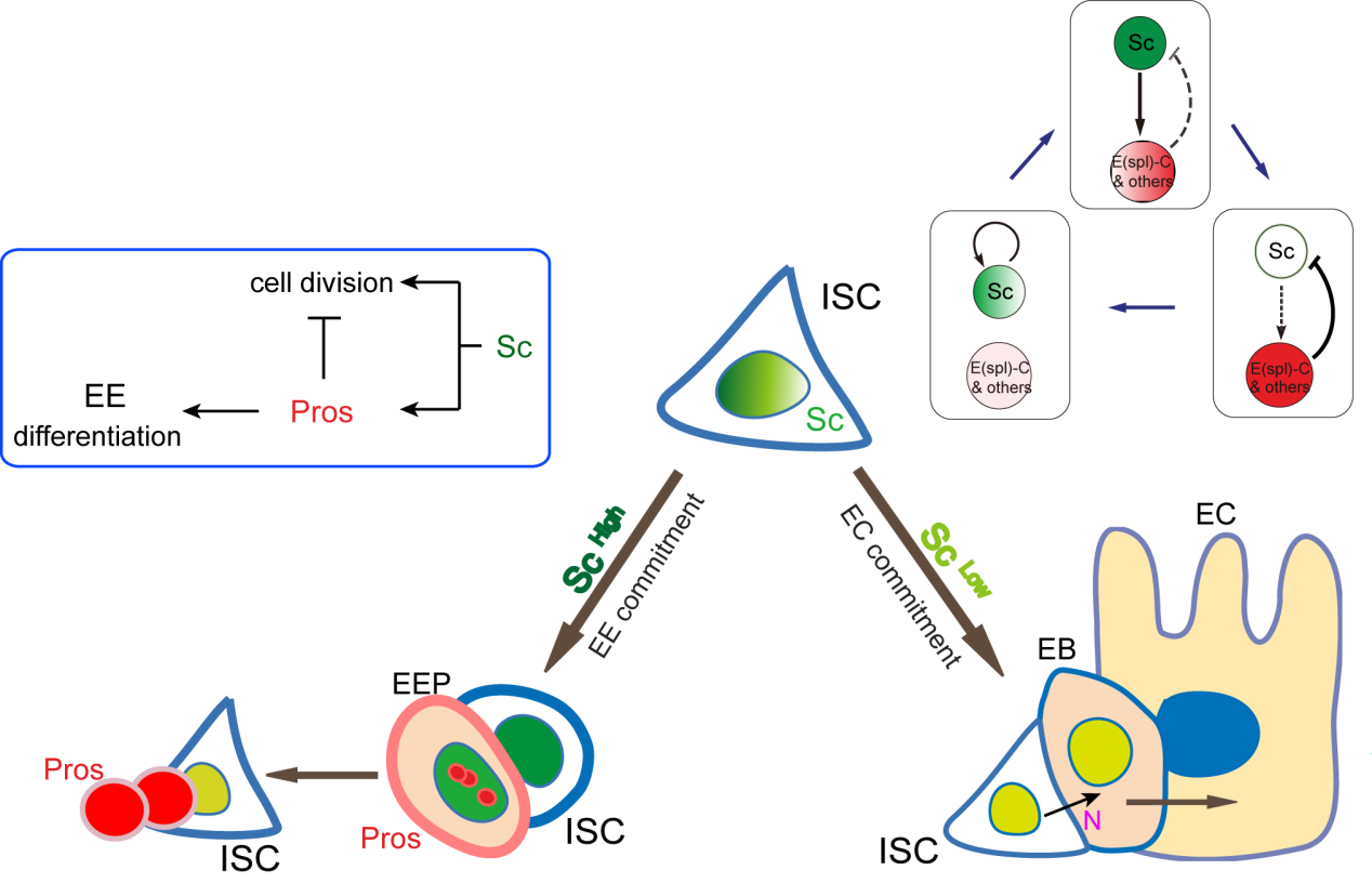
Figure 1. A graphic model to describe how ECs and EEs are respectively generated from ISCs. Notch-signaling-guided EC generation from ISCs acts as the default mode, while transient expression of Sc triggers EE generation from ISCs. Oscillatory expression of Sc in ISCs is achieved by transcriptional self-stimulation combined with a negative feedback regulation between Sc and E(spl) proteins & other Notch targets. During the generation of EEs, increased Sc expression induces asymmetric cell division that generates a new ISC and an EEP; residual Sc activity in the newly formed EEP is then able to induce one round of cell division and precisely generate a pair of EEs.
A cell fate is determined by a transiently expressed protein
To better understand the process of EE specification in ISCs, we set up an EE regeneration assay and examined de novo EE regeneration. This assay was first beautifully set up by Na Xu and Pin Huang. Based on the finding that Sc is required for EE generation from ISCs, we temporally knocked down sc starting from the pupal stage, and this process produced flies with midguts lacking EE cells. We then used these EE-less midguts to examine the process of EE production by using temperature shift to re-introduce Sc expression in the midgut. With this assay, we discovered that (i) ISCs actually undergo an initial division to generate a new EE progenitor cell (EEP), and (ii) the EEP then undergoes one final round of cell division to produce a pair of EEs (Figure 2).
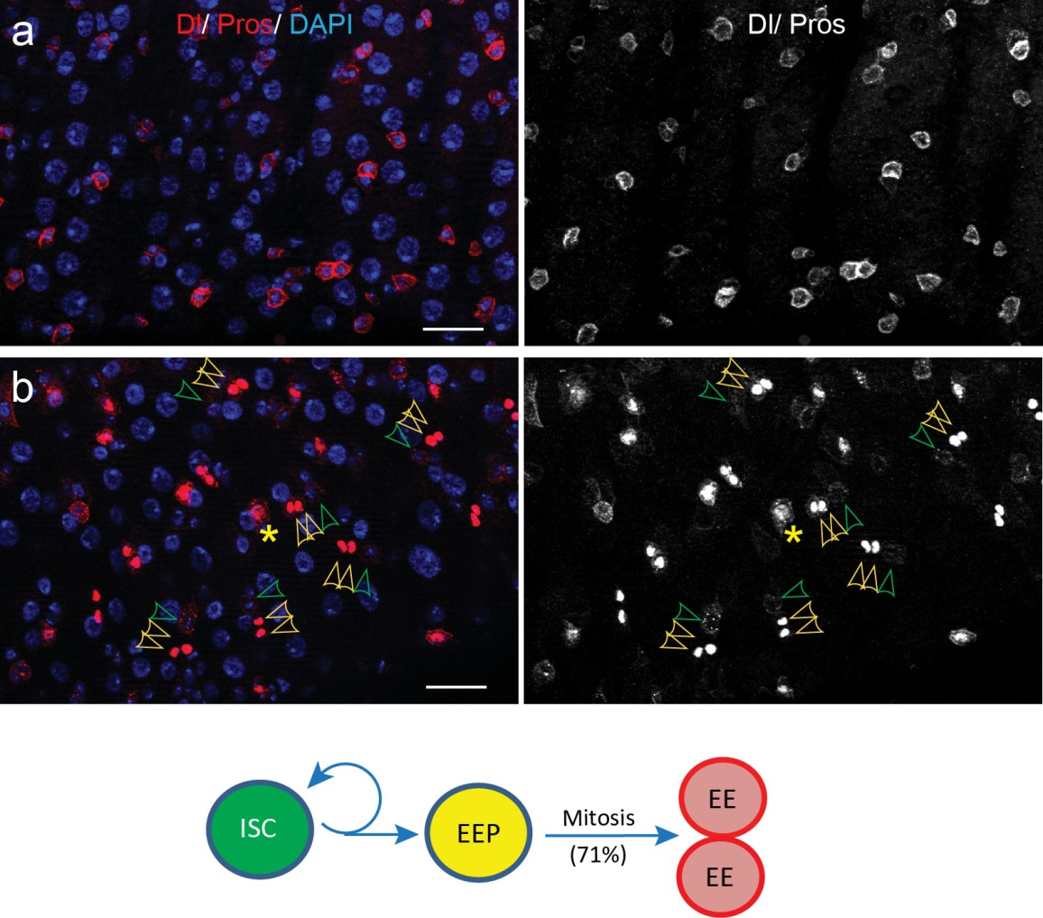
Figure 2. An EE-regeneration model reveals that ISCs self-renew during the generation of EE pairs. (a-b) Patterns of ISC (marked by anti-Dl, red on membrane) and EE cells (marked by anti-Pros, red in nuclear) during sc-RNAi mediated EE depletion (a) and the following EE regeneration (b). An ISC undergoes self-renewal before generating an EEP, and 71% of EEPs undergoes one round of mitosis to generate a pair of EEs, and the rest directly differentiate into a single EE.
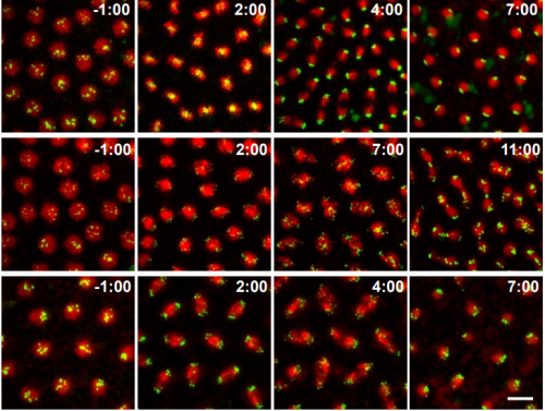
To further analyze this two-step cell division process, Chenhui Wang genetically overexpressed sc in ISCs and monitored the cellular events in a time-course experiment. Chenhui found that transient sc expression caused a rapid cell division response, and also induced expression of the EE-marker gene Pros, which is known as a potent cell-cycle inhibitor. These findings and subsequent experiments enable us to precisely define the regulatory circuitry that directs the formation of a pair of EEs from each ISC (Figure 1). Here a concern still exists that we have not given a “seeing is believing” results for cellular events of EE generation because we have not established long-term live imaging technique for fly midgut yet. To solve this problem, I expressed a UAS-RedStinger reporter in sc overexpression system. RedStinger is relatively stable and can serve as a lineage marker to trace the progeny of the originally marked ISCs. The number of cell divisions of the initially labeled ISCs could be deduced based on the mitotic marker PH3 and the number of RedStinger+ cells in a single cluster. In this experiment, I observed a tightly ordered process: The first cell division following sc overexpression occurred in ISCs (PH3+ in a one-cell clone), and at telophase of the first cell division, one of the two daughter cells began to show cytoplasmic Pros accumulation; the second cell division (PH3+ in a two-cell clone) always occurred in the Pros+ daughter cell, that is EEP; the third cell division (PH3+ in a three-cell clone) occurred again in ISCs. These observations suggest that EEs are generated from ISCs via two rounds of cell divisions: an asymmetric division of ISC to generate an EEP, and then the EEP division to produce an EE pair (Figure 3).
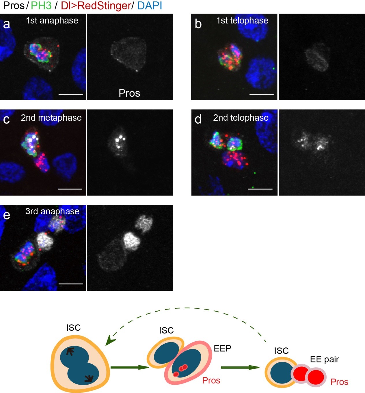
Figure 3. The process of sc-overexpression-induced EE generation from ISCs. (a-e) Expression of Dl>RedStinger (red), PH3 (green) and Pros (white) during sc-overexpression-induced mitosis. Sc induction in ISCs promotes asymmetric cell division that generates EEPs, which begin to show punctate nuclear Pros expression. Each EEP immediately divide once prior to terminal differentiation, yielding a pair of EEs.
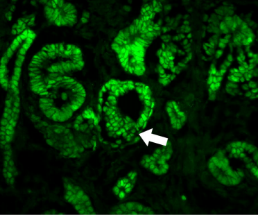
Next, to visualize the expression of Sc in midgut, Pin generated a green fluorescent protein (GFP) tagged line for Sc in collaboration with Zhongsheng Yu and Renjie Jiao from the Institute of Biophysics of the Chinese Academy of Sciences. In this line, the GFP was fused to 3’ of the Sc coding region. Initially we were a little bit disappointed as the GFP signal was too weak to visualize and all the researchers had to immunostain with anti-GFP antibody, which effectively amplified the Sc-GFP signal. Immunostaining results revealed that Sc-GFP could be observed in virtually all ISCs but the expression level is largely indistinguishable among ISCs. With improved microscopy technology, I managed to capture GFP signal in unstained samples and found that the Sc-GFP fusion protein is expressed at higher levels in ~15% of the ISCs (Figure 4). This result was exciting because it indicated that Sc may be expressed in a dynamic manner in ISCs, and in a snap shot, you may see a weak expression level in most ISCs, and increased expression levels in a small subset of ISCs.
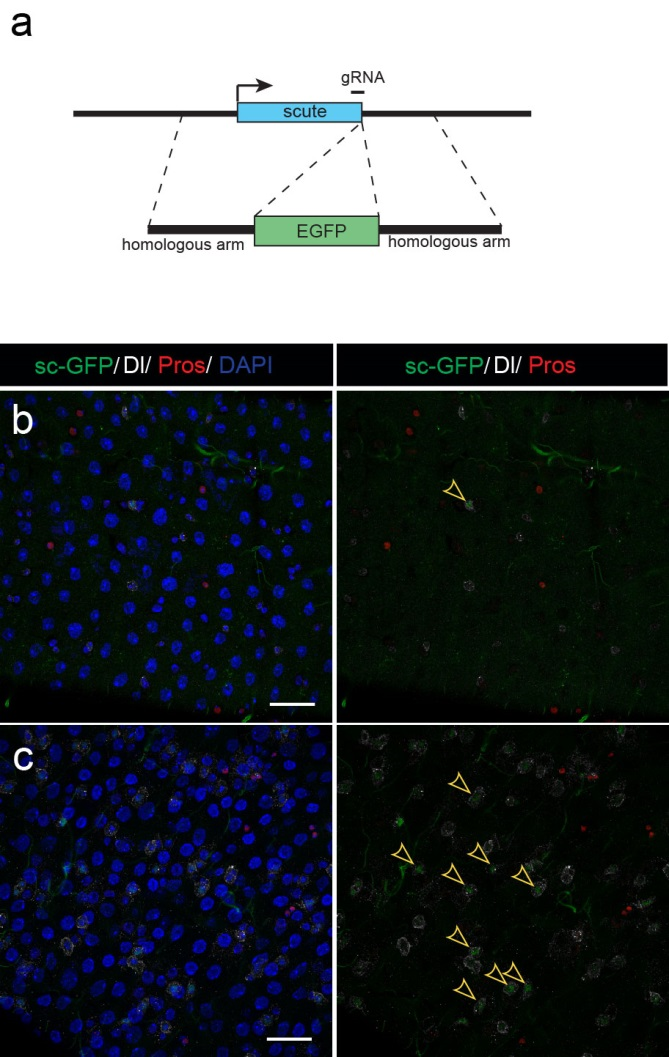
Figure 4. Sc is expressed in a small subset of ISCs. (a) A diagram showing genomic information for C-terminal insertion of EGFP in sc gene region. (b-c) Expression of Sc-GFP (green), Dl (white) and Pros (red) in midgut of 5-7 day old flies.
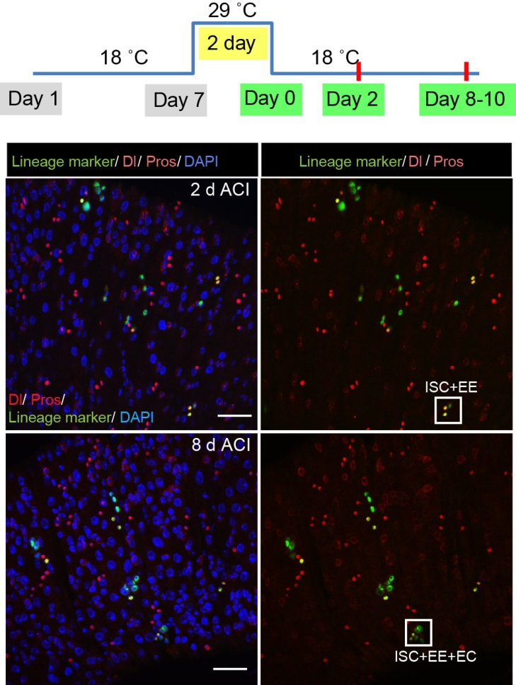
The next step was to test the cell lineage fate of these Schigh ISCs. The follow-up cell lineage tracing studies with a Sc-Gal4 line will help to do that, but there was no available Sc-Gal4 line at that time. Fortunately, from 23 (upstream and downstream) glass multiple reporter (GMR) enhancer-GAL4 lines generated for sc, I identified one GAL4 line that drove UAS-GFP expression in some diploid cells in the midgut epithelium. The density (also in ~15% ISCs), distribution, and individual variability of the GFP+ cells were largely similar to those of Sc-GFP+ cells, and about half of RFP reporter driven by this GAL4 line recapitulates Sc-GFP expression, suggesting that this GAL4 line is driven by the enhancer element for sc expression in the midgut. Cell lineage tracing studies with this GAL4 line revealed that the immediate daughter cells of Sc-GAL4+ ISCs were mainly EEs; however, these ISCs re-assume their default EC-producing fate once Sc expression is downregulated (Figures 1&5).
Figure 5. The cell lineage tracing results with the Sc-Gal4 line.
The knotty problem
With these exciting new observations, we inevitably faced a mechanistic question, “How does such transient upregulation of Sc in ISCs occur?” This question comes like a boss in video games, and has always been difficult to tackle. Studies over the decades on proneural genes have revealed that the AS-C genes in the neural cell lineages are regulated by highly-complex-cis-regulatory regions, and these regulatory regions are considered to constitute an integrating device for multiple signaling regulators and chromatin factors. Firstly came to our minds was to avoid such “net” and to set out from the reported signals that regulate EE specification in Drosophila midgut. Previous studies suggest that the Slit molecules secreted from EEs activate the Robo2 receptors of ISCs to prevent EE generation, thereby establishing a negative feedback to coordinate EE production with tissue demand. However, Sc expression pattern was unaltered in Robo2 mutants, in which the excessive EE phenotype was prominent. Considering Robo2 activation in ISCs is not sufficient to prevent EE production from ISCs, this mechanism appears to be a modulator rather than a key component in the EE fate decision process. Thus, I had to go back to hit the core of the question, the transcriptional control of As-c genes.
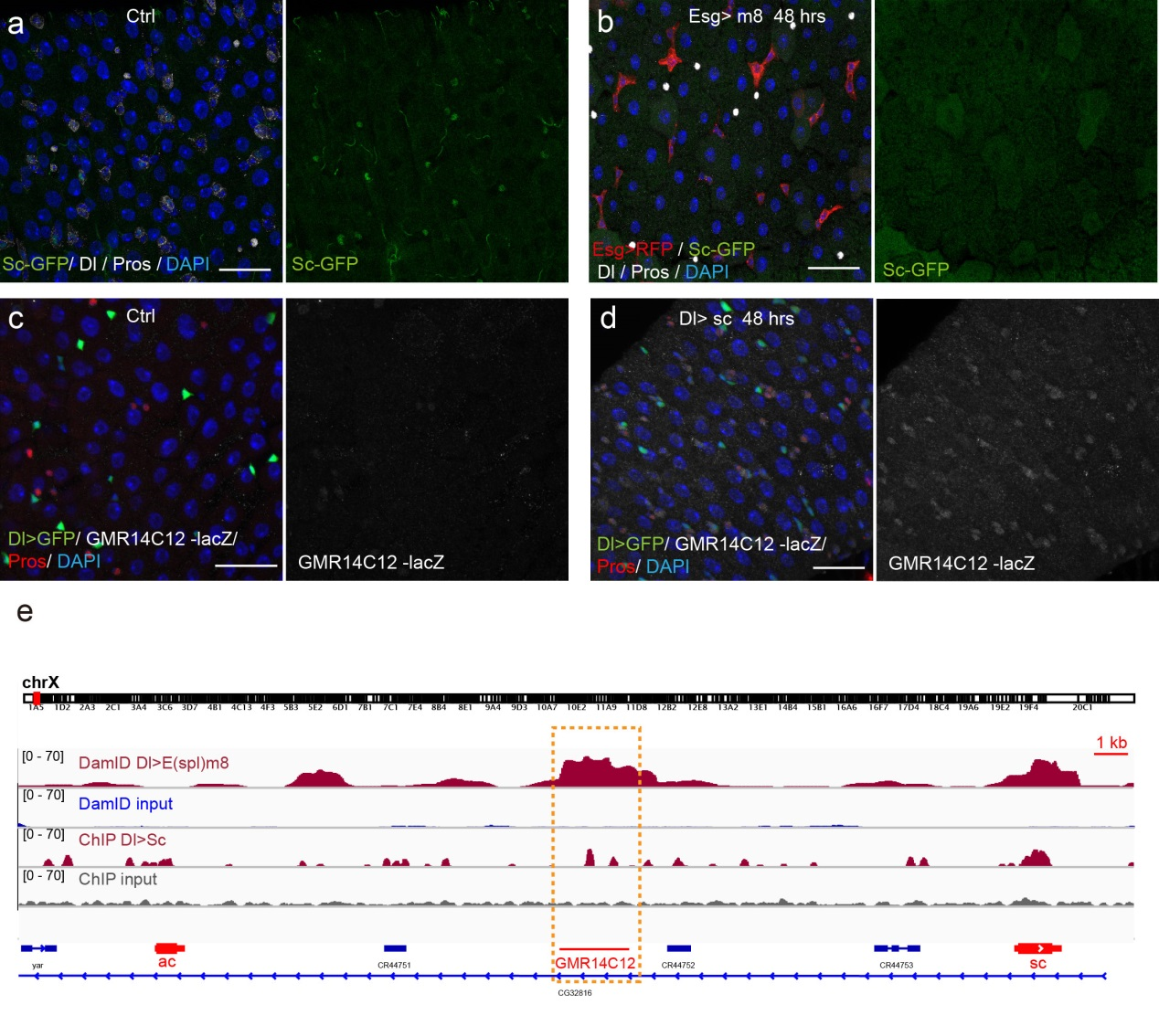
Previous studies on early Drosophila development have suggested reciprocal regulatory relationships between AS-C genes and the enhancer of split complex (E(spl)) genes, which are known as the Notch target genes. Inspired by these reports, I screened a number of candidate reporters for individual E(spl) genes, and identified a single reporter, m8-lacZ, which showed a weak, but similar expression pattern to Sc in wild type guts. To characterize the regulatory relationship between Sc and E(spl)m8, I transiently overexpressed sc in ISCs, and surprisingly saw robust upregulation of m8-lacZ expression in all ISCs. Notably, co-expressing Notch-RNAi did not prevent the upregulation of m8-lacZ expression caused by sc overexpression, suggesting that E(spl)m8 expression is independent of Notch activity in ISCs. To test whether such regulatory relationship similarly applies to other E(spl) genes, I sorted out sc-overexpressed ISCs for mRNA profiling by RNA-seq analysis. Strikingly, in addition to m8, many other E(spl) genes, including m4, m6, m7, mγ, and mδ were strongly upregulated upon sc overexpression. By combining genetic assays and ChIP-seq analysis, we showed Sc could bind to the enhancer regions of many E(spl) genes, and directly upregulate these E(spl) genes in ISCs (Figure 6).
It’s then instinctive to consider whether these E(spl) genes, also known as neural fate repressors, would in turn negatively regulate Sc expression. By combining genetic assays and targeted DamID analysis using a E(spl)m8-Dam fusion line, we showed that E(spl)m8 suppresses sc expression by directly binding to the enhancer region of sc. The direct two-way regulation between Sc and E(spl)m8 form a typical negative feedback regulatory loop, which may explain the transient activation pattern of Sc in ISCs (Figure 6).
The question still has half part unanswered, “how does sc initially build up?” Searching for other transcriptional activators, like other bHLH activators as reported, would make this question a “chick and egg” issue. Interestingly, Sc has been reported to transcriptionally self-stimulate itself, which acts as an essential mechanism for proneural protein accumulation during sensory organ development. To test whether self-stimulation of Sc also occurs in ISCs, we constructed LacZ transcriptional reporter for sc using the Sc-Gal4 enhancer fragment that we had identified. This lacZ reporter was barely detectable in WT midgut epithelium, but effectively induced in ISCs when sc was transiently induced. ChIP-seq data analysis also revealed two Sc binding peaks within this Sc-Gal4 enhancer region (Figure 6). Thus, Sc is able to stimulate its own transcription directly by binding to sc enhancer. Together, our results suggest that two feedback regulatory loops control the transient upregulation of Sc in ISCs prior to EE fate commitment. There is a transcriptional self-stimulation loop that allows Sc to gradually build up and eventually reach a high level to induce EEP specification, and there is a negative feedback regulation loop between Sc and E(spl) genes that returns sc expression back to the baseline level (Figure 1).
Figure 6. Regulatory feedback loops control Sc expression in ISCs. (a-b) Overexpression of m8 rapidly reduced sc-GFP and Dl expression in all ISCs. (c-d) GMR14C12-lacZ (LacZ reporter for Sc-Gal4 line) was nearly undectable in normal midgut epithelium. Overexpression of sc in ISCs led to GMR14C12-lacZ expression in progenitor cells and newly formed EEs. (e) DamID analysis for E(spl)m8 and ChIPseq analysis for Sc in ISCs revealed binding activities for both E(spl)m8 and Sc at the GMR14C12 region.
The beginning of the end
Given that negative feedback is a common mechanism underlying biochemical oscillations in virtually all organisms, the feedback loops between Sc and E(spl) genes could plausibly be the driver of an oscillatory expression pattern for Sc in ISCs; in theory such oscillatory expression could potentially serve as an internal timer for periodic production of EEs from ISCs. This clock mechanism would be similar to what is known about the circadian clocks, a biological research field that was recently honored with the 2017 Nobel Prize for Physiology or Medicine. We are obviously very excited about the findings and potential implications. However, this is just a tip of iceberg, future cellular and molecular analysis, likely in combination with in vivo live imaging work will allow further testing and refining of the oscillation model proposed in our study, and such experiments will determined whether and how any internal timer is regulated by certain endogenous and/or environmental cues, and whether the oscillation model is generally applicable in other tissue stem cells, including that in humans.
Finally, I want to say that I am very fortunate and grateful to be a part of such a wonderful research team and work on such an exciting project. This work would not be possible without the contribution and help from our past and current lab members, especially Na Xu, Chenhui Wang, and Pin Huang, as well as informaticians Huanwei Huang and Tao Cai at NIBS. I especially want to thank my mentor Dr. Xi for his great guidance and trust, as well as his helpful advice on the writing of this article. As you can imagine, in addition to the “high” moments when the exciting results were first observed, I also had many upset and head-scratching moments during the course of this study. These experiences have endowed me a lot on how to explore, to observe, to cooperate, to write, and to persevere. I believe that no matter how hard it seems like, if you continue to stay focused and think hard, great things may eventually happen, in an instant.
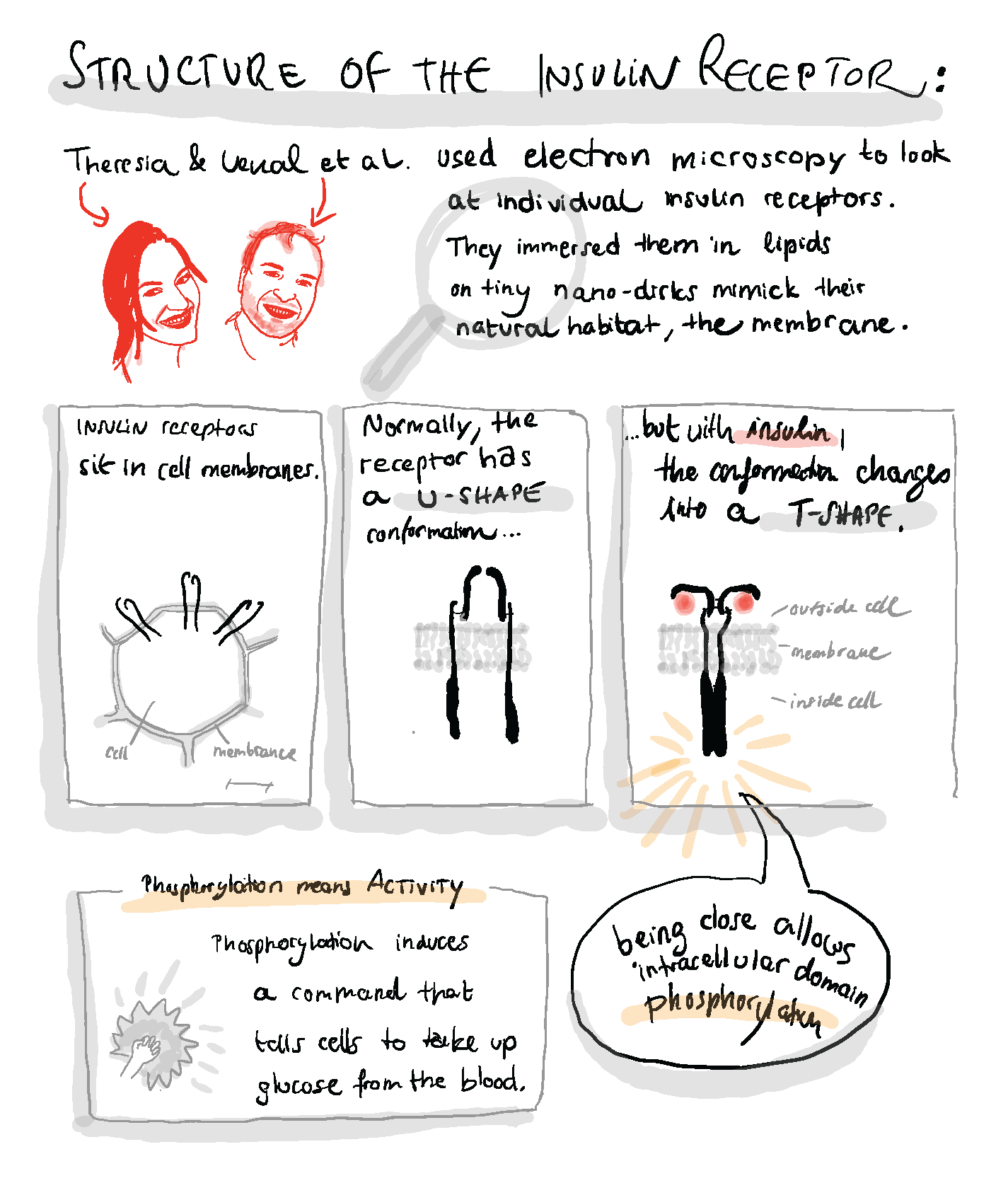
A few days back over dinner at a CNV gathering, Theresia Gutmann from the Coskun lab casually told me about her PhD work. In collaboration with the Rockefeller University NYC, Theresia had visualized the changing conformation of the human insulin receptor upon insulin binding (paper). I made a sketchnote summarizing their discovery of a conformational switch that could explain how the insulin receptor transforms information about extracellular ligand binding into an intracellular activity to react by taking up glucose!
A postdoc position is available in the Lehoczky Lab (Brigham and Women’s Hospital/Harvard Medical School). The lab is focused on understanding the molecular basis of mouse digit tip regeneration, with the ultimate goal of teasing apart the genetic pathways necessary for this process. For more information about the lab see LehoczkyLab.org
Applicants with a strong background in regenerative biology, genetics, developmental biology, and/or molecular biology are encouraged to apply. Prior experience with mouse genetics is preferred. Experience with RNAseq analysis is a plus.
Interested candidates should provide: 1) cv, 2) a brief letter detailing your interest in the lab and relevant past research experience, and 3) contact information for three references who can comment on your research
Application materials and any questions regarding the position should be sent to Jessica: jlehoczky@bwh.harvard.edu
Our latest monthly trawl for developmental biology (and other cool) preprints. Let us know if we missed anything.
On February 20th, The Company of Biologists launched preLights, a community-led preprint highlighting service. A panel of early career researchers (the ‘preLighters’) select and comment on recent preprints that caught their eye, and encourage preprint authors to answer any questions about the work that they had. So far it looks great, and the developmental biology content has been especially good (see the dedicated subjectcategory). We’d love to know what you think: you can contact the team via the site or the Twitter feed.
The idea behind the site was influenced in part by this list – as it got longer and longer (reflecting increased preprint usage), we were wondering how else we could encourage and promote the discussion of preprints, and the preLights idea took form. Rest assured that this list will live on, at least until the point at which it gets impossibly long!
And here’s the list – all the developmental biology I could find, plus relevant and cool other preprints thrown in for good measure.
The preprints were hosted on bioRxiv, PeerJ, andarXiv. Use these links to get to the section you want:
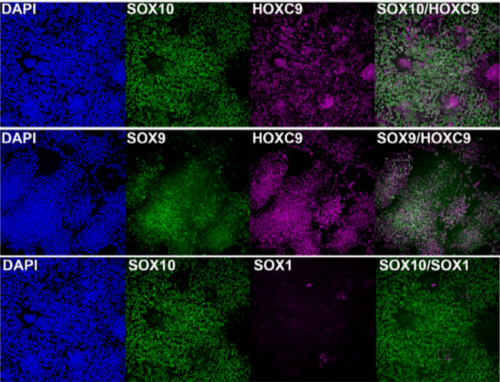
Axial progenitors after 8 days of differentiation, from Frith, et al.’s preprint
Human axial progenitors generate trunk neural crest cells. Thomas J.R. Frith, Ilaria Granata, Erin Stout, Matthew Wind, Oliver Thompson, Katrin Neumann, Dylan Stavish, Paul R Heath, James O.S. Hackland, Konstantinos Anastassiadis, Mina Gouti, James Briscoe, Valerie Wilson, Mario R Guarracino, Peter W Andrews, Anestis Tsakiridis
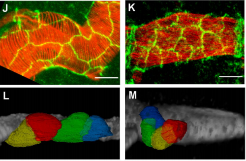
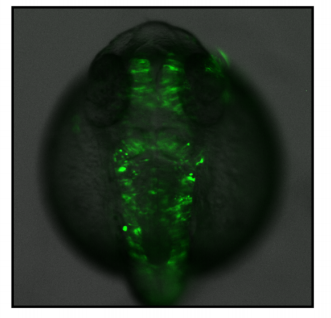
Zebrafish embryogenesis from Hess, et al.’s preprint
A conserved regulatory program drives emergence of the lateral plate mesoderm. Christopher Hess, Karin Dorien Prummel, Susan Nieuwenhuize, Hugo Parker, Katherine W. Rogers, Iryna Kozmikova, Claudia Racioppi, Sibylle Burger, Eline C. Brombacher, Alexa Burger, Anastasia Felker, Elena Chiavacci, Gopi Shah, Jan Huisken, Zbynek Kozmik, Lionel Christiaen, Patrick Mueller, Marianne Bronner, Robb Krumlauf, Christian Mosimann
Hedgehog signaling controls progenitor differentiation timing during heart development. Megan Rowton, Andrew D. Hoffmann, Jeffrey D. Steimle, Xinan Holly Yang, Alexander Guzzetta, Sonja Lazarevic, Chul Kim, Nikita Deng, Emery Lu, Jessica Jacobs-Li, Shuhan Yu, Erika Hanson, Carlos Perez-Cervantes, Sunny Sun-Kin Chan, Kohta Ikegami, Daniel J. Garry, Michael Kyba, Ivan P. Moskowitz
Neutralizing Gatad2a-Chd4-Mbd3 Axis within the NuRD Complex Facilitates Deterministic Induction of Naive Pluripotency. Nofar Mor, Yoach Rais, Shani Peles, Daoud Sheban, Alejandro Aguilera-Castrejon, Asaf Zviran, Dalia Elinger, Sergey Viukov, Shay Geula, Vladislav Krupalnik, Mirie Zerbib, Elad Chomsky, Lior Lasman, Tom Shani, Jonathan Bayerl, Ohad Gafni, Suhair Hanna, Jason Buenrostro, Tzachi Hagai, Hagit Masika, Yehudit Bergman, William J. Greenleaf, Miguel A. Esteban, Yishai Levin, Rada Massarwa, Yifat Merbl, Noa Novershtern, Jacob H. Hanna
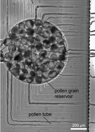
Adaptive Reduction of Male Gamete Number in a Selfing Species. Takashi Tsuchimatsu, Hiroyuki Kakui, Misako Yamazaki, Cindy Marona, Hiroki Tsutsui, Afif Hedhly, Dazhe Meng, Yutaka Sato, Thomas Stadler, Ueli Grossniklaus, Masahiro M. Kanaoka, Michael Lenhard, Magnus Nordborg, Kentaro K. Shimizu
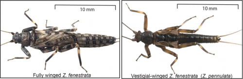
Firefly genomes illuminate parallel origins of bioluminescence in beetles. Timothy R Fallon, Sarah E Lower, Ching-Ho Chang, Manabu Bessho-Uehara, Gavin J Martin, Adam J Bewick, Megan Behringer, Humberto J Debat, Isaac Wong, John C Day, Anton Suvorov, Christian J Silva, Kathrin F Stanger-Hall, David W Hall, Robert J. Schmitz, David R Nelson, Sara Lewis, Shuji Shigenobu, Seth M Bybee, Amanda M Larracuente, Yuichi Oba, Jing-Ke Weng
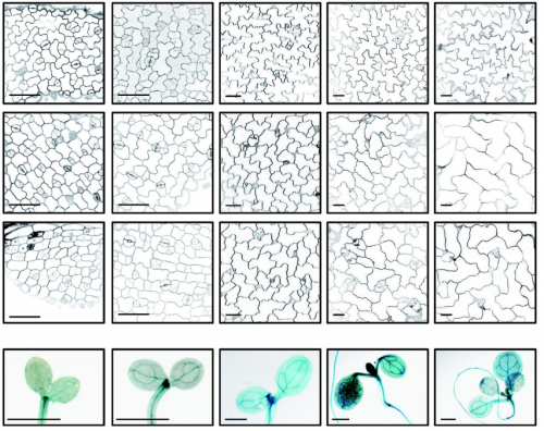
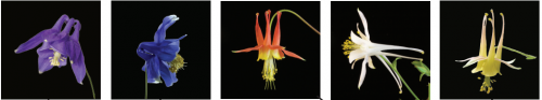
10 Aquilegia species species from Filiaut, et al.’s preprint
The genome of the water strider Gerris buenoi reveals expansions of gene repertoires associated with adaptations to life on the water. David Armisen, Rajendhran Rajakumar, Markus Friedrich, Joshua B Benoit, Hugh M Robertson, Kristen A Panfilio, Seung-Joon Ahn, Monica F Poelchau, Hsu Chao, Huyen Dinh, HarshaVardhan Doddapaneni, Shannon Dugan-Perez, Richard A Gibbs, Daniel ST Hughes, Yi Han, Sandra L Lee, Shwetha C Murali, Donna M Muzny, Jiaxin Qu, Kim C Worley, Monica Munoz-Torres, Ehab Abouheif, Francois Bonneton, Travis Chen, Li-Mei Chiang, Christopher P. Childers, Andrew G Cridge, Antonin JJ Crumiere, Amelie Decaras, Elise M Didion, Elizabeth Duncan, Elena N Elpidina, Marie-Julie Fave, Cedric Finet, Chris GC Jacobs, Alys Jarvela, Emily J Jennings, Jeffery W Jones, Maryna P Lesoway, Mackenzie Lovegrove, Alexander Martynov, Brenda Oppert, Angelica Lilico-Ouachour, Arjuna Rajakumar, Peter N Refki, Andrew J Rosendale, Maria Emilia Santos, William Toubiana, Maurijn van der Zee, Iris M Vargas Jentzsch, Aidamalia Vargas Lowman, Severine Viala, Stephen Richards, Abderrahman Khila
In vivo CRISPR-Cas gene editing with no detectable genome-wide off-target mutations. Pinar Akcakaya, Maggie L. Bobbin, Jimmy A. Guo, Jose Malagon Lopez, M. Kendell Clement, Sara P. Garcia, Mick D. Fellows, Michelle J. Porritt, Mike A. Firth, Alba Carreras, Tania Baccega, Frank Seeliger, Mikael Bjursell, Shengdar Q. Tsai, Nhu T. Nguyen, Roberto Nitsch, Lorenz Mayr, Luca Pinello, Mohammad Bohlooly-Y, Martin J. Aryee, Marcello Maresca, J. Keith Joung
Multiple laboratory mouse reference genomes define strain specific haplotypes and novel functional loci. Jingtao Lilue, Anthony G Doran, Ian T Fiddes, Monica Abrudan, Joel Armstrong, Ruth Bennett, William Chow, Joanna Collins, Anne Czechanski, Petr Danecek, Mark Diekhans, Dirk-Dominic Dolle, Matt Dunn, Richard Durbin, Dent Earl, Anne Ferguson-Smith, Paul Flicek, Jonathan Flint, Adam Frankish, Beiyuan Fu, Mark Gerstein, James Gilbert, Leo Goodstadt, Jennifer Harrow, Kerstin Howe, Mikhail Kolmogorov, Stefanie Koenig, Chris Lelliott, Jane Loveland, Richard Mott, Paul Muir, Fabio Navarro, Duncan Odom, Naomi Park, Sarah Pelan, Son K Phan, Michael Quail, Laura Reinholdt, Lars Romoth, Lesley Shirley, Cristina Sisu, Marcela Sjoberg-Herrera, Mario Stanke, Charles Steward, Mark Thomas, Glen Threadgold, David Thybert, James Torrance, Kim Wong, Jonathan Wood, Fengtang Yang, David J Adams, Benedict Paten, Thomas M Keane
Reproducible big data science: A case study in continuous FAIRness. Ravi K Madduri, Kyle Chard, Mike D’Arcy, Segun C Jung, Alexis Rodriguez, Dinanath Sulakhe, Eric W Deutsch, Cory Funk, Ben Heavner, Matthew Richards, Paul Shannon, Ivo Dinov, Gustavo Glusman, Nathan Price, John D Van Horn, Carl Kesselman, Arthur W Toga, Ian Foster
 (2 votes)
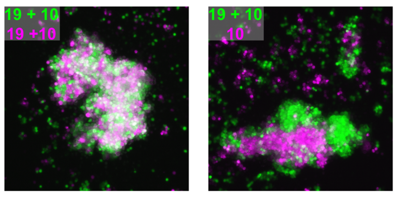
 (3 votes)
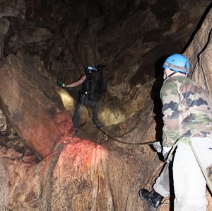
 (8 votes)