PhD student position – Max Planck Institute for Molecular Biomedicine
Posted by bedzhov, on 15 December 2017
Closing Date: 15 March 2021
TheMax-Planck-Institute for Molecular Biomedicine in Muenster, Germany has an opening for a PhD student (position-code 15-2017).
The position
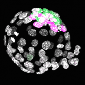
The position is available in the group of Dr. Ivan Bedzhov that is focused on understanding the self-organization of early mammalian embryos and stem cells. The successful candidate will investigate the mechanisms of spatiotemporal organization and cell fate transitions of the early lineages. Technical approaches cover 3D cell and embryo culture techniques, genetic and genomic engineering in stem cells and embryos, cell transplantation studies, embryo micromanipulations, live-imaging, molecular biology and next generation sequencing techniques. Supervision by senior scientists and technical assistance from a laboratory technician will be provided.
Your profile
We are looking for a talented and highly motivated PhD student with strong interest in stem cell biology and mouse embryonic development. Previous research background in epithelial polarity or mouse development and embryonic stem cells is an advantage, but not a requirement. Excellent organizational skills, ability to work effectively as part of a team and to plan and execute experimental research independently are required.
Our offer
The position is available immediately. This is a fully funded 4 years position, part of the Collaborative Research Center (CRC) 1348 “Dynamic Cellular Interfaces”. The income will be according to 65% of level E13 TVöD (the regulations of the contracts for the civil service – Tarifvertrag für den öffentlichen Dienst).
The Max Planck Institute for Molecular Biomedicine offers dynamic, multidisciplinary environment with state-of-the-art transgenic, imaging, robotics, genomics and proteomics equipment and core facilities. The working language in the institute is English, knowledge of the German language is not required. A childcare facility is situated in the guesthouse of the institute next to the main building. The institute is located in Muenster that has been awarded LivCom-Award for ‘The World’s Most Liveable City’ by the UN.
The Max-Planck Society is committed to increasing the number of individuals with disabilities in its workforce and therefore encourages applications from such qualified individuals.
Furthermore, the Max Planck Society seeks to increase the number of women in those areas where they are underrepresented and therefore explicitly encourages women to apply.
Your application
Please send your application (with the position-code 15-2017), letter of motivation, CV, the contact information of 2 referees and (optional) the Masters degree’s thesis to:
Max Planck Institute for Molecular Biomedicine
Roentgenstrasse 20
48149 Muenster
Germany
 (1 votes)
 (No Ratings Yet)