The Wellcome-Warwick Quantitative Biomedicine Programme Wellcome-Warwick Quantitative Biomedicine Programme was established to enhance the world-class interdisciplinary research environment at the University of Warwick by driving further development of our existing centres of excellence, including the Centre for Mechanochemical Cell Biology (www.mechanochemistry.org) and the Zeeman Institute for Systems Biology& Infectious Disease Epidemiology Research.
Warwick Medical School is seeking to appoint three outstanding early career scientists as Assistant Professors in Quantitative Biomedicine, aiming to expand our research on cell dynamics at the molecular, cellular and/or tissue scales. We are eager to recruit candidates using quantitative and/or interdisciplinary approaches and with an exemplary track record in cell biology, developmental biology, structural biology, computational biology or biophysics.
Successful candidates must have a strong track record with first class publications,together with the enthusiasm and expertise to contribute to our innovative undergraduate taught programme in Interdisciplinary Science. Evidence of being able to attract funding and/or fellowships would be a further advantage. You will also contribute to the Public Engagement interface of the QBP.
Successful candidates will receive a start-up package, laboratory space in the brand new extension to our mechanochemical cell biology building, access to state-of-the-art infrastructure, including light and electron microscopy, advanced proteomics, and support from our thriving and dynamic research community.
The posts will be in the Division of Biomedical Sciences, Warwick Medical School and the successful candidates will be expected to play an active role in advancing the mission of the QBP. The posts will be subject to a five year probation period and once successfully completed, promotion to Associate Professor will follow, subject to criteria set out by the University of Warwick being met.
Potential candidates are encouraged to make informal contact with the Directors of the Wellcome Warwick Quantitative Biomedicine Programme, Profs. Mohan Balasubramanian (M.K.Balasubramanian@warwick.ac.uk) and Andrew McAinsh (A.D.McAinsh@wawick.ac.uk).
To be considered, please fill out an online application, including a CV, names of three expert referees who are able to comment on your readiness to embark on an independent career, a one-page cover letter and a two-page research proposal describing an exciting research program in cell dynamics.
A postdoctoral position is available in the laboratory of Dr. Sophie Astrof to study roles of cell-extracellular matrix interactions in cardiovascular development and disease using cell biological approaches and mouse model system. The project will involve investigation of signaling by extracellular matrix in development and differentiation, utilizing state-of-the art imaging and genetic approaches. In our lab, we use genetics, conditional mutagenesis, and transgenic approaches to explore roles of tissue microenvironment during organogenesis and disease. Experience with genetic manipulation, embryology and cell biology is desirable. My laboratory is a part of the Center for Translational Medicine at Jefferson Medical College (http://www.tju.edu/jmc/medicine/translational_medicine/faculty/astrof.cfm?detail=0) located in the heart of Philadelphia. To apply, send a letter of interest, CV and names and contact information of three references to sophie.astrof@gmail.com…
The Woods Hole Embryology Course, which will celebrate its 124th birthday this year, is a continual source of beautiful images (and videos) of development. Since 2011 the Node has run a competition for the community to pick the best images from a given year – the winning pictures become immortalised as Development covers!
Below you will find 4 images from the 2015 course, Round 1. Choose the one you would like to see on the cover of Development by voting on the poll at the end of the post (you can see full size versions by clicking on the images).
The poll is set up to allow only one vote per person, and closes at 12.00 GMT, Friday 14th April. Results will be announced Tuesday, 18th April.
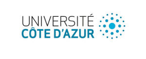
Chicken
Stage 10 chick embryo with noggin coated beads (blue). Imaged with cell phone.
Theodora Koromila
CalTech, USA
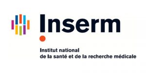
Clathria
10 hours post-dissociation aggregate of cells of the marine sponge, Clathria prolifera. A subset of the cells contains autofluorescent vesicles excited by a 405nm laser (shown in green). The nuclei are labeled with Hoechst staining (shown in purple), and the two signals were separated by spectral imaging and linear unmixing on a Zeiss LSM 780 confocal.
Shun Sogabe
The University of Queensland, Australia
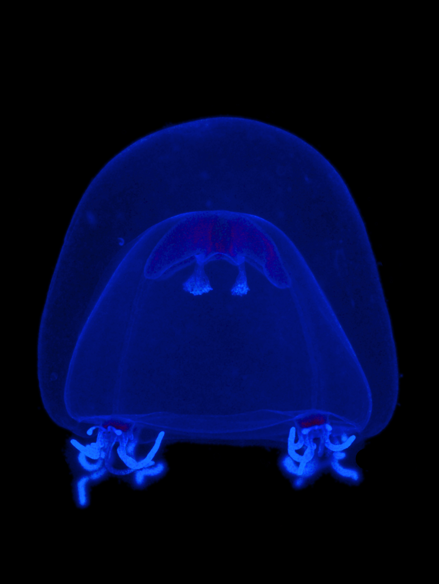
Jellyfish
Juvenile jellyfish Nemopsis bachei collected in a plankton tow. Stained with DAPI (nuclei). Imaged with a Zeiss AxioImager and processed with Photoshop.
Chiara Sinigaglia
Observatoire Océanologique de Villefranche sur Mer/ CNRS, France.
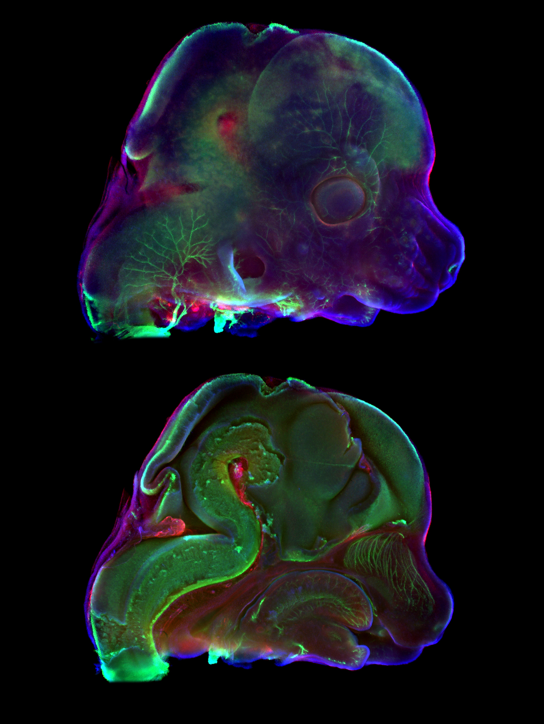
Mice
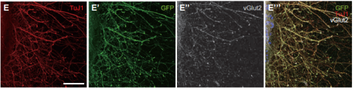
Midsagittal section through E12.5 mouse embryo head showing muscle fibers (MF20, red), nerve fibers (TUJ1, green) and nuclei (DAPI, blue). Head surface on the top, surface of the section plane on the bottom. Captured using Zeiss Axio Zoom V16 stereomicroscope. The final image was composed in GIMP.
This year marks the centenary of D’Arcy Thompson’s On Growth and Form, an attempt to outline the physical and mathematical principles underpinning the generation of biological form. Modern day developmental biologists, bolstered by new technologies, have taken up Thompson’s cause to try to understand the mechanics of development, particularly with regard to morphogenesis. While the generation of forces by the actomyosin cytoskeleton has received a lot of attention, how the material properties of developing tissues influence morphogenesis is less well understood. Today’s paper was recently published in eLife, and investigates the relationship between forces and tissue stiffness in the elongation of the C. elegans embryo. We caught up with lead author Thanh Vuong-Brender and hersupervisor Michel Labouesse of the Institut de Biologie Paris-Seine, to hear the story behind the work.
The Labouesse lab, with Thanh second from left and Michel third from left.
Michel, can you tell us your scientific biography and the questions that your lab is trying to answer?
ML I did my undergrad in Maths/Physics, but chose to do a PhD in Genetics, which appealed to the mathematical neuron I had. I fell in love with C. elegans through a series of seminars by Sydney Brenner – I like the concept of the lineage – and I went to get worm training with Bob Horvitz at MIT. Initially interested by cell fate specification, I rapidly moved to analyse epithelial morphogenesis, and progressively realized that I could thereby feed my second physically-oriented neuron.
Broadly speaking, in the lab we want to understand how mechanical forces impact on cellular processes. Indeed, a cellular phenotype corresponds to its global fate and its 3D organisation; so the challenge is to understand how mechanical forces can modify gene expression programs and/or cell shape determinants, which are defined by junction and cytoskeleton organisation, plus trafficking. Addressing these issues is not as trivial as it may look. At the molecular level, one can think that identifying the structure that senses the force, and the signal transduction that can next modify cell fate or shape should be enough. But it is unlikely to be so. First, the effect or forces is rarely an isolated one-time event, but is often repeated such that the question of timing/periodicity becomes more central than for a chemical signal. Second, understanding how a force can have an effect generally requires thinking, not (only) in biochemical terms, but chiefly in physical terms. Entities to be considered should be energy, entropy, elasticity.
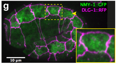
Myosin distribution in embryonic epidermal cells, from Figure 1, Vuong-Brender et al., 2017, eLife
And Thanh, how did you come to work in Michel’s lab?
TV-B I was trained as a physicist. My PhD was about the use of automated imaging and fluorescent markers for diagnosis of cervical cancer. After that, I went to work for a small company that developed automated imaging systems. I realised that I was more interested in academic research to explore and understand natural phenomena. During my PhD, I learnt some biology but not as much as I wanted. So I looked for a postdoc during which I could learn more about biology and came across Michel lab’s papers on mechanical problems of C. elegans embryonic elongation. I found the subject attractive, maybe because it presented to me a mechanical problem to solve. I did not understand all the biology but I thought it was really interesting to learn and to work on it. I sent my postdoc application to Michel and was really lucky to be accepted.
In an interview with Current Biology in 2005, you said you were excited by the challenge of understanding the mechanics of development. I wonder what you think of the progress the field has made in the 12 years since then?
ML The field has evolved tremendously, in part due to progress in imaging and data processing, and in part because a new generation of scientists with strong background in physics has entered the area. In my field, papers that brought key paradigmatic changes, which in retrospect seem quite common sense, include the demonstration that a morphogenetic event requires small increments that progressively modify the cell (Martin et al, Nature; Rauzi et al, Nature; Solon et al, Cell). Another one is that two apposed tissues with distinct mechanical properties will twist (Savin et al, Nature).
You write that the material properties of developing tissues have received less attention than the forces that act on them. Why do you think this is?
ML There are two probable reasons: in vertebrate embryos, the field has more frequently focused often on global movements (although the issue of stiffness has been pointed out more than 30 years ago in Xenopus), whereas in fly embryos, the field has focused on processes dependent on cells having a homogenous behaviour.
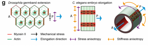
TV-B Deformation depends both on forces and material properties, so in theory, one can have as many regulatory pathways of shape formation through the regulation of material properties as through forces. The role of forces has been intensively investigated through the studies of non-muscle myosin and its regulatory pathways. Many studies have suggested the role of anisotropic material properties, like the elongation of Drosophila developing eggs or trachea. The role of material properties has received more attention in plants, but studies and mechanical measurements of material properties in animal morphogenesis are scarce. Our aim was to bring attention to this important parameter.
Overview of C. elegans embryo elongation, from Figure 1, Vuong-Brender et al., 2017, eLife
What makes the C. elegans embryo a good model for developmental mechanics?
ML The worm is very simple and many of its past successes have been linked to cell biology (PAR proteins, netrin, centrosome assembly, apoptosis, EGF/Ras signalling, to name a few). Its only drawback is that the embryo is quite small, very fragile and not easily amenable to approaches available in other species.
TV-BC. elegans embryonic elongation is very simple and different from other animal models, since it does not involve cell division or cell migration, but is mostly driven by cell shape changes. The number of cells is limited so that one can investigate at the cell resolution and the whole embryo. Other advantages are the easy genetics and worm cultivation.
Can you give us the key results of the paper in a paragraph?
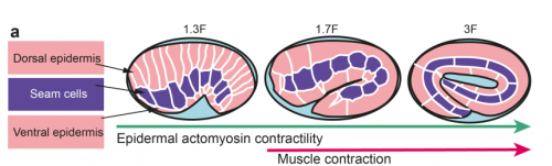
TV-B In the last step of embryonic development, the embryos of C. elegans transform from a ball of cells into the characteristic cylindrical shape of a worm. This process is powered by the association of the molecular motor myosin II, and the actin cytoskeleton in the embryonic epidermis. The epidermis is made up of six strips of cells running along the head-to-tail axis. Myosin II is mostly active in two strips of cells on the two sides of the embryo (lateral cells), but has low activity in the upper and lower strips of cells (dorsally and ventrally to the lateral cells). It is unclear how this distribution of myosin causes embryos to elongate only along the head-to-tail axis. Using laser nano-ablation, we have probed the forces exerted and the material properties in the embryonic epidermal cells. The results show that myosin’s activity in the lateral cells induced constriction around the embryo, sort of similar to the effect of a boa constrictor tightening around its prey. At the same time, the actin filaments in the dorsal and ventral strips form rigid bundles oriented along the circumference. They form a “belt” preventing the constriction from causing the cells at the dorsal and ventral strips embryo to expand. Finally, the only direction the embryo can elongate is along the head-to-tail axis.
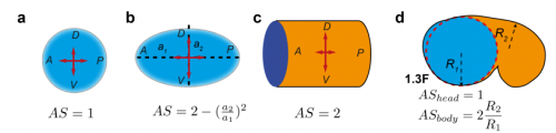
Modelling anisotropic stresses in different shapes, from Figure 4, Vuong-Brender et al., 2017, eLife
How important was the modelling to complement and reinforce your experimental data?
ML It was critical in two ways. First to guide the interpretation of experimental data; second as suggested in the answer to the first question, because understanding a process involving a mechanical force should include a physical dimension whenever possible.
TV-B The mathematical modelling helps us to understand mechanistically what we observed. It can also predict the behaviours of the system in another situation or the role of another component in the process. So it can be your initial hypothesis or feedback, which combines with the experiment data to make the work evolve.
Are the anisotropies of stress and stiffness established by particular signals in the cell, and could this be this linked to cell fate choice in the embryo?
ML These are key mostly unsolved questions for the future.
TV-B I think that the anisotropies of stress and stiffness are linked to the cell specific myosin/actin regulator distribution, which have been shown to be different between lateral and dorso-ventral epidermal cells. It is likely to be linked to the lateral/dorso-ventral cell fate, but remains to be proved.
Comparing models of fly and worm elongation, from Vuong-Brender et al., 2017, eLife
When doing the research, was there a particularly exciting result or eureka moment that has stayed with you?
TV-B I did not really have a “eureka”, but for me, everyday is like an adventure. There were problems (technical or theoretical) to solve and challenges to overcome because we always tried new things. For more routine stuff, I tried to make it better or quicker. There are small victories like “yes ! it (laser ablation) works”, “I got my CRISPR knock-in strain”, “ the experiment data matches (the theoretical one))”… which led me through the days and frustrations sometime. Well, like every scientist, I hope to have a “bigger eureka” in the future.
And what about the flipside: any moments of frustration or despair?
TV-B Yes, I was trying desperately to make optogenetically-controlled gene expression work in worms. I have tried 3 different systems, none of these worked. Finally I tried a different optogenetic method controlling protein aggregation, but did not have the time to finish it. One consolation is that my preliminary results were used by someone else. I told myself that the failures are part of the quest and became now more resilient to them.
Where next for you following this work?
TV-B I am trying to shift (again) to another domain of research. During my postdoc, my interest for microbiology, symbiosis between microbes and the origin of eukaryotes has grown. So I decided to go to study the microbial diversity. It will be totally different but surely I will learn a lot of things.
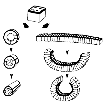
30 year old foresight about embryo mechanics – from Jim Priess and David Hirsch’s Developmental Biology paper.
And what is the next step for the Labouesse lab?
ML I am quite proud of the work we did with Thanh and our two other co-authors, as I think it accounts in physical terms for a big part of the early phase of elongation. Incidentally, I want to pay tribute to a visionary landmark paper of our field written in 1986 by Jim Priess, a PhD student at the time, who had foreseen almost everything in early worm elongation. Now, I want to account for the second phase of C. elegans elongation that requires the mechanical input of muscle contractions (which Jim had not touched upon), and want to reach a similar level of understanding. We are nearing this phase.
STEM Graduates is a graduate recruitment agency and jobs board. We offer permanent salaried roles to students and graduates from Science, Technology, Engineering and Mathematics disciplines. We believe these candidates have a unique set of career needs that can only be met by a specialist within this field. We launched STEM Women in 2016 to provide a specific place for female careers advice, profiles of women in STEM and a dedicated job board.
We are always looking to expand what we can offer STEM students to make them more employable in their highly competitive markets. That is why we are excited to launch our recent partnership with the Science Council. This partnership will include a variety of activities and exciting content.
The Science Council will host a careers advice blog topic each month written by STEM Graduates (most recent blog post here) and we will be focusing on educating our candidates on the benefits on offer from joining the Science Council. We will also be listing the numerous specific professional bodies under the science umbrella including the Royal Society of Biology, the Institute of Science and Technology and the Institution of Environmental Sciences. We will achieve this through our social media channels, a new dedicated section on our website and with weekly articles.
We are proud to endorse the ‘working towards registered scientist’ (Registered Scientist (RSci)) initiative that has been launched by the Science Council. This will focus on the conduct, competence and professional development of early years’ scientists. For many graduates this initiative will be the first step towards becoming a chartered scientist.
The Science Council is a membership organisation for professional bodies and learned societies across the disciplines of science. They are in a unique position, bringing together a range of disciplines and sectors to reflect the multi-disciplinary practice of science in today’s society.
We are looking to expand this section further with other societies, associations and communities so please let us know if you have any ideas.
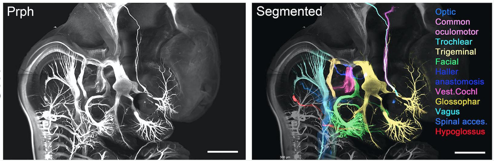
View of the head of a transparent 7 week embryo labelled with Peripherin. Cranial nerves have been pseudocoloured. From Fig1. Belle et al., Cell, 2017
Throughout history, the desire of scientists to understand physiology and disease by thoroughly studying anatomical features, has always faced an intractable limitation: they cannot simply see through the tissue! Dissection has therefore been the modus operandi of anatomists: from Galen’s pioneering studies, to modern day biologists who routinely section tissues to label structures for microscopic analysis.
Whilst these methods have informed a wealth of knowledge linking anatomical form to function, they are inherently flawed due to a 3-Dimensional appreciation of structures being lost. This has been especially problematic for the study of human development, where structures are continually evolving, and therefore a precise visualisation has been impossible to achieve through traditional methods and anatomical atlases. Coupled with the difficulties in tissue access, our understanding of human development has progressed perhaps the slowest of any biological process since the 1930’s; whilst in some cases observations of lower vertebrates have subsequently been erroneously applied to humans.
“Birth defects of structural or functional origin currently affect more than 3% of births”
Without a good understanding of physiological development, we lack the fundamental knowledge required for clinicians and researchers to tackle a healthcare issue that inflicts a severe healthcare and emotional burden. Recent advances in non-invasive, in vivo imaging techniques, have shown great promise in detecting congenital abnormalities as well as providing information on gross topological features of fetal development; however, they lack sufficient resolution in order to inform developmental biologists of currently unknown features of organogenesis. This has recently been most strikingly highlighted by the surge in Zika virus infections and reports of its detrimental effects on cephalic development.
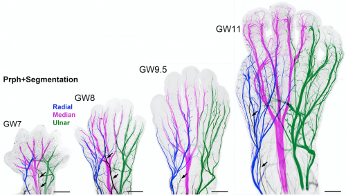
The developing innervation of the human hand from 7-11 weeks gestation. From Fig2. Belle et al., Cell, 2017
In order to begin rapidly addressing the gaping holes in our knowledge, we have begun to form a cutting edge research consortium of developmental biologists, to build a detailed human cell atlas. Here we have expanded the use of clearing techniques to human tissues and immunolabeled with over 40 antibodies, intact human embryos during the first trimester of gestation (from 6 to 14 gestational weeks).
In recent years, several techniques have been developed for tissue clearing, in which whole organs are rendered macromolecule permeable and optically transparent. Among tissue-clearing techniques, the process called 3D imaging of solvent-cleared organs, or 3DISCO has been proved to be a simple, robust and inexpensive method for 3D analysis of immunolabeled transparent organs in embryonic and postnatal mice.
Currently organised along seven organ systems, the project aims to expand and evolve as more data is added. Current data in the atlas examines molecular organogenesis based on over 40 samples comprising 1,500,000 optical sections – making it the most complete 3D analysis of early human development currently available.
Constructed by imaging intact tissues and whole embryos, the atlas will be an invaluable tool for researchers with the ability to explore cell distributions, count proliferating cells in each organ etc. whilst also being useful for didactic purposes, with the ability to 3D print models to inform health science teaching programs.
This has long been the dream of developmental biologists, which has finally been realised by the use of organic solvent based clearing techniques (3DISCO/iDISCO) combined with light sheet imaging. Together, these powerful approaches allow inexpensive labelling of any cell population of all developing organ systems during development and imaging at cellular resolution whilst fully maintaining structural relationships. To further highlight the robustness and power of this technique to analyse human development, it should be noted that these data were generated in just over one year by a small team of researchers.
And this is just the beginning, as the online resources are open access – available for researchers all over the world to analyse and contribute to, along with new data we will acquire. The goal of the project is to make a continuously updated 3D molecular reference atlas of human cells during development, paramount to a better understanding of human development during health and disease.
The online video series of immunolabeled tissues along with the original data sets is available at https://transparent-human-embryo.com/ . The 3D database was developed with Keen eye technologies with support from the “fondation voir et entrendre”
University of Oregon biologists have figured out how zebrafish perfectly regenerate amputated fins with a precisely organized skeleton.
Adult zebrafish fins, including their complex skeleton, regenerate exactly to their original form within two weeks after an amputation. The process, they found, is driven by clusters of specialized skin cells that migrate over reforming bones, known as rays, and escort bone cells into the right positions to form individual bones of a branched skeleton.
These skin cells produce a protein called Sonic hedgehog, which interacts with bone-building cells called osteoblasts to promote bone patterning during fin regeneration.
“The orderly reconstruction of zebrafish fins is amazing to see,” said Kryn Stankunas, a professor in the Department of Biology and member of the Institute of Molecular Biology. “Zebrafish fins, which are akin to our limbs, regenerate perfectly. The zebrafish bony rays re-branch just like the original structure. This would be like losing your arm and watching it progressively regenerate complete with a hand and fingers — all the bones restored in their original configuration.”
The findings will not lead to humans re-growing lost limbs, Stankunas said, but such advances in understanding the fundamental processes of regeneration in related vertebrate organisms will inform innovative and targeted therapeutic strategies to improve the repair of broken bones.
“The mechanism — how the skin and bone cells dynamically move and interact using the signaling pathway — is elegant and unexpected, broadening the project’s impact on regenerative medicine,” Stankunas said.
Hedgehog signaling, he added, is also linked to several human cancers.
“The zebrafish fin provides a tractable and simple model to decipher mechanisms of regenerative skeletal patterning,” the researchers wrote in their paper in the March 28 issue of the journal Development, a publication of the non-profit Company of Biologists in the United Kingdom.
Benjamin E. Armstrong, who earned a doctorate in biochemistry in 2016, was the study’s lead author. Scott Stewart, a research professor in the Institute of Molecular Biology, co-directed the project.
Green fluorescent proteins show where bone-building is occurring in the regeneration of a zebrafish caudal fin that had been amputated. Complete repairs begin at the tail’s base and gradually proceed to the tip, a process that is completed within two weeks. Courtesy of Kryn Stankunas
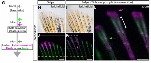
The research team used genetically modified zebrafish that produces a fluorescent protein that helps identify the subset of skin and bone cells that respond to Hedgehog signals. The fluorescent marker appears green under the microscope until illuminated with ultraviolet light to photo-convert the green protein to red.
This photo-conversion method revealed that repairing skin cells collectively move towards the tip of the regenerating fin. At particular times, Sonic hedgehog is induced in skin cell clusters that then split into two pools. Simultaneously, the skin cells activate a Hedgehog response in adjacent osteoblasts. That drives them to associate with the skin cells and co-migrate into split groups. The now separated bone cells continue to regenerate replacement bone, but now forming two rays instead of one – a branched skeleton.
“We could see that the bone cells responding to the skin-produced Sonic hedgehog become physically attached to the migrating skin cells”
“We could see that the bone cells responding to the skin-produced Sonic hedgehog become physically attached to the migrating skin cells,” Stewart said. “The pathway is quickly turned off but the now split groups of bone cells will then form two separated mature bony rays connected at a branch point.”
To define the functions of the Hedgehog signaling pathway, the researchers used a new chemical inhibitor, BMS-833923, to turn off Hedgehog signaling in their experimental fish. With Hedgehog blocked, the skin and bone cells failed to interact, and the fin regenerated with stick-like rays rather than forming a branched skeleton.
The inhibitor used in the study is in clinical trials against some forms of human cancers, but it had not been used in zebrafish. The Hedgehog pathway is most associated with basal cell carcinoma and medulloblastoma, Stankunas said.
“The Hedgehog response is absolutely required for branching and not essential for any other aspect of regeneration,” Stankunas said. “Instructions that drive the branching come from the skin cells moving into two groups and likewise dividing the osteoblasts. This is new information. It is the traffic pattern generated by the signaling that regenerates the fin. It is skin and bone working together.”
###
Astra Henner, lab manager and research assistant, was the fourth co-author of the paper.
The National Institutes of Health funded the project through a training grant to Armstrong and research grants to Stankunas and Stewart.
Source: Kryn Stankunas, associate professor of biology, 541-346-7416, kryn@uoregon.edu
Note: The UO is equipped with an on-campus television studio with a point-of-origin Vyvx connection, which provides broadcast-quality video to networks worldwide via fiber optic network. There also is video access to satellite uplink and audio access to an ISDN codec for broadcast-quality radio interviews.
We are seeking an enthusiastic, highly motivated and productive postdoctoral research associate to join a BBSRC-funded research project investigating the formation of primordial follicles in the developing mammalian ovary, led by Dr Andrew Childs, Lecturer in the Department of Comparative Biomedical Sciences (Royal Veterinary College, University of London, UK).
The successful candidate will use a combination of in vitro and in vivo techniques to investigate how growth factor signalling converges with the transcriptional machinery in fetal ovarian somatic cells to regulate the timing and extent of follicle assembly in the mammalian ovary. The post-holder will be an active member of the research team, contributing to experimental design, data collection, analysis and dissemination, and student supervision.
The successful candidate should have a PhD in reproductive or developmental biology (or a closely related discipline). They will be proficient in a range of research techniques, including molecular biology, immunohistochemistry, and cell or organ culture. Excellent organisational and communication skills are essential, as is the ability to work independently and as part of a team. Experience of Chromatin Immunoprecipitation (ChIP), large scale transcriptomic analyses, and/or working with animal models would be advantageous.
Applicants should be available to start no later than 1st May 2017.
Prospective applicants are encouraged to contact Dr Andrew Childs (Lecturer, Comparative Biomedical Sciences) at achilds@rvc.ac.uk.
Here are the highlights from the new issue of Development:
Making thalamic neurons in vitro
In recent years, methods to derive multiple differentiated neuronal types from embryonic stem cells (ESCs) in vitro have been reported. Three-dimensional (3D) culture methods not only support differentiation but also recapitulate spatial aspects of brain development.
Such studies were pioneered by the late Yoshiki Sasai, and on p. 1211, his colleagues Atsushi Shiraishi and Keiko Muguruma adapt the original 3D culture conditions – which supported rostral neural fate – to derive thalamic neurons from mouse ESCs for the first time. They find that addition of insulin and FGF pathway inhibitors can specify caudal forebrain identity, and that subsequent treatment with BMP7 can promote thalamic fate. Within the neuroepithelial-sphere structure that forms in these cultures, there is significant spatial organisation: early progenitors are found by the apical cavity, while more mature cell types are located towards the outside, and the spheres display rostral-caudal regionalisation. The derived neurons can extend axons that – both in culture and in transplantation experiments in vivo – show projection patterns consistent with thalamic identity. Not only does this work allow the generation of thalamic neurons in vitro, but it also provides insights into the signalling mechanisms regulating thalamus development in vivo.
Bone regeneration in the fish fin
Zebrafish can fully regenerate their fins, a process that involves the reconstitution and patterning of multiple tissue types. New bone is regenerated via the de-differentiation, proliferation and re-differentiation of osteoblasts, which occur in a spatially organised manner to recapitulate the original fin shape and skeleton. How skeletal patterning – including outgrowth and bifurcation of new rays – is controlled in this context is incompletely understood, though is thought to involve Hedgehog signalling.
Kryn Stankunas, Scott Stewart and colleagues (p. 1165) now define distinct roles for the two Hedgehog ligands expressed in the regenerating fin: shha and ihha. shha is expressed in epidermal cells immediately adjacent to osteoblasts at the site of ray branching, and is required for branching. Intriguingly, it appears to act at short range, through direct contact with osteoblast progenitors via cellular protrusions, to promote splitting of the ray through cell movements. ihha, on the other hand, is expressed in the osteoblasts, where it promotes differentiation via a non-canonical signalling route. These data clarify the role of Hedgehog signalling in ray regeneration and shed light onto the mechanisms underlying skeletal patterning in regenerative contexts.
Turning off translation in germ cells
Stem cell quiescence has been reported in many systems, and typically involves the slowing or stalling of the cell cycle and low transcriptional activity. Primordial germ cells (PGCs) of sea urchin are known to enter a quiescent state prior to gastrulation, before re-activating later in development.
Now (p. 1201), Gary Wessel and co-workers show that this quiescence also involves a significant reduction in translational activity. Two potential mechanisms are uncovered. Firstly, Nanos2, which is expressed specifically in PGCs, binds to and downregulates the critical translation factor eIF1A. Secondly, mitochondrial number and activity is low in PGCs, which might induce a switch to glycolytic metabolism and hence an acidification of the cytoplasm. Increasing cellular pH promotes translational activity specifically in PGCs. This work raises many intriguing questions. For example, how is translational activity re-activated at later stages? How are the metabolic changes in PGCs orchestrated? How general might this be in quiescent stem cell populations? Thus, the identification of this previously unrecognised phenomenon of transient translational quiescence in sea urchin PGCs opens up many new avenues for investigation.
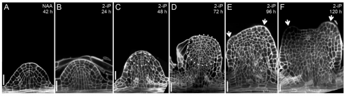
Timing is key to turn root into shoot
Plant cells show remarkable plasticity. For example, lateral roots can be converted into shoots by supplementing the culture medium with cytokinin, which induces shoot fate. When properly controlled, this conversion does not involve callus formation, and so allows a detailed analysis of the processes directing the switch of organ identity.
Using this system, Philippe Rech and colleagues (p. 1187) find that competence for root-to-shoot conversion is restricted to a narrow time window of lateral root development, coinciding with the stage at which the stem cell niche is formed in the new root. Furthermore, conversion can be reversed during this period – auxin treatment can switch the tissue back to a root – confirming that organ identity is not immediately fixed. Importantly, the authors provide evidence that root-to-shoot conversion does not occur via dedifferentiation, but rather via a direct transdifferentiation process. Transcriptome and methylome profiling provide insights into the gene expression and epigenetic changes occurring during conversion. This atypical mode of organogenesis may lead to novel methods for the vegetative multiplication of valuable plant cultivars.
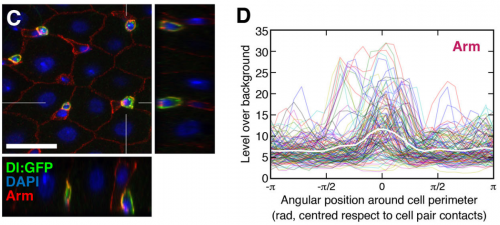
How cell-cell contact defines fate
In many systems, stem cell fate is regulated by Notch signalling. One such example is the Drosophila midgut, where intestinal stem cells (ISCs) can divide either asymmetrically, generating a Notch-positive enteroblast (EB) and a Notch-negative ISC, or symmetrically, either forming two EBs or two ISCs. But what determines the outcome of ISC division, and how does Notch signalling influence this?
Joaquín de Navascués, Jordi Garcia-Ojalvo and co-workers address this question on p. 1177 using a combination of experimental and modelling approaches. Their key insight is that contact area between the two daughter cells correlates with cell fate: where the contact area is small, both cells tend to remain ISCs, where it is larger, one or both cells differentiate. Since Delta-Notch signalling involves direct contact between the two cells, the area of contact can influence the effective signalling threshold. Both the computational and experimental analyses support the idea that the pattern of cell fates following ISC division can be at least partly explained by variability in cell contact area, and hence in the levels of Notch-Delta signalling between the two daughter cells. That such a model might also apply in other stem cell systems is an intriguing possibility.
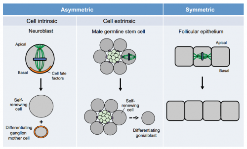
Spindle orientation: a question of complex positioning
Dan Bergstralh and colleagues discuss key features of the spindle-orientating complex and reviews how this complex is regulated and localized to allow correct mitotic spindle orientation.
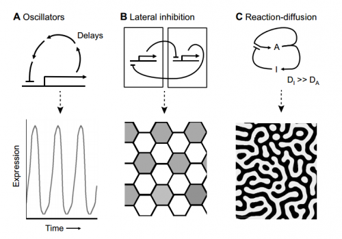
Using synthetic biology to explore principles of development
Jamie Davies explores how synthetic biology-based approaches have been used to explore the principles underlying patterning, differentiation and morphogenesis during development.
Featured movie
In our featured movie, Naoto Ueno, Makoto Suzuki and colleagues show how two patterns of calcium fluctuation in the Xenopus neural plate control epithelial folding, with extracellular ATP and N-cadherin also participating in calcium-induced apical constriction.
Two years postdoctoral position at INSERM U1065 (C3M)-team 3, Nice, France
on the study of cell death of motor neurons. Starting ASAP.
A two-year postdoctoral position starting ASAP, funded by the French National Research Agency is available in the ‘Metabolic control of cell death’ team (INSERM U1065), located at the Archet Hospital in Nice, south of France.
Title: How mitochondrial dysfunction leads to motor neuron disease?
Recently, in close collaboration with Pr. Paquis-Flucklinger, we showed that mitochondrial dysfunction can have a causative effect in motor neuron degeneration. We reported a large family with a mitochondrial myopathy associated with motor neuron disease and cognitive decline looking like frontotemporal dementia (FTD). We identified a missense mutation (p.Ser59Leu) in the HCHD10 gene coding for a mitochondrial protein whose function was unknown (Genin EC et al. EMBO Mol Med 2015 Dec 14:58-72).
We and others reported CHCHD10 mutations in patients with dementia-amyotrophic lateral sclerosis (FTDALS) and familial or sporadic pure ALS.
Project: Amyotrophic lateral sclerosis is a devastating disease affecting upper and lower motor neurons leading to progressive failure of the neuromuscular system and death from respiratory failure. Among all factors involved in ALS pathogenesis, mitochondrial dysfunction has always been recognized as a candidate major player. However, whether mitochondria have a causative role in ALS has been always debated. Our results open a new field to explore the pathogenesis of motor neuron disease by showing that mitochondrial dysfunction may be at the origin of some of these phenotypes.
Our goals are:
(i) to better characterize the role of the CHCHD10 protein on cell death and to compare the effects of
different CHCHD10 mutations leading to different clinical phenotypes,
(ii) to understand how CHCHD10 mutations lead to motor neuron cell death by generating specific human cellular (IPS) and characterizing in vivo models,
Candidate profile:
The candidate should hold a PhD in physiology, pharmacology or related disciplines and have previous expertise in cell culture / characterization of primary neuronal cells.
Practice or knowledge of in vivo animal experimentation techniques as well as in cellular and molecular biology techniques would be appreciated.
How to apply?
Candidates should send a curriculum vitae with publication list, a short summary of research achievements, and the names and email addresses of at least two references to ricci@unice.fr
Dr. J-E Ricci
INSERM U1065, C3M
Directeur de l'équipe- 3
Batiment Universitaire Archimed
151 Route de Ginestière
BP 2 3194
06204 NICE Cedex 3
Tel 33+ (0)4 89 06 43 04
Fax 33+(0)4 89 06 42 21
Email: ricci@unice.fr
 (No Ratings Yet)
 (8 votes)
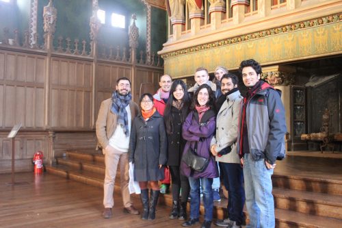

 (3 votes)
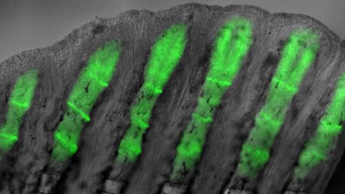
 Now (p.