Our latest monthly trawl for developmental biology (and other cool) preprints. See June’s post for background, and let us know if we missed anything
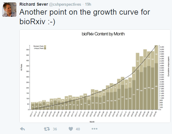
It was another bumper month for preprints, as bioRxiv’s Richard Sever celebrated:
This month we found three pieces on the development of the spinal cord, three investigations into gene expression networks in the early fly embryo, some in vivo cell biology featuring centrosomes and kinesins, as well as a lot of evolutionary content and some very cool sounding tools, perhaps the most striking of which is a hand-powered centrifuge modelled on a whirligig! bioRxiv provided the bulk of the preprints, with one coming from PeerJ.
Happy preprinting…
Developmental Biology
Unraveling the differential dynamics of developmental fate in central and peripheral nervous systems. Dola Sengupta, Sandip Kar
PAPC couples the Segmentation Clock to somite morphogenesis by regulating N-cadherin dependent adhesion. Jerome Chal, Charlene Guillot, Olivier Pourquie
Eph/ephrin signaling controls progenitor identities in the ventral spinal cord. Julien Laussu, Christophe Audouard, Anthony Kischel, Poincyane Assis-Nascimento, Nathalie Escalas, Daniel Liebl, Cathy Soula, Alice Davy
Evo-engineering and the Cellular and Molecular Origins of the Vertebrate Spinal Cord. Benjamin Steventon, Alfonso Martinez Arias
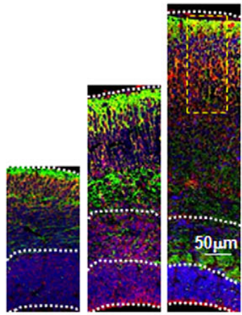
Accelerated cell divisions drive the outgrowth of the regenerating spinal cord in axolotls. Fabian Rost, Aida Rodrigo Albors, Vladimir Mazurov, Lutz Brusch, Andreas Deutsch, Elly M Tanaka, Osvaldo Chara
Transcriptomic Signatures for Ovulation in Vertebrates. Dongteng Liu, Michael Brewer, Shixi Chen, Wanshu Hong, Yong Zhu
A conceptual view at microtubule plus end dynamics in neuronal axons. André Voelzmann, Ines Hahn, Simon Pearce, Natalia P Sánchez-Soriano, Andreas Prokop
Activated Ras expression in eye discs with altered levels of hsrω lncRNA causes JNK-induced Dilp8 secretion and reduces post-pupal ecdysone leading to early pupal death in Drosophila. Mukulika Ray, Subhash C. Lakhotia
Dynamic Maternal Gradients Control Timing and Shift-Rates for Gap Gene Expression. Berta Verd, Anton Crombach, Johannes Jaeger
A damped oscillator imposes temporal order on posterior gap gene expression in Drosophila. Berta Verd, Erik Clark, Anton Crombach, Johannes Jaeger
Odd-paired controls frequency doubling in Drosophila segmentation by altering the pair-rule gene regulatory network. Erik Clark, Michael Akam
Different combinations of ErbB receptor dimers generate opposing signals that regulate cell proliferation in cardiac valve development. Ryo Iwamoto, Naoki Mine, Hiroto Mizushima, Eisuke Mekada
An RNA-binding tropomyosin recruits kinesin-1 dynamically to oskar mRNPs. Imre Gaspar, Vasily Sysoev, Artem Komissarov,Anne Ephrussi
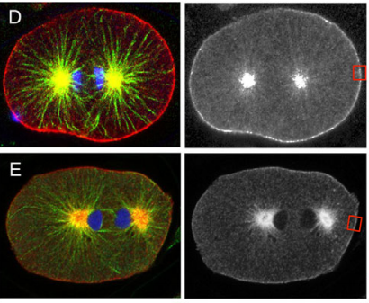
Polo kinase phosphorylation determines C. elegans centrosome size and density by biasing SPD-5 toward an assembly-competent conformation. Oliver Wueseke, David Zwicker, Anne Schwager, Yao Liang Wong, Karen Oegema, Frank Julicher, Anthony A Hyman, Jeffrey Woodruff
Divergence of gene regulatory network linkages during specification of ectoderm and mesoderm in early development of sea urchins. Eric M Erkenbrack, Eric H Davidson
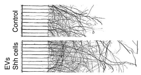
Hypergravity hinders axonal development of motor neurons in Caenorhabditis elegans. Saraswathi Subbammal Kalichamy, Tong Young Lee, Kyoung-hye Yoon, Jin Il Lee
Evaluating the Stability and Flexibility of DNA Methylation Patterns from Stem to Differentiated Cells. Minseung Choi, Diane P Genereux, Jamie Goodson, Haneen Al-Azzawi, Shannon Q. Allain, Stan Palasek, Carol B. Ware, Chris Cavanaugh, Daniel G. Miller, Winslow C. Johnson, Kevin D. Sinclair, Reinhard Stoger, Charles D. Laird
Stem Cell Plasticity and Niche Dynamics in Cancer Progression. Noemi Picco, Robert Gatenby, Alexander Anderson
Dynamics of lineage commitment revealed by single-cell transcriptomics of differentiating embryonic stem cells. Stefan Semrau, Johanna Goldmann, Magali Soumillon, Tarjei S. Mikkelsen, Rudolf Jaenisch, Alexander van Oudenaarden
Strigolactone regulates shoot development through a core signalling pathway. Tom Bennett, Yueyang Liang, Madeleine Seale, Sally P Ward, Dorte Mueller, Ottoline Leyser
Modifications to a LATE MERISTEM IDENTITY-1 gene are responsible for the major leaf shapes of Upland cotton (Gossypium hirsutum L.). Ryan J Andres, Viktoriya Coneva, Margaret Frank, John R Tuttle, Sang-Won Han, Luis F Samayoa, Baljinder Kaur, Linglong Zhu, Hui Fang, Daryl T Bowman, Marcela Rojas-Pierce, Candace H Haigler, Don C Jones, James B Holland, Daniel H Chitwood, Vasu Kuraparth
Evolution etc
Promoter architecture and sex-specific gene expression in Daphnia pulex. R. Taylor Raborn, Ken Spitze, Volker P Brendel, Michael Lynch
Increased taxon sampling reveals thousands of hidden orthologs in flatworms. Jose M Martin-Duran, Joseph F Ryan, Bruno Cossermelli Vellutini, Kevin Pang, Andreas Hejnol
Clustered brachiopod Hox genes are not expressed collinearly and are associated with lophotrochozoan novelties. Sabrina M. Schiemann, Jose M. M. Martin-Duran, Aina Borve, Bruno C. Vellutini, Yale J. Passamaneck, Andreas Hejnol
Expression of segment polarity genes in brachiopods supports a non-segmental ancestral role of engrailed for bilaterians. Bruno Cossermelli Vellutini, Andreas Hejnol
Conserved traits of spiralian development in the bryozoan Membranipora membranacea. Bruno Cossermelli Vellutini, Jose M Martin-Duran, Andreas Hejnol
Comparative transcriptomics reveals candidate genes involved in the adaptation to non-marine habitats in panpulmonate mollusks. Pedro Eduardo Romero, Barbara Feldmeyer, Markus Pfenninger
Evolution of simple multicellularity increases environmental complexity. Maria Rebolleda-Gomez, William C. Ratcliff, Fankhauser Jonathon, Michael Travisano
Tools and resources
Assembly of Radically Recoded E. coli Genome Segments. Julie E. Norville, Cameron L. Gardner, Eduardo Aponte, Conor K. Camplisson, Alexandra Gonzales, David K. Barclay, Katerina A. Turner, Victoria Longe, Maria Mincheva, Jun Teramoto, Kento Tominaga, Ryota Sugimoto, James E. DiCarlo, Marc Guell, Eriona Hysolli, John Aach, Christopher J. Gregg, Barry L. Wanner, George M. Church
CIDR: Ultrafast and accurate clustering through imputation for single-cell RNA-Seq data. Peijie Lin, Michael Troup, Joshua W. K. Ho
Highly parallel direct RNA sequencing on an array of nanopores. Daniel R Garalde, Elizabeth A Snell, Daniel Jachimowicz, Andrew J Heron, Mark Bruce, Joseph Lloyd, Anthony Warland, Nadia Pantic, Tigist Admassu, Jonah Ciccone, Sabrina Serra, Jemma Keenan, Samuel Martin, Luke McNeill, Jayne Wallace, Lakmal Jayasinghe, Chris Wright, Javier Blasco, Botond Sipos, Stephen Young, Sissel Juul, James Clarke, Daniel J Turner
Paperfuge: An ultra-low cost, hand-powered centrifuge inspired by the mechanics of a whirligig toy. M. Saad Bhamla, Brandon Benson, Chew Chai, Georgios Katsikis, Aanchal Johri, Manu Prakash
Genetically targeted 3D visualisation of Drosophila neurons under Electron Microscopy and X-Ray Microscopy using miniSOG. Julian Ng, Alyssa Browning, Lorenz Lechner, Masako Terada, Gillian Howard, Gregory Jefferis
Direct determination of diploid genome sequences. Neil I Weisenfeld, Vijay Kumar, Preyas Shah, Deanna Church, David B Jaffe
Biotinylation by antibody recognition – A novel method for proximity labeling. Daniel Zvi Bar, Kathleen Atkatsh, Urraca Tavarez, Michael R Erdos, Yosef Gruenbaum, Francis S. Collins
The impact of chromatin dynamics on Cas9-mediated genome editing in human cells. Rene M. Daer, Josh P. Cutts, David A. Brafman, Karmella Haynes
DNAmod: the DNA modification database. Ankur Jai Sood, Coby Viner, Michael M. Hoffman
The ExAC Browser: Displaying reference data information from over 60,000 exomes. Konrad J Karczewski, Ben Weisburd, Brett Thomas, Douglas M Ruderfer, David Kavanagh, Tymor Hamamsy, Monkol Lek, Kaitlin E Samocha, Beryl B Cummings, Daniel Birnbaum, The Exome Aggregation Consortium, Mark J Daly, Daniel G MacArthur
Bright photoactivatable fluorophores for single-molecule imaging. Luke D Lavis, Jonathan B Grimm, Brian P English, Anand K Muthusamy, Brian P Mehl, Peng Dong, Timothy A Brown, Zhe Liu, Timothée Lionnet
Thousands of primer-free, high-quality, full-length SSU rRNA sequences from all domains of life. Soeren M Karst, Morten S Dueholm, Simon J McIlroy, Rasmus H Kirkegaard, Per H Nielsen, Mads Albertsen
Is there anything we missed for August? Anything in the list that catches your eye? Let us know in the comments section
 (2 votes)
 Loading...
 (No Ratings Yet)
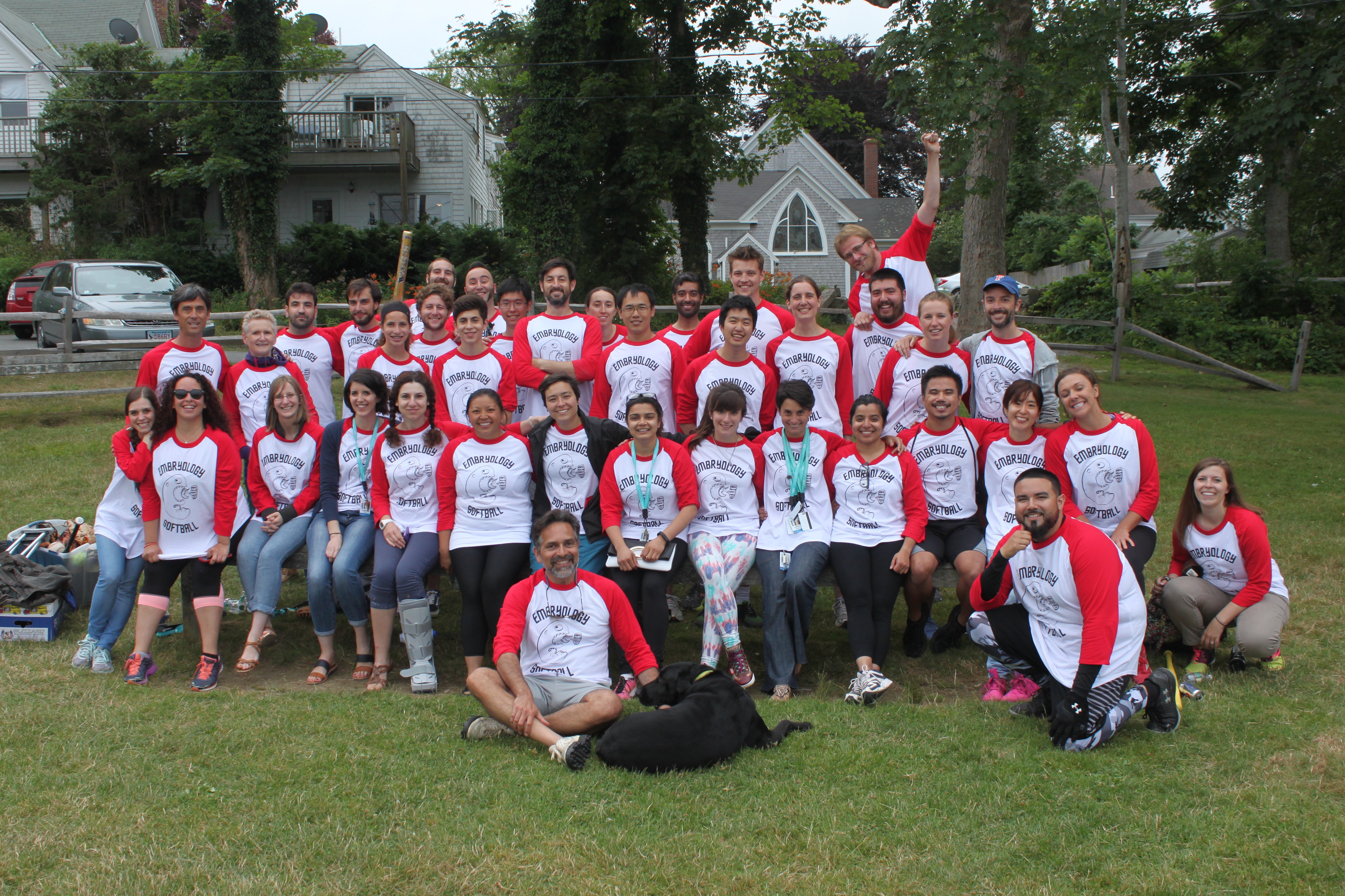
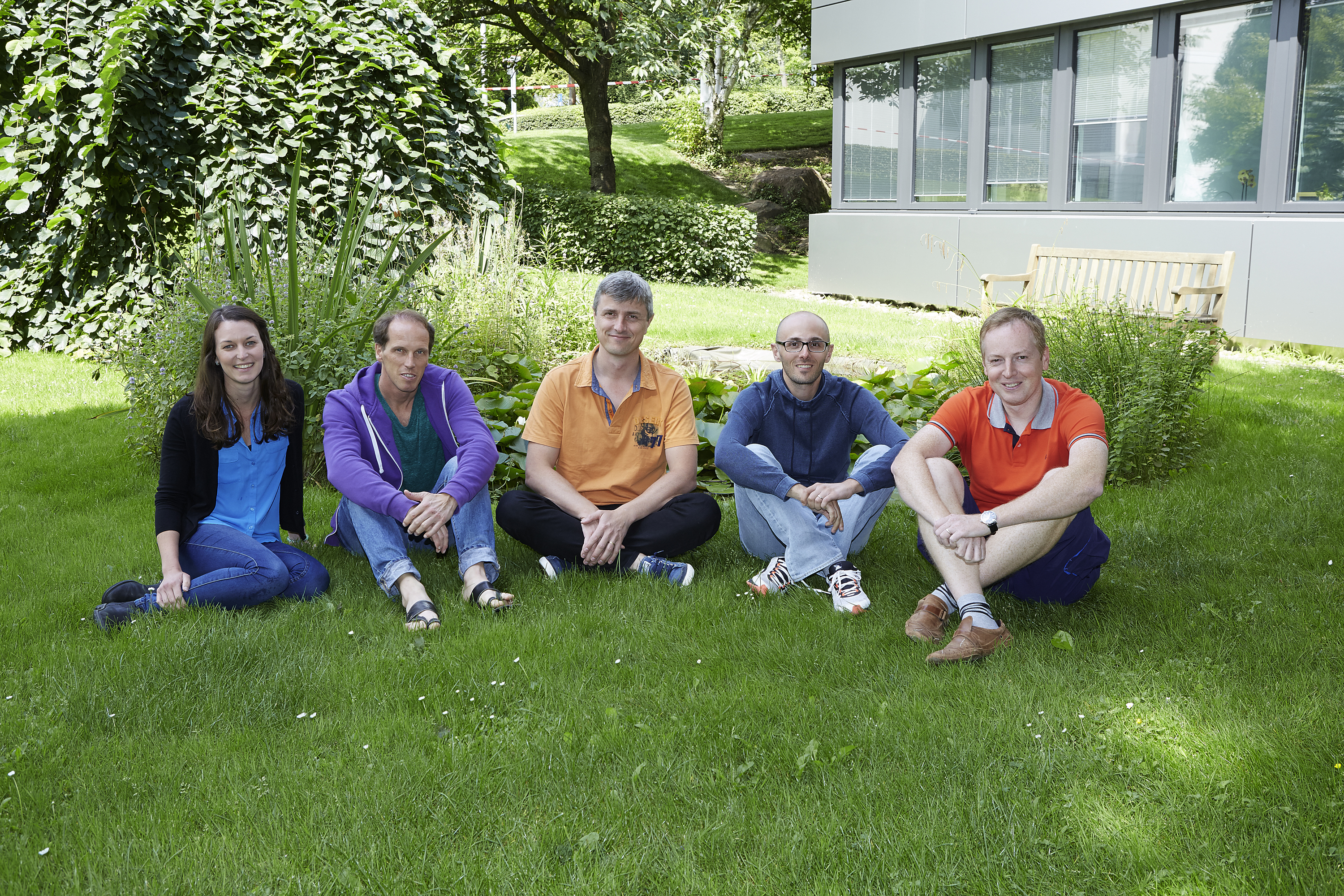
 The Tanentzapf lab