Professors Graham Williams and Duncan Bassett have 3 full-time posts (5 years each) available. One technical post in skeletal biology is funded by the Horizon 2020 grant: Resetting the THYRoid axis for prevention of AGE-related diseases and co-morbidities. One post-doc and one technician post in molecular and cellular biology are funded by the Wellcome Trust: Cellular thyroid hormone availability: regulation of development and tissue repair, and pathogenesis of degenerative disease.
Please see our website for this exciting opportunity to join our cutting-edge research program based at Imperial College London.
The aim of this initiative is to capitalise on MRC’s (and others’) investments in the International Mouse Phenotyping Consortium (IMPC) (https://www.har.mrc.ac.uk/research/large-scale-functional-genomics/international-mouse-phenotyping-consortium-impc) and make best use of the resources, data and tools available. The expectation is to provide small, flexible awards to allow the take-up and use of the existing phenotyped and banked knockout lines and resources to support UK research. It is expected that this will catalyse more ambitious studies in the future.
Applications are invited from UK-based research groups to receive, via the MRC’s Mary Lyon Centre (http://www.har.mrc.ac.uk/about/mary-lyon-centre), knockout mice produced by the IMPC, and undertake early biological investigations that build on existing research expertise and underpin longer-term research goals.
This Call will run in May 2016 and 2017, with 10-25 awards made in September 2016 and 2017. Awards will be of 1-2 years’ duration and 20-40k in value.
The deadline for this year’s applications is 4pm on 8th July 2016.
Last year, the BSDB carried out a student and postdoc survey (see here), and the results clearly highlighted the need for more dedicated program items for young researchers on BSDB conferences. In response, the BSDB student rep Alex Ashcroft, the BSCB postdoc rep Alexis Barr, and the BSDB postdoc rep Michelle Ware took initiative and organised a serious of highly successful events at the 2016 BSCB/BSDB joint Spring Meeting. Read below what happened.
Career workshop
From the BSDB student/postdoc survey results last year, it was evident that most people wanted to find out more about ‘alternative’ careers other than those on the traditional route of academia. With more PhDs being awarded and few top level jobs there is a need to provide more information as to what else can you do with your PhD. For this reason, we chose to focus this year’s careers session on alternative careers to academia. The highly attended session took the format of roundtable discussions and covered a plethora of topics including but not limited to, consulting, publishing, academic fellowships and engaging with the media. We would like to thank all the table leaders who provided stimulating discussions. This event wouldn’t have been possible without you!
Obtaining a lectureship/fellowship
Claudia Barros (Peninsula School of Medicine, Plymouth University)
Paul Conduit (Henry Dale Fellow, Department of Zoology, University of Cambridge)
James Wakefield (University of Exeter)
Careers outside of academia
Katherine Brown (Editor, Development)
Anne Wiblin (Research Collaborations Manager, Abcam)
From the feedback, we realise how valuable it is for young scientists to talk to other scientists who have trained as cell or developmental biologists and go on to have successful ‘alternative’ careers. For future workshops, we intend to build on this theme and invite an even more diverse selection of speakers.
Some selected comments from the participants:
‘Open and honest speakers, Enough time to discuss and explore career prerequisites, responsibilities and prospects’
‘Great organisation and table choices, Thank you! I feel quite optimistic now!’
‘Table leaders were friendly, easy to talk to and answered all questions’
‘Learning about career paths, Variety of careers amongst speakers’
Click here for a more in depth summary of the workshop.
Science Breakfasts
This was the first year that we ran science breakfasts, whose goal was to facilitate informal discussions between junior researchers and scientists at the top of their field. A small number of students and postdocs got to participate in this event, discussing everything from research, careers and life in general with Abigail Tucker, Ottoline Leyser, Jordan Raff, Lidia Vasilieva and Thomas Surrey – who we are really grateful for giving up their time.
Student social
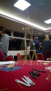
This year the reps decided it would be fun to do something more interactive for the student/postdoc social. We randomly attached a name to the back of every guest, where each name was one-half of a famous pair, such as Romeo and Juliet. Each guest could not see their own name but could see the names of other guests. Using yes-no questions, each guest had to work out who they were and then find the other half of their pair. Each pair were given the task to build the tallest possible tower using marshmallows and spaghetti. The pairs were then grouped into teams of ten which competed against each other in a round of science pictionary.
Thank-you to all the sponsors who donated gifts. For the pair that found their pair first won a bound notebook each from Horizon and sweets. Class Learning provided a voucher for two books up to £100, which was awarded to the winners of spaghetti towers (Erik Clark, Gautham Dey and their winning tower pictured here).
Chocolates and sweets from the BSDB committee were awarded to the winning teams after the Pictionary round. Molecular biology of the Cell (Garland Science) was given to the winner of the best image (Rachna Narayanan with a drawing of WALL-E).
Student Symposium
This year the graduate student symposium was moved to the middle of the meeting, resulting in excellent attendance. This was a truly excellent event – the speakers covered a diverse range of topics in an engaging manner. Some talks even got mentioned in the twitterverse!
The format was also altered so that there were six fifteen minute presentations and six five minute presentations. All the speakers did an excellent job – particular mention must be made for everyone who managed to describe their complex research in just five minutes!
We hope to see many of you next year. If you have any comments or ideas please get in touch with Alex (students@bsdb.org), Alexis (Alexis.Barr@icr.ac.uk) or Michelle (postdocs@bsdb.org); especially if you have ideas for games to play in the student social, know someone who would be a great table leader for the careers workshop or if there is someone with whom you would really like to have breakfast.
For the first time, a reference mouse embryo atlas has been created using HREM image data. For other embryo imaging methods such as micro-CT, a reference embryo atlas has previously been shown to be the basis of automated phenotyping (Wong et al, 2014). This new work (a collaboration between the DMDD programme and the Mouse Imaging Centre in Toronto) is a proof of principle for automated phenotyping of HREM data, which can now be tested in more detail.
COMPARING LIKE WITH LIKE
In order to phenotype an embryo, we need to compare it to another embryo that we consider to be normal. With histology this can be very difficult, as it’s virtually impossible to take exactly the same slice through two embryos.
So this leads to the question ‘have I really observed a phenotype, or am I simply looking at a different part of the embryo?’
The ideal scenario for a phenotyper is to have exactly equivalent slices of anatomy to compare. They can then do a direct visual comparison, or an algorithm can be used to do a statistical comparison, therefore automating the process to some degree.
A REFERENCE EMBRYO ATLAS
Using a technique previously employed with micro-CT data (Wong et al, 2012), the team have created a reference embryo atlas from HREM data. The atlas can be used to find these equivalent sections of anatomy.
21 wild-type embryo image stacks were merged together to give an unbiassed ‘average embryo’. Every possible pair of embryos was compared in terms of displacement. The embryos were then combined together so that the displacement for all embryos to join the atlas was a minimum. This technique avoids giving undue weighting to any one embryo in the average.
As HREM data is so large, the resolution was scaled back first to make the computation feasible.
For each individual embryo, a different deformation field is needed to bring the image into alignment with the average. The inverse of this field can then be used to propagate back from any plane in the reference embryo to a section of the original input embryo.
This means it’s possible to find equivalent embryo sections to compare, ensuring that any differences observed are much more likely to be genuine phenotypes.
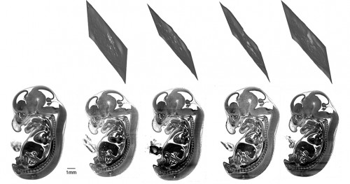
Bottom left, the merged reference embryo where the plane of interest is defined. Bottom right, the equivalent data from four individual mouse embryos. Top, the crumpled sections show the effective ‘cut’ plane through the original HREM data that is homologous to the plane of interest in the reference embryo. (Image recreated with the permission of Elsevier).
It’s important to note that these sections are never planes but crinkly slices through the embryo. This means that the sections couldn’t have been identified by manually scrolling through the image stacks.
AUTOMATED PHENOTYPING
Automated selection of equivalent embryo sections is the starting point for automated phenotyping. Given the time-consuming nature of phenotyping embryos, and the large task of phenotyping many mouse lines, automated phenotyping is an attractive prospect to many. It can act as a guide – a primary screen to highlight areas of interest to manual phenotypers.
The next step, then, is to compare other HREM embryo data with this reference atlas.
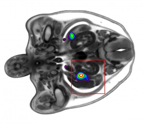
The results of automated primary screens are typically presented as a heatmap (Wong et al, 2014), highlighting areas of interest. The authors showed a proof of principle for an automated primary screen of HREM data by separating the embryos into male and female groups. After selecting equivalent sections through the gonads to compare between the two groups, they were able to automatically distinguish statistically significant differences in this region.
The coloured regions show statistically significant differences between males and females in the region of the gonads.
This is an exciting result for the future of automated phenotyping of HREM mouse embryo data, which will be tested much more extensively over the coming months.
This post originally appeared on the DMDD blog, Annotations (blog.dmdd.org.uk).
DMDD is a systematic study of embryonic-lethal mouse gene knockouts, and provides a database of high-resolution image and phenotype data – a valuable resource for both developmental biologists and clinicians. Visit the website to find out more: dmdd.org.uk
1Mouse Imaging Centre (MICe), Hospital for Sick Children, University of Toronto, Toronto, Canada 2 The Francis Crick Institute Mill Hill Laboratory, London, UK
1Mouse Imaging Centre (MICe), Hospital for Sick Children, University of Toronto, Toronto, Canada 2 The Lunenfeld-Tanenbaum Research Institute, Mount Sinai Hospital, Toronto, Canada
1Department of Medical Biophysics, University of Toronto, Toronto, Canada 2Mouse Imaging Centre (MICe), Hospital for Sick Children, University of Toronto, Toronto, Canada 3 Sunnybrook Hospital, Toronto, Canada 4 Regeneron Pharmaceutecals, Tarrytown, USA
A 15 months postdoctoral position is available at the Observatoire Océanologique de Villefranche-sur-Mer -LBDV (UPMC – Sorbonne Universités). We are seeking a highly motivated and passionate researcher to join a 5-years international project, DEVODIVERSITY, funded by the French Agence Nationale de la Recherché (ANR) and the São Paulo Research Foundation (FAPESP).
By using molecular and cell biology, NGS transcriptomic, genomic, and ecological approaches, a multidisciplinary consortium led by two teams, the Tiozzo Lab at the Villefranche-sur-Mer Developmental Biology Laboratory in France (CNRS-UPMC), and the Brown Lab at the Istituto de Biociências in Brazil (USP), will study the evolution of regeneration, asexual reproduction, and clonality in several species of ascidians (Urochordata), and examine how ecological factors affect distribution ranges, evolution of life cycles and developmental strategies. DEVODIVERSITY has the following main aims:
To resolve the phylogenetic relationships and evolutionary transitions between strict sexual reproduction to budding and high regenerative abilities among Styelidae (Ascidiacea).
To provide a coupled morphological and ecological understanding of asexual propagation (budding). We will generate detailed anatomical and developmental descriptions of budding processes, and explore if and which environmental conditions are associated with the use of particular budding modes.
To compare gene pathways involved in stem cell functions or trans/de-differentiation of budding and regenerative processes by the generation and in silico analyses of transcriptomic data.
To launch a comparative genomic approach study in order to better understand the evolution of a major life history transition in marine chordates, in particular the evolutionary transition from sexual to asexual propagation.
The candidate will be mainly based at the LBDV (Villefranche sur Mer, France) and may have the opportunity to do laboratory and fieldwork in both countries.
It is ESSENTIAL:
A doctoral degree in animal biology, evolutionary biology or related disciplines.
Demonstrated experience in molecular biology, organismal biology and at least bases of bioinformatics and biostatistics.
Enthusiasm and genuine commitment to research work.
Demonstrated ability to undertake independent research, flexibility and problem solving skills.
Excellent communication skills in English spoken/written.
Ability to collaboratively work on a team.
Expertise in phylogenomics and transcriptomics is highly desirable but not mandatory. The LBDV has an international working environment, speaking French is not required but it would be a plus.
The applicant should contact directly Dr. Stefano Tiozzo (tiozzo@obs-vlfr.fr) providing a letter of interest, a CV and the contact of three potential referees. Please, add “DD_postdoc” on the subject of your email. Applications review will continue until the position is filled. Starting data could be any time from September 2016.
The Carthew and Mani Labs are looking to hire a joint postdoc with familiarity in live-imaging and quantitative analysis. Building on the expertise of both labs, the goal of the project is to construct a live imaging platform for eye development with a view to explore the coupled dynamics of cell-cell signaling, mechanical forces, and gene regulation using state-of-the-art techniques. The postdoc will benefit from the highly collaborative and interdisciplinary environment of the two labs that already share many common interests and students. Both investigators are committed to supporting the development of soft skills, in addition to the experimental and quantitative skills that are required for success in science. Competitive salary, computing resources, and travel money will be provided.
Interested? Contact us at madhav.mani@northwestern.edu
I am hugely grateful to the Journal of Cell Science and the Company of Biologists for awarding me a traveling fellowship to visit the laboratory of Dr Guillermo Velasco at Complutense University in Madrid, Spain. During this visit I gained a greater understating of the skills and techniques used to manipulate autophagy regulatory pathways within melanoma stem-like cells, as well as knowledge of autophagy regulation within cancer.
Cutaneous malignant melanoma is the most aggressive form of skin cancer and despite the recent introduction of targeted therapeutics to treat the disease, relapse is common and there remains no effective cure for metastatic melanoma. Therefore, an improved understanding of the underlying biological mechanisms mediating development and progression of melanoma is desperately required. A potential therapeutic approach may be to target melanoma stem-like cells that sustain tumour growth and contribute to drug resistance. Stem-like progenitor cells from other cancers have been shown to use autophagy, the principle lysosomal mediated mechanism for the breakdown and recycling of damaged proteins, for survival. Therefore, as autophagy is also known to be important for melanoma growth, the current project aims to test the hypothesis that melanoma subpopulations that display a stem-like phenotype use autophagy for tumour maintenance, invasion and survival.
The trip to Dr Velasco’s lab allowed me to learn specific practical techniques for the identification and characterisation of cancer stem cells, as well as gain knowledge of autophagy regulation from experts in the field. During my time in Madrid I met a number of very interesting people who I hope will remain close collaborators in the future. Also, the experience of living and working in such a cosmopolitan city as Madrid is one that I can whole heartedly recommend to anyone!! It certainly is a fantastic city and I can’t wait until the next time I can visit.
2nd Postdoc Position in Evo-Devo and Macroevolution
Hiring Organization:
Howard University
Date Posted:
2016-05-19
Position Description:
Postdoctoral Position in Evo-Devo and Macroevolution
Apart from the position posted a few days ago, we are happy to announce that a 2nd postdoctoral researcher is sought to join the Rui Diogo lab (www.ruidiogolab.com), at the Howard University College of Medicine, Department of Anatomy (Washington DC). For more details on the research done at the lab, and the papers/chapters/books published in recent years, see also:
https://www.researchgate.net/profile/Rui_Diogo
Diogo’s books: http://www.amazon.com/Rui-Diogo/e/B001JS2K96
The main difference between this position and the one previously announced is that the position will not be related to anatomical networks nor building websites. We are therefore interested in a candidate that will have the ability to:
1) contribute to uncover evolutionary and developmental mechanisms underlying both hard tissue (cartilages and bones) and soft tissue (mainly muscles) formation and patterning during ontogeny of a wide range of vertebrate taxa. Some of the issues and broader questions in which we are particularly interested include: the parallelism between ontogeny and phylogeny, the remarkable similarity between the hard and particularly the soft tissues of the upper and lower limbs of tetrapods, the importance of evolutionary reversions/neotenic events, the study of birth defects in human and non-human primates and their implications for medicine and for the understanding of evolutionary biology, and the regeneration of hard and soft tissues in key vertebrate taxa. For more information about these subjects and about other issues being studied in the lab, please see www.ruidiogolab.com.
2) help write review papers and books on the broader macroevolutionary topics covered above and other topics, and, by doing this you can relatively rapidly get a substantial experience in publishing in top journals and monographs.
In summary, this will be a position in a very productive lab, in which there is a relatively high independence, where the drive of the postdoc is essential to make her/him more productive, taking advantage of the broader scope and numerous collaborations of the lab. Contrary to the previously announced position, you will thus more involved in lab evo-devo research and in writing and publishing.
There are funds available for one year, starting fall (September) 2016, with the hypothesis of extending the position for two years, in case of excellent results and fit within the lab, i.e. the second contract depending on the productivity, interest and dedication of the candidate. There are possibilities to continue being part of the lab after the two-year period of the post-doc position. The post-doc will also have the opportunity to learn, and potentially to then become an instructor/faculty of human gross anatomy; this will further allow him/her to also postulate for faculty positions in medical schools in the DC area (including Howard University) as well as in other regions.
Interested candidates should send a CV including research interests, a list of publications and the names and contact information for three references to Rui Diogo, at rui.diogo@howard.edu. Please write “post-doc in Diogo’s lab” followed by your last name in the email subject.
Howard University is a historical University situated in the center of Washington DC, which is a beautiful, green and enjoyable city with numerous cultural and outdoor activities. The Department of Anatomy provides a prosperous, resourceful and multidisciplinary environment for biomedical research, includes faculty with a broad experience in developmental biology, paleontology, neurobiology, comparative anatomy and medicine. We have strong ties with surrounding institutions, particularly with George Washington University, and the candidate will probably have the opportunity to do part of his/her research at those institutions and thus to expand his/her knowledge and academic connections.
Qualifications/Experience:
The successful candidate will have a PhD degree with a broad experience in developmental biology (e.g., doing/using developmental techniques such as antibody staining, in situs, and cell tracing, among others) and/or evolutionary biology, and hopefully both, backed by publications in peer-reviewed journals, and ideally also some experience in comparative anatomy. He/She will have the skills and motivation to pursue a career in research, be interested in studying and comparing a wide range of taxa and various model organisms and in discussing various evolutionary and developmental issues, and be able to fulfill at least some, and ideally all four, items listed above.
How on earth do we turn into a living, breathing, singing, speaking being, from just the fusion of two cells? Development is fascinating and mysterious. Stem cells are one component of how this intricate process unfolds, allowing the embryo to not have every type of cell from the start.
Imagine if the only way our bodies could form a brain, a liver, a heart, is if the embryo started out with little batches of each type of cell. Instead, stem cells make it possible to start with just one ‘flavour’ of cell, and turn that into the many cell types needed to make a fully formed human.
In order to take full advantage of stem cells, we need to understand development. What signals are these stem cells sending and receiving to tell them to become brain or liver or heart?
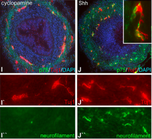
In a collaboration between Harvard University and Semmelweis University in Budapest, Nandor Nagy and his team used bird embryos to study the development of a special part of nervous system devoted to controlling our gut. The literal manifestation of the term, ‘gut instinct,’ the so-called enteric nervous system is an extensive network of ‘gut-brain’ cells wrapped around our gut.
To help answer the huge question of how this ‘second brain’ of ours develops, Nagy and his colleagues honed in on the role of one specific molecule, called sonic hedgehog (Shh), which is crucial to development in humans and many other creatures. Specifically, they wanted to look at how Shh influences the cells around the gut to turn into ‘gut-brain’ cells or not.
To visualize the effect of Shh, they took thin slices of the bird embryo’s gut and added fluorescence molecules that showed whether the ‘gut-brain’ cells were in early or late stages of development. The later stage they were in, the more Shh would be shown to push their development forward.
The red fluorescence shows early stage, whereas the green shows late. Adding Shh, in the right column, shows more green fluorescence, but the same amount of red, indicating that Shh pushes the cells into later stages.
In the span of just a few hundred words we have narrowed in on the effect of one molecule on one type of cell, and how we can visualize this. This story attests to the power of images in answering big questions, by zooming way in to the level of microscopic cells.
Nagy, Nandor, et al. “Sonic hedgehog controls enteric nervous system development by patterning the extracellular matrix.” Development 143.2 (2016): 264-275.
Sonnenburg, Justin and Erica Sonnenburg. “Gut feelings- the “Second Brain” in our gastrointestinal systems.” Scientific American (2015).
POSTDOCTORAL POSITIONS are available to study different aspects of lymphatic biology in health and disease, including the cellular and molecular mechanisms controlling the development of the of the lymphatic vasculature in mice, endothelial cell plasticity and reprogramming, as well as different aspects of lymphatic function in metabolic diseases. Highly motivated individuals who recently obtained a PhD. or MD degree and have a strong background in molecular, vascular and developmental biology are encouraged to apply. Interested individuals should send their curriculum vitae, a brief description of their research interests, and the names of three references to:
 (No Ratings Yet)
 (1 votes)
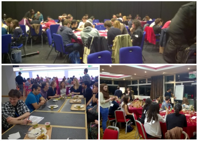
 This year the reps decided it would be fun to do something more interactive for the student/postdoc social. We randomly attached a name to the back of every guest, where each name was one-half of a famous pair, such as Romeo and Juliet. Each guest could not see their own name but could see the names of other guests. Using yes-no questions, each guest had to work out who they were and then find the other half of their pair. Each pair were given the task to build the tallest possible tower using marshmallows and spaghetti. The pairs were then grouped into teams of ten which competed against each other in a round of science pictionary.