Here is August’s round-up of some of the interesting content that we spotted around the internet!
News & Research
– Ian Sussex, one of the founding fathers of plant developmental biology, recently passed away. Developmental Biology published an obituary.
– Nature jobs published several articles providing advice to new PIs: how to manage your lab budget, to serve (or not to serve) in committees, and the testimony of Samantha Morris, who has just successfully applied for her first PI position. Steve Royle also shared his tips.
– ‘Pay very careful attention to unexpected results’- Shinya Yamanka shared his thoughts in Science Careers.
– The organoids boom and what they are teaching us about human development- in Nature.
– The first iPSC clinical has been halted due to genomic issues. Paul Knoepfler covered this story here and here.
– Do biases keep LGBT scientists from coming out? Nature investigates.
– Thinking about leaving the lab? The first step is to know yourself.
– And the LMB sponsored an online exhibition on the life and work of Fred Sanger.
Weird & Wonderful
– London is currently hosting a series of DNA sculptures, to raise funds for CRUK. Do the DNA trail to see them all!
– The winning image of this year’s Wellcome Trust Image Awards was a pregnant pony uterus. This video shows some of the other similar historical specimens housed at the Royal Veterinary College in London.
– Which 10 scientists would you want by your side in a bar brawl? Here’s a potential list!
– And here is the human body reimagined as a metro map!
The protein E-Cadherin (E-Cad) is a kind of adhesive that keeps cells tightly bound together, thus favouring the organisation of tissues and organs. Scientists at the Institute for Research in Biomedicine (IRB Barcelona) now reveal a new function for E-Cad, one that contrasts with its accepted role in impeding cell movement. The researchers have published an article in Nature Communications in which they report that this protein is crucial for the coordinated movement of diverse cell types.
This new function of E-Cad may explain why tumours that express intermediary levels of this protein have a poorer prognosis.
Coordinated cell movement
E-Cad facilitates the movement of heterogeneous groups of cells—understanding as heterogeneity cells that exert a range of activities because they have different genes activated: some may divide many times, others trigger certain hormones, while others interact with the membrane, etc…
Thanks to E-Cad, this group of diverse cells moves in a coordinated manner to its destination. Once there, the cells distribute where they are needed; their moderate levels of E-Cad keep them bound but not immobile during this migration. IRB Barcelona researchers Kyra Campbell and Jordi Casanova have addressed this phenomenon in the development of the embryonic digestive system of the fly Drosophila melanogaster, a model that allows them to study cell migration in a growing organism.
“Cell migration is a common and necessary process for an embryo and also for the correct function of the adult organism. What has been most surprising is the observation that E-Cad is a key component in cell movement, when its role was previously assumed to be that of keeping cells static,” explains Jordi Casanova, head of the Development and Morphogenesis in Drosophila Lab at IRB Barcelona and CSIC research professor.
Cell migration is also of great biomedical relevance, and research into this phenomenon sheds light on how, for example, cancer metastasis and other processes such as wound healing and inflammation arise.
Cell migration and metastasis
According to Casanova, intermediary levels of E-Cad are often associated with aggressive tumours, precisely those which are capable of metastasising. He also reveals that, “the more we learn about metastases, the more evidence emerges that they are formed by groups of cells and not by individual ones”.
E-Cad would facilitate highly diverse heterogeneous groups of cells to migrate together from the original tumour. “A cell that migrates alone is much easier to eliminate that a group of cells with different functions,” explains the researcher.
“Our results in Drosophila are clinically relevant because they offer an explanation of the role that may be played by E-Cad in tumours with metastasis,” says Casanova.
The study has involved the participation of researchers from Advanced Digital Microscopy Core Facility at IRB Barcelona, headed by Julien Colombelli. Sébastien Tosi, Senior Research Officer with this facility, set up the programmes to monitor cells in vivo during their migration.
A postdoctoral position is available to study the mechanisms regulating skeletal development and their relationship to skeletal birth defects using mouse models, molecular biology, and next-generation sequencing. Highly motivated candidates who recently obtained a Ph.D. and have a strong background in developmental biology are encouraged to apply. Preference will be given to those with model organism experience who have a first-author publication as a result of their graduate work. Interested candidates should send their CV, a brief description of their research, and names of three references to:
Funds, especially for travel have been tightly squeezed in recent times and here at Abcam we know of how difficult it is for scientists, especially those starting out in their careers to attend conferences. Abcam is committed to bringing scientists together to share information, strengthen relationship and forge new collaborations.
With this ethos, we’re very pleased to offer travel grants to people wishing to attend the Epigenetics, Obesity & Metabolism conference on 11-14 October 2015 at the Wellcome Genome Campus Conference Centre in Hinxton, Cambridge, UK.
This meeting will brings together key scientists from around the world to discuss:
– Epigenetics, metabolism and the links with circadian rhythms
– Early developmental origins of metabolic disorders
– Epigenetic linkages between nutrition and longevity
– Transgenerational consequences of metabolic dysfunction
– Mechanisms of signalling to the epigenome and inheritance
Each grant is valued up to £300 (GBP) each and are available to students and young postdocs (who have received their PhD within the last 4 years). Grants can be used to help cover travel and accommodation expenses associated with the conference.
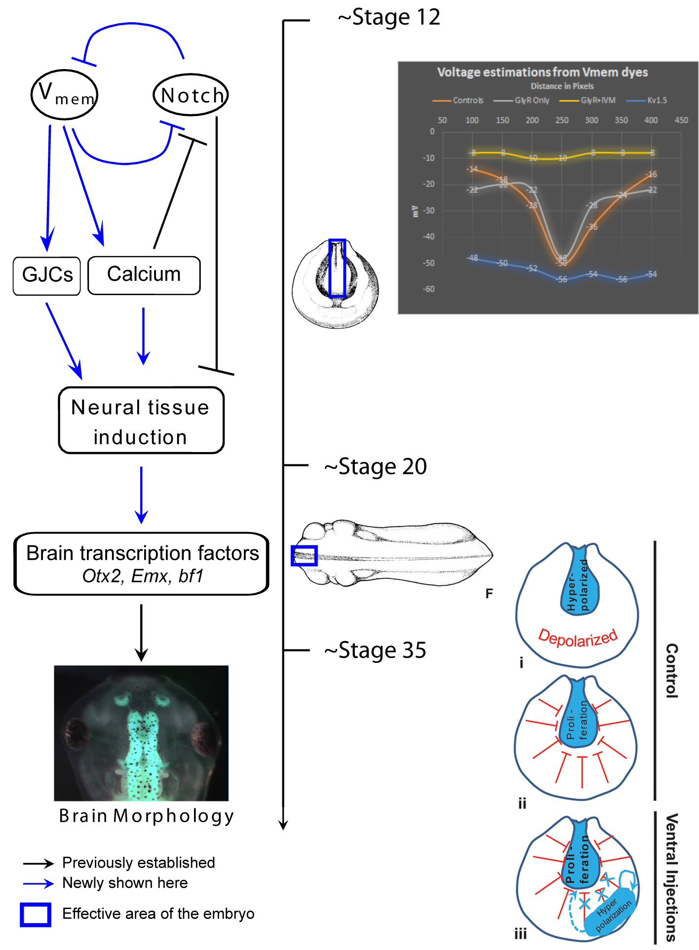
My lab works on developmental bioelectricity, studying how cells communicate via endogenous gradients of plasma membrane resting potential (Vmem) in order to coordinate their activity during pattern regulation (Levin, 2013; Levin, 2014b; Tseng and Levin, 2013). It is well-known that resting potential is an important regulatory parameter for individual cells’ proliferation, differentiation, and oncogenic potential (Blackiston et al., 2009; Sundelacruz et al., 2009). Voltage itself is an important “master control knob” because the same morphogenetic phenotype (e.g., inducing eye formation or metastatic conversion) can be induced by using sodium, potassium, chloride, or even proton flows to achieve a particular Vmem level. The chemical nature of the ion (and the genetic identity of the channel) often does not matter, as long as the voltage gradient is established correctly for a particular downstream outcome. In this Node post, I wanted to briefly mention a few of our recent studies which highlight an exciting new aspect of this field: long-range signaling via gap junctions.
Gap junctions (GJs) are electrical synapses – direct conduits for small molecules between cells, which can be used to form isoelectric compartments in vivo; they have numerous roles in normal development and disease (Levin, 2007; Sohl and Willecke, 2004; Wong et al., 2008). Most importantly, they are extremely versatile signaling elements (Palacios-Prado and Bukauskas, 2009; Pereda et al., 2013), because they both regulate cellular resting potential and are themselves voltage-gated. GJs are able to function as a kind of transistor, allowing voltage to control current flow. Because they are ideally-suited to process information in physiological cell networks, is no surprise that gap junctional communication is a key regulator of brain activity, developmental patterning, and carcinogenesis.
One of our recent studies investigated the role of endogenous bioelectric gradients in brain formation in the Xenopus laevis embryo (Pai et al., 2015). Early frog embryos exhibit a characteristic hyperpolarization of cells lining the neural tube; disruption of this spatial gradient of the transmembrane potential (Vmem ), using misexpression of depolarizing channels, diminishes or eliminates the expression of early brain markers, and causes anatomical mispatterning of the brain. Conversely, forced establishment of the brain-specific voltage pattern (using expression of select ion channels) was able to rescue brain defects induced by mutant Notch protein (a potent regulator of neurogenesis), and even induce ectopic brain tissue in posterior regions of the tadpole.
In addition to cell-autonomous effects, we showed that hyperpolarization of transmembrane potential (Vmem ) in ventral cells, well-outside the brain, induced upregulation of neural cell proliferation. These long-range effects were mediated by gap junctional communication, and another recent paper extended such long-range regulation of cell division to similar non-local control of apoptosis (Pai, 2015). We suggested a model in which brain cells coordinate growth and sculpting decisions with the remaining tissues (to determine appropriate location, size, and boundaries of the nascent brain) via electrical signals mediated by GJ paths.
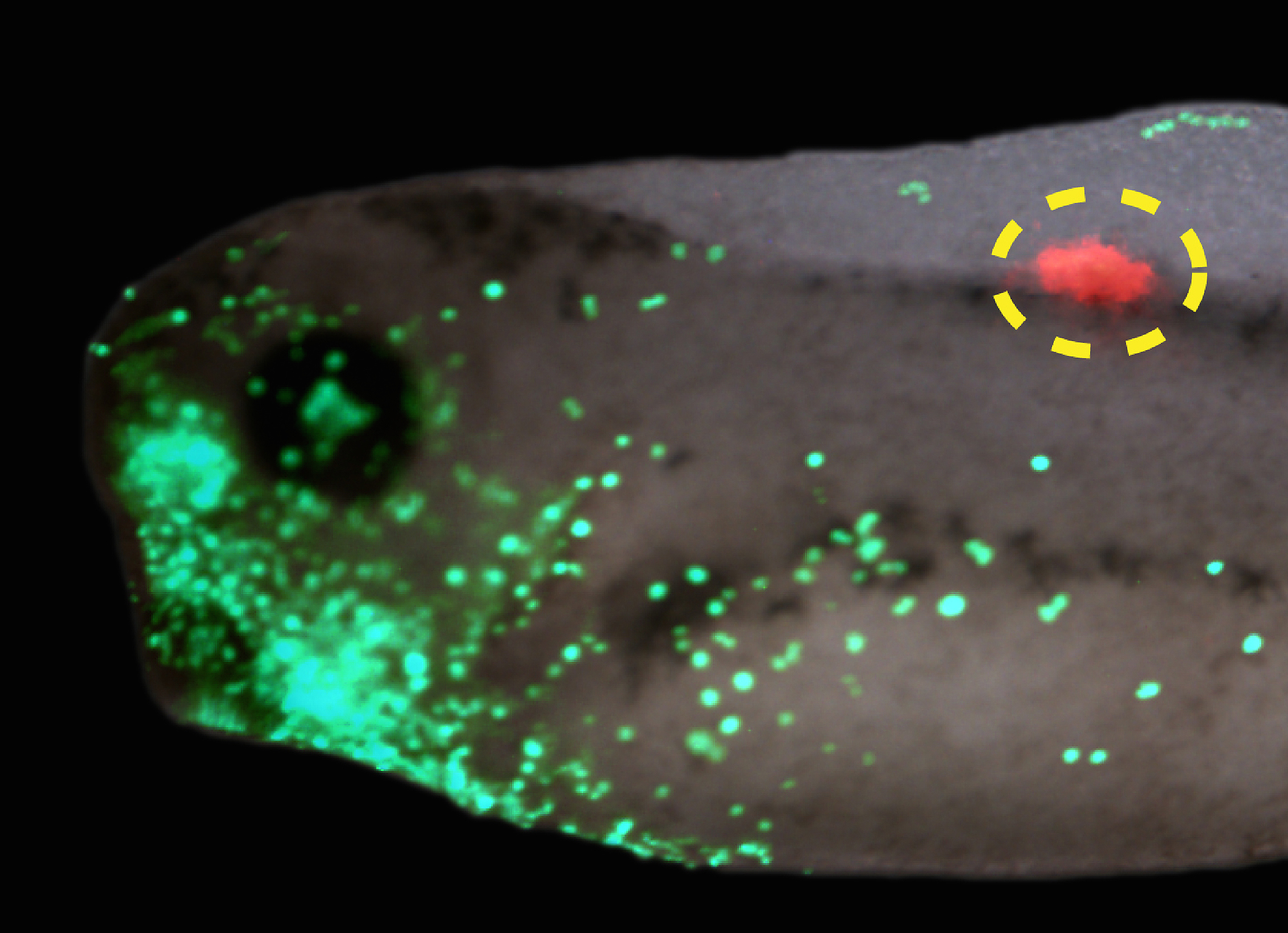
Interestingly, a similar story was found for tumorigenesis in Xenopus (Chernet et al., 2014). mRNA encoding mutant KRAS induces tumors in a zebrafish cancer model (Le et al., 2007). We showed that the same thing happens in Xenopus (complete with induced angiogenesis, overproliferation, expression of tumor markers, and immune response); remarkably, a specific bioelectric state of cells at a considerable distance (on the other side of the body) can suppress tumor formation, despite strong expression of the oncogene. The effect is mediated by butyrate signaling (Chernet and Levin, 2014), which links voltage regulation to chromatin modification, and – GJs. These data are part of a growing body of evidence (Bizzarri and Cucina, 2014; Chernet and Levin, 2013; Soto and Sonnenschein, 2011; Tarin, 2011) highlighting aspects of cancer as a “disease of geometry” – a disorder of patterning cues and cell:cell communication that normally harnesses cell activity towards specific morphogenetic goals and away from tumorigenesis.
It appears that in diverse contexts, such as embryonic establishment of pattern and tumor suppression, GJs link bioelectric and biochemical pathways to regulate events at considerable distance. Thus, future work must focus not only on ever-more detailed dissection of biophysical signaling events within single cells, but also address group dynamics and large-scale emergent properties of physiological networks linked by electrical synapses (Donnell et al., 2009; Levin, 2014a; Saraga et al., 2006; Schiffmann, 2008; Steyn-Ross et al., 2007). Multicellular models of GJ signaling will surely contribute to the understanding of patterning and deviations from normal growth and form.
References
Bizzarri, M. and Cucina, A. (2014). Tumor and the microenvironment: a chance to reframe the paradigm of carcinogenesis? Biomed Res Int2014, 934038, http://www.ncbi.nlm.nih.gov/pubmed/25013812
Chernet, B. and Levin, M. (2013). Endogenous Voltage Potentials and the Microenvironment: Bioelectric Signals that Reveal, Induce and Normalize Cancer. J Clin Exp OncolSuppl 1, http://www.ncbi.nlm.nih.gov/pubmed/25525610
Chernet, B., and Levin, M., (2014), “Transmembrane voltage potential of somatic cells controls oncogene-mediated tumorigenesis at long-range”, Oncotarget, 5(10): 3287-3306
Chernet, B. T., Fields, C. and Levin, M. (2014). Long-range gap junctional signaling controls oncogene-mediated tumorigenesis in Xenopus laevis embryos. Front Physiol5, 519, http://www.ncbi.nlm.nih.gov/pubmed/25646081
Levin, M. (2013). Reprogramming cells and tissue patterning via bioelectrical pathways: molecular mechanisms and biomedical opportunities. Wiley Interdisciplinary Reviews: Systems Biology and Medicine5, 657-676, http://www.ncbi.nlm.nih.gov/pubmed/23897652
Levin, M. (2014a). Endogenous bioelectrical networks store non-genetic patterning information during development and regeneration. The Journal of Physiology592, 2295-2305, http://jp.physoc.org/content/592/11/2295.abstract
Levin, M. (2014b). Molecular bioelectricity: how endogenous voltage potentials control cell behavior and instruct pattern regulation in vivo. Mol. Biol. Cell25, 3835-3850, http://www.ncbi.nlm.nih.gov/pubmed/25425556
Pai, V. P., Lemire J. M., Chen Y., Lin G., and Levin M. (2015). Local and long-range endogenous resting potential gradients antagonistically regulate apoptosis and proliferation in the embryonic CNS. Int. J. Dev. Biol., in press
Pai, V. P., Lemire, J. M., Pare, J. F., Lin, G., Chen, Y., & Levin, M. (2015). Endogenous Gradients of Resting Potential Instructively Pattern Embryonic Neural Tissue via Notch Signaling and Regulation of Proliferation Journal of Neuroscience, 35 (10), 4366-4385 DOI: 10.1523/JNEUROSCI.1877-14.2015
Pereda, A. E., Curti, S., Hoge, G., Cachope, R., Flores, C. E. and Rash, J. E. (2013). Gap junction-mediated electrical transmission: regulatory mechanisms and plasticity. Biochimica et biophysica acta1828, 134-146, http://www.ncbi.nlm.nih.gov/pubmed/22659675
Saraga, F., Ng, L. and Skinner, F. K. (2006). Distal gap junctions and active dendrites can tune network dynamics. J. Neurophysiol.95, 1669-1682, http://www.ncbi.nlm.nih.gov/pubmed/16339003
Schiffmann, Y. (2008). The Turing-Child energy field as a driver of early mammalian development. Prog Biophys Mol Biol98, 107-117, http://www.ncbi.nlm.nih.gov/pubmed/18680762
Soto, A. M. and Sonnenschein, C. (2011). The tissue organization field theory of cancer: a testable replacement for the somatic mutation theory. Bioessays33, 332-340, http://www.ncbi.nlm.nih.gov/pubmed/21503935
Steyn-Ross, M. L., Steyn-Ross, D. A., Wilson, M. T. and Sleigh, J. W. (2007). Gap junctions mediate large-scale Turing structures in a mean-field cortex driven by subcortical noise. Phys Rev E Stat Nonlin Soft Matter Phys76, 011916, http://www.ncbi.nlm.nih.gov/pubmed/17677503
Tarin, D. (2011). Cell and tissue interactions in carcinogenesis and metastasis and their clinical significance. Semin Cancer Biol21, 72-82, http://www.ncbi.nlm.nih.gov/pubmed/21147229
First Announcement
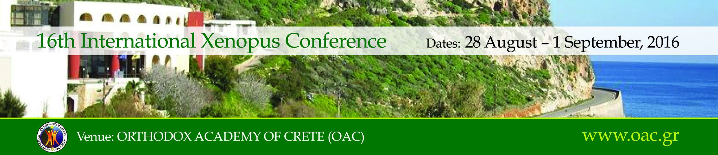
The 16th International Xenopus Conference
Sunday 28 August – Thursday 1 September, 2016
Venue: Orthodox Academy of Crete, Chania
Please join us for the 16th International Xenopus Conference to be held 28 August – 1 September 2016 at the Orthodox Academy of Crete. This conference has been held biennially since 1984 and brings together researchers from diverse fields, all of whom use Xenopus as a model system. The format of the conference provides outstanding opportunities for scientific exchange with over three hundred poster presentations and talks. In particular, junior faculty are encouraged to speak. There will also be numerous opportunities for postdocs and students to present their work and participate in career development activities. Finally, this meeting has traditionally been an ideal forum to learn about the latest technological advances and resources in the Xenopus system.
Xenopus as a model system spans many fields, including subjects such as cell cycle and cytoskeletal regulation, developmental biology and stem cells, immunology, neurobiology and systems biology. This diversity of topics is a major advantage of the Xenopus community, and this meeting seeks to highlight and reinforce these interdisciplinary interactions.
For this meeting, we expect over 50 talks and 200 posters, as well as plenary sessions, with an outstanding line up of speakers from around the world.
Check back regularly at the website for updates about the conference, including information on registration, abstract submission, the scientific and social programme as well as details of our sponsors and exhibitors, whose essential support and contribution to the conference are very much appreciated.
We hope you can join us and look forward to welcoming you to Crete on 28 August to 1 September, 2016.
The Department of Developmental Biology at Washington University School of Medicine invites applications for a Biomedical Informatics Assistant. The Department of Developmental Biology is a dynamic research community, with interests spanning multiple model organisms and disease paradigms.
We are seeking an outstanding applicant to join at the level of Biomedical Informatics Assistant to provide bioinformatics expertise and develop data analysis pipelines. The applicant is expected to have a B.A./B.S. or M.S. in a relevant area, including, but not limited to, computer science, statistics, and biology. Knowledge of developmental biology and/or regenerative medicine would be a plus. The successful candidate will report to the head of the Bioinformatics Research Core (http://brc.wustl.edu/) and assist on fundamental next generation sequencing analysis (ChIP-seq, RNA-seq, genome/exome sequencing), utilizing state of the art bioinformatics tools. Additional duties include the development of pipelines and novel analysis software. Proficiency with statistics and common scripting languages and analysis tools (e.g., Perl, R, and/or Python) is expected. In addition, the candidate should have excellent writing, communication and interpersonal skills. This is an excellent opportunity to gain additional experience with biomedical data analysis, interact with multiple research paradigms, and author scientific publications.
Review of applications will start September 1, 2015. Interested applicants should email a single PDF file consisting of a cover letter and curriculum vitae, and should arrange for submission of three letters of reference to:
Biomed Informatics Search Committee (c/o Bo Zhang, Ph.D.)
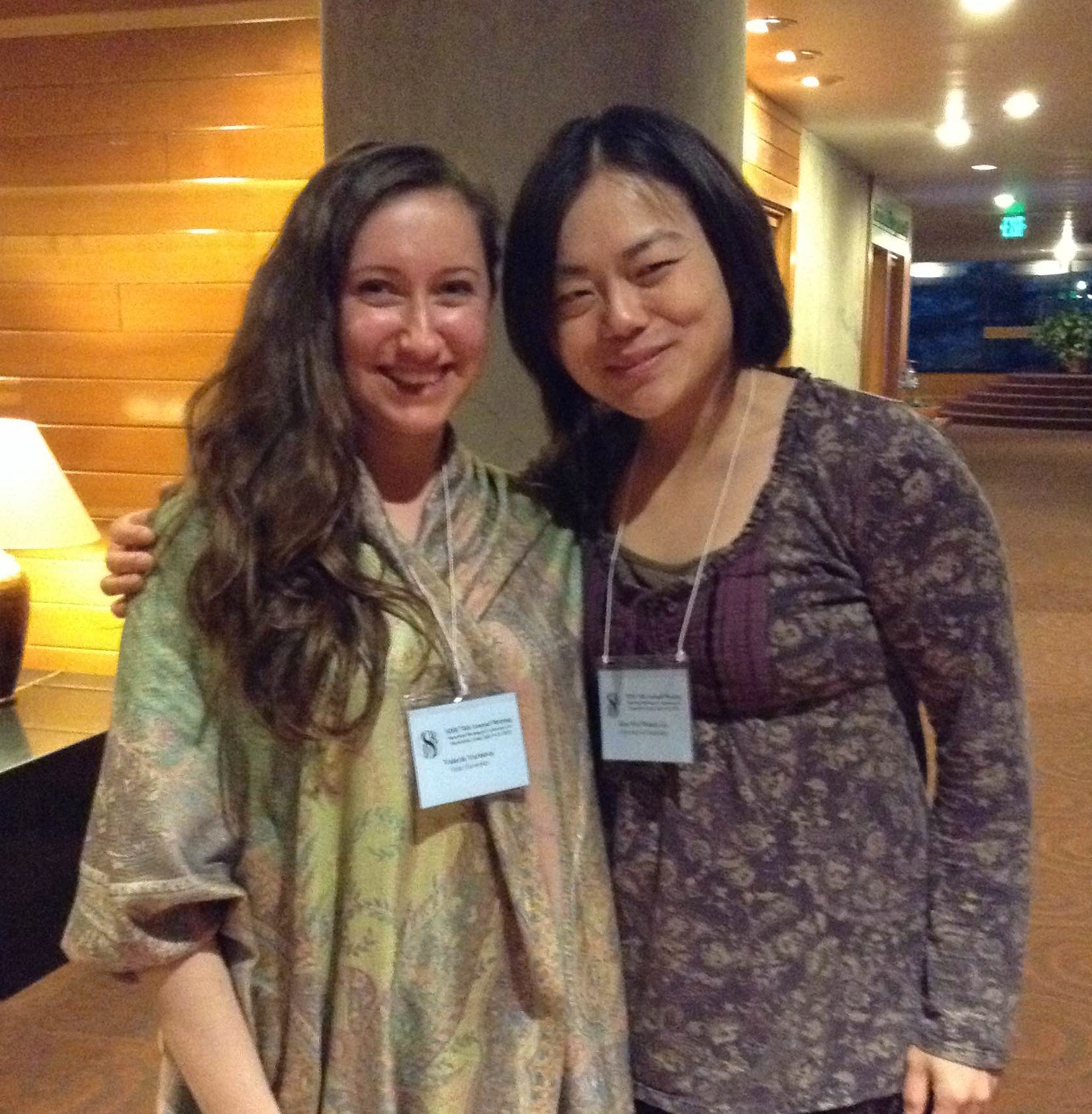
Wendy Gu was the winner of this year’s British Society for Developmental Biology (BSDB) meeting poster competition. Her prize was to attend the annual meeting of the Society for Developmental Biology (SDB), which took place in Snowbird, Utah, in July. Continuing the interview chain, Wendy interviewed Valeria Yartseva, who won the poster prize at the meeting. Valeria’s prize will be to attend the BSDB meeting at the University of Warwick, UK, next year.
WG- Valeria, congratulations on winning the 2015 SDB poster prize! The readers of the Node would be interested in knowing a bit more about you, and specifically about your lab. Can you tell us a bit more about that?
VY: Thank you Wendy, I’m really thrilled to be the recipient of this award! I am a graduate student at Yale University, in the lab of Antonio Giraldez. Our lab has a deep interest in understanding how a single cell embryo becomes a multicellular organism. Life in all animals begins as a single cell that contains all the information required to make a mature organism, and understanding how this is possible has been the driving force of our work. My work specifically addresses how the maternal instructions, which are so critical for the earliest stages of development, are actively degraded during the maternal to zygotic transition, to facilitate the transition from being an oocyte to becoming a pluripotent embryo.
WG: What was your poster about?
VY: The critical results of my project were obtained using a high throughput reporter assay that we developed in the lab. We first used high throughput sequencing methods to identify the transcripts that are degraded over time. Once we knew the identity of these transcripts, we were able to zoom in on the specific sequences within these mRNAs that were regulating their fate during the maternal to zygotic transition. From these snippets of sequences we were able to build consensus motifs that explain the degradation of groups of these regulated transcripts. It was a nice way to go from the whole transcript to the regulatory element, and then building a motif that explains the behavior many transcripts.
WG: What is the biological relevance of the degradation of these transcripts?
VY: We believe the turnover of maternal RNAs during the maternal to zygotic transition is critical for the transition to the totipotent embryonic state. Life begins as a union between two terminally differentiated cells, the oocyte and a spermatozoon, which triggers the reprogramming of the oocyte into a totipotent zygote. Clearance of maternal mRNAs is a universal feature of embryonic development in metazoans and evidence to support that this process is instructive for development is the striking parallel between somatic cell reprogramming in vitro and the maternal to zygotic transition. The microRNA miR-430 is a major regulator of maternal mRNA clearance and its mammalian orthologue miR-302 enhances reprogramming efficiency to pluripotency in vitro. It is our belief that uncovering additional mechanisms that eliminate the maternal program of development will advance our understanding of developmental reprogramming to pluripotency as it occurs in vivo within the embryo.
WG: Which experiment or data are you most proud of?
VY: We tried many different directions to answer this question, but ultimately the experiment that made the project was this high throughput reporter assay. It is a funny story actually. We finally figured out how to build this assay and were getting ready to submit the experiment for sequencing. I was on the phone with Antonio, who was traveling at that time, to discuss the last details of this experiment. I was saying to him ‘I am not sure this is going to work. This might fail, that might fail, etc’. He said ‘Just close your eyes and do the experiment’. It was very eye opening. I learned that sometimes it pays off to just go for an experiment blindly.
WG: Have you won a poster prize at a meeting before?
VY: The first time I won a poster prize was at the annual retreat of the Genetics Department at Yale. I was just having such a great time talking about my science; I never thought about winning an award for it. I am also extremely grateful to have received the Northeast SDB best poster award, which allowed me to come to this meeting.
WG: So you have a track record of prize winning! What is next for you?
VY: I’m starting my 5th year in graduate school and I am really enthusiastic about completing this project. Of course, I will be really sad to leave next year, because I have invested half a decade of my life to understanding this one problem and Antonio has been such an amazing mentor to me over the years. I don’t anticipate to stray far from developmental biology in my future career; it’s just too much fun. I also deeply believe that by understanding developmental principles one can understand how these processes go awry in disease states. This motivates me to go to the lab every day.
WG: Your prize is attendance of the 2016 British Society of Developmental Biology meeting. Will you be attending this meeting?
VY: That is an absolute dream come true. I have always wanted to be a developmental biologist, and to be honored in this way is most thrilling to me. I am very excited to attend the BSDB meeting next year.
WG: On behalf of the British developmental biology community we welcome you with open arms and we look forward to hearing more about your work!
A Postdoctoral Fellow position is available in Lee laboratory at Johns Hopkins University. This position is NOT for stem cell expert, rather for who has Outstanding expertise on either extensive RNA-seq analysis, skeletal muscle biology, optogenetics or single-cell studies.
We have established novel methodologies to direct human induced pluripotent cell (hiPSC) into peripheral neuron lineages, Schwann cells and skeletal muscle cells using multiple genetic reporter systems. Now we are broadening our research topics into specific experiments using new techniques, mentioned above. In addition, the Lee lab continues to study several developmental and degenerative disorders (pain disorders, myelination disorders, and muscular dystrophies) to unravel the underlying cellular/molecular mechanism.
Previous experience on human pluripotent stem cells is not necessarily required, and we value more on the ‘non-stem cell’ experience.
Applicants can send a CV and the names of three references to the address listed below:
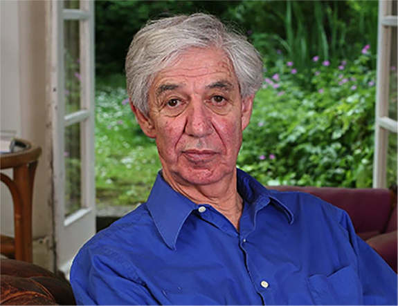
Lewis Wolpert is a retired developmental biologist who, over his long career, has made many important contributions to the field, from his French Flag model and the concept of positional information to the famous quote that it is “not birth, marriage or death, but gastrulation which is truly the most important time in your life.” In addition to his scientific contributions, Lewis is also a prolific writer, from the textbook ‘Developmental Biology’ to books about popular science, religion and his battle with depression. Although born in South Africa, it was in the United Kingdom that Lewis spent most of his scientific career. We met Lewis at the Spring Meeting of the British Society for Developmental Biology, where he was awarded the Waddington Medal.
What does it mean to you to receive the Waddington Medal?
It is wonderful. I was very excited indeed to receive it. I knew Conrad Waddington, who was a friend of mine, so it makes it personal. And of course I am a developmental biologist and I am old, so it is very nice to receive it!
This award recognises your career contributions to developmental biology, but you were originally a civil engineer. How did you make this transition?
I studied civil engineering during my undergraduate studies because I wanted to do science. I qualified as a civil engineer, and worked as a soil mechanic for a building research institute for two years. However, I had been involved with Mandela and was a bit frightened, so I decided to leave South Africa to get away from the politics…and also from my parents! I hitchhiked up Africa and eventually ended up in Europe. I did another course in soil mechanics at Imperial College but was very unhappy: I didn’t want to spend the rest of my life working on soil mechanics! A friend of mine knew this, and he had read some papers where people were looking at the mechanics of cell division. So I went to King’s College in London where I did a PhD on this topic. Having a civil engineering background gave me a very different perspective, and I applied my mechanics knowledge to the cells. And this is how I got involved in biology.
You have switched fields and models multiple times in your career – from cell mechanics to developmental biology, from sea urchins and amoebae to Hydra and the vertebrate limb. What motivated these changes and how easy was it to make these moves?
I did my cell mechanics PhD mainly on sea urchin eggs. I became interested in sea urchin development, and that’s how I got into developmental biology. However, to work on sea urchins you needed to visit marine stations and this became difficult once I got married. I therefore switched to working on Hydra for a time. Later, I moved to Middlesex Hospital Medical School, now part of University College, London, and I felt that Hydra was not an appropriate model to work on in a medical school. So I started working on the development of the limb. Changing so many times, from civil engineering to biology and then to different systems, required me to work very hard. For example, during my PhD I had to learn quite a lot and pass exams in zoology. It was a little difficult but interesting.
You’re perhaps best known for your French Flag model of positional information. This is now considered a landmark publication but wasn’t widely accepted at first. Why do you think that was?
Conrad Waddington had organised a theoretical biology meeting at Serbelloni, on Lake Como in Italy, and the idea was well received there. However, when I gave a big lecture on it at Woods Hole nobody would speak to me afterwards. I don’t know what it was, but the Americans didn’t like my ideas. The next morning, while I was bathing, Sydney Brenner found me crying in the water and said “Lewis, pay no attention. We like your ideas. Pay no attention to people who don’t like it.” And he’s the one who saved me. He gave me total encouragement, so I didn’t care that all these Americans didn’t like what I was doing. If Sydney liked it, that’s what mattered, because Sydney is an amazing man. I knew him from South Africa of course, since I went to university with him. He is funny and brilliant, the only genius I know. And Francis Crick liked my ideas too. So that lecture was a bit of a disaster but I recovered because of Sydney. It took quite a long time for the Americans to come around and accept my ideas.
It’s now over 45 years since you proposed this model. Where do you think the French Flag model fits with our current understanding of positional information, and what do you think are the exciting questions at the moment?
There are problems we haven’t solved. It is terrible, but we still don’t have a molecular basis for it. If I still had an active lab, finding the molecular basis for positional information would be my objective, but would be quite tricky, since I’m not a biochemist or molecular biologist. There is one case of a molecule that might encode positional information, Prod 1, which is graded along the amphibian limb and was discovered by Jeremy Brockes. But it would be nice to find similar molecules in other systems.
You have said in earlier interviews that you are not interested in the details, but rather in the overall principles, and also that you don’t like doing experiments. How has this influenced your career?
I’m really a theoretician – I’m not very good at doing experiments! Fortunately I had PhD students, postdocs and research workers to do the experiments for me. I had, for example, a German technician, Amata Hornbruch, who was my hands for 20 or 30 years!
Over the years many of your PhD students and postdocs moved on to become successful developmental biologists in their own right – including previous winners of the Waddington Medal, Julian Lewis and Jim Smith. Are you proud of your mentoring?
Absolutely. I was very fortunate to work with very nice people. I’m very pleased with them – I like them very much and we got on very well. I encouraged them, and we had good discussions. It is very satisfying to see them doing well.
How do you think science has changed since your early days, and do you have any advice for young scientists?
Oh, it has changed tremendously. I would always say “don’t be frightened of changing your career as a scientist.” It is hard for young scientists to know what they should work on. When I look at journals these days I find them very boring. It’s just detail and very little general theory at all. Find something really exciting and don’t be afraid. If you are going to spend your life just studying little details it won’t be terribly interesting. At least for me! Study something like the brain, where we understand the least. It is a very exciting and complicated area.
Over the years you have written several popular science books, presented the RI Christmas Lectures and have generally been an advocate for science education. Do you consider yourself passionate about science outreach? Do you encourage other scientists to communicate their science?
I would, but it’s not terribly rewarding. Rather, it is quite rewarding and I enjoyed it, but I don’t think people pay much attention! I was quite involved in outreach because I was the Royal Society chairman of the committee for the public understanding of science, but I don’t think we made much progress. And I wrote a textbook, of course. Funnily enough I’m just writing a book at the moment, asking ‘What has science done for us?’ It addresses the way science has affected our lives, not just from a biology perspective but also physics, chemistry, the lot.
You have been very open about your battles with depression. What advice would you give to young scientists struggling with mental health issues?
They should be open about it and try to get cognitive therapy. I found professional therapy very helpful. You know, when I came out of my depression, out of hospital – I was in hospital for three weeks – my wife hadn’t told anybody that I was depressed. She just said that “he’s very tired and not feeling well.” The stigma related to depression is still very severe and that makes it much worse. That’s why I wrote a book about it. I wanted to understand what depression was, so I researched it a bit. I still lecture on depression every now and then. In fact I’m shortly going to Abu Dhabi to give a lecture on depression.
What would people be surprised to find out about you?
Just how bad my memory is! My favourite story is that I was once in my son’s house in Cambridge, and they were away. I went downstairs for breakfast and I found a young woman in the kitchen, doing some washing up. I said, “Hello, and where are you from?” It was my granddaughter! I didn’t recognise her!