The community site for and by
developmental and stem cell biologists

GUDMAP – an online resource for genitourinary research

Posted by , on 26 February 2014

GUDMAP (Harding et al., 2011, McMahon et al., 2008) is an open-access atlas-based on-line resource developed by a consortium of laboratories to provide the scientific and medical community with resources to facilitate research and teaching focussed on the murine genitourinary (GU) system. Gene expression data, descriptors of new mouse lines, and other tools can be freely accessed at the project website (www.gudmap.org). The GUDMAP expression database includes large-scale in situ (hybridization and immunohistochemistry) screens and microarray gene expression data and is currently in the process of making available 3D imaging (OPT) & next generation sequencing data of the developing mouse genitourinary (GU) system.

GUDMAP Website Homepage

The database contains over 10,500 in situ assays, the majority of which are in situ hybridisation (ISH), although the resource also contains immunohistochemistry (IHC) and transgenic reporter assays. The ISH assays cover in excess of 3,600 genes – ~2,900 unique genes have been studied by wholemount in situ analysis and ~1,400 by section in situ analysis. The resource also contains over 400 microarray samples, the majority of which have been isolated and prepared using either laser capture or FACS. These data types are now being extended to include next-gen sequencing such as RNA-Seq and full 3D image data mapped onto reference models.

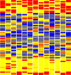
In addition to gene expression data, the website also provides detailed tutorials that describe genitourinary development. These are supplemented with schematic diagrams that serve to illustrate the developing components of the mouse genitourinary system over different developmental stages (Kylie Georgas, University of Queensland). The GUDMAP consortium has also generated a resource of novel transgenic mouse strains carrying genetic markers, with characterization, verification and the new strategy for production of the strains (GUDMAP Mouse Marker Strains). A further additional feature, which continues to be developed within the resource, is access to large-scale data analysis over aggregated GUDMAP genomic profiling data. This provides a series of compartment-specific genelists that reflect cell type and stage specific gene expression. These lists are viewable in technology and sample specific heatmaps, and are also integrated with the ToppGene analysis suite.

GUDMAP data is freely accessible via both simple and advanced query mechanisms. Querying by gene returns ‘gene expression summaries’, which provide a simple visualisation of all data available per gene with links to in situ assays, in situ histological images, microarray data and disease associations. The GUDMAP consortium has developed a high-resolution anatomy ontology (Little et al., 2007) to describe in detail the sub-compartments of the developing murine genitourinary tract. It is against this ontology that gene expression is annotated, describing both the presence and strength of expression in different sub-compartments.

Funding for the GUDMAP consortium was initially established in 2005 and has ongoing support from the NIH. The GUDMAP Project page on the website gives details of all contributing laboratories, both past and present, summarising their scientific focus and contribution to the GUDMAP effort. The GUDMAP Publications page list all publications related to GUDMAP. For any further information about GUDMAP please contact the GUDMAP Editorial Office: gudmap-editors@gudmap.org.

References:

Harding SD et al. 2011. The GUDMAP database – an online resource for genitourinary research. Development. 138(13):2845-53.

McMahon AP et al. 2008. GUDMAP: the genitourinary developmental molecular anatomy project. J Am Soc Nephrol. 19, 667-671.

Little MH et al. 2007.  A high-resolution anatomical ontology of the developing murine genitourinary tract. Gene Expr Patterns. 8(1):47-50.

Thumbs up (1 votes)
Loading...

Tags: , , ,
Categories: Research, Resources

Navigate the archive

Use our Advanced Search tool to search and filter posts by date, category, tags and authors.

the Node at the CSHL conference on Avian Model Systems

Posted by , on 25 February 2014

The Node is on the road again! Next week, from the 5th to the 8th of March, Cold Spring Habor Laboratories will be hosting a conference on Avian Model Systems, and the Node will be there!

Are you attending this meeting? Then say hello to Cat, our community manager, who would love to hear your thoughts and suggestions about the Node. If you are attending this meeting, then why not write a meeting report about it for the Node? Get in touch if you are interested- we can help you get started!

If you are not attending this meeting, keep an eye on our twitter account. We will be tweeting from the meeting if an internet connection is available, and look out for a meeting report here on the Node. In the meanwhile, if you are interested in developmental biology research on birds, do check ‘A day in the life of a chick lab‘, part of our ongoing model organisms series.


Node avian 4 (cropped)


Thumbs up (2 votes)
Loading...

Tags:
Categories: Events, News

On the origins of species-specific size

Posted by , on 25 February 2014

by Jennifer L. Fish and Richard A. Schneider


“For every type of animal there is a most convenient size, and a large change in size inevitably carries with it a change of form.”
Haldane 1926.


As articulated most eloquently by Haldane (1926) in his classic essay on “Being the Right Size”, every animal has it’s own particular size, which is ultimately linked to form, function, and fitness. Consequently, size is tightly regulated during development. To achieve proper structural integration, organs must keep track of size, as exemplified by the equal length of limbs. Right and left sides of the body develop independently, yet right and left limbs consistently reach comparable length (Allard & Tabin 2009). Mounting evidence suggests that developmental mechanisms regulating size are highly conserved and buffer variation (Leevers & McNeill 2005). So then how do size differences evolve?

In our recent manuscript, (Fish et al. 2014), we have addressed the question of organ size evolution from the perspective of the jaw. Utilizing two avian species, duck and quail, that exhibit remarkably different jaw size (Figure 1), we asked when, where, and how do duck acquire their long bills compared to quail who make short beaks? We began with the simple analogy that building a bigger structure such as a wall might involve using more bricks (rather than bigger bricks). Thus, we focused on the number of cellular precursors, which in this case is the cranial neural crest that gives rise to the jaw skeleton.


Figure 1: Species-specific differences adult quail (A) and duck (B) skulls showing species-specific differences in jaw size

Figure 1: Species-specific differences adult quail (A) and duck (B) skulls showing species-specific differences in jaw size


We started by counting neural crest cells at several embryonic time points. We found that at a very early stage (HH8) the number of neural crest cells that are specified along the length of the neural folds is the same in quail and duck. But slightly later (HH10) when these cells begin to accumulate dorsally, duck have significantly more cells (15%) in the midbrain and rostral hindbrain region, which ultimately enables more duck cells to migrate into the presumptive jaw region by HH13. Remarkably, by HH20, duck have twice as many cells in their jaw primordia as do quail. To understand how an initial 15% difference could result in a doubling of the population by HH20, we analyzed cell proliferation and cell cycle length. We found that cell cycle length is longer in duck than quail, but when developmental rate is taken into account over absolute time, duck neural crest proliferate relatively faster than quail, which can explain the progressive increase in jaw size in duck embryos.

To uncover a mechanism through which duck increase the number of precursor cells that come out of the midbrain, we assayed for species-specific differences in the expression of brain regionalization markers. We compared Pax6 (forebrain), Otx2 (fore- and midbrain), Fgf8 (midbrain-hindbrain boundary), and Krox20 (r3 and r5 of the hindbrain) in duck and quail embryos at HH10. We found that duck and quail embryos have divergent brain shapes and spatial domains of gene expression. For example, duck embryos have a shorter and broader midbrain, which is also evidenced by a unique pattern of Otx2 expression (Figure 2A,B). Presumably, this broader duck midbrain congregates more neural crest cells in the region that will ultimately populate the jaw primordia. Interestingly, the Otx2 expression domain is already distinct in duck and quail embryos at HH6 (Figure 2C,D), suggesting that critical species-specific patterning mechanisms that affect jaw size may be in place from the earliest developmental stages.


Figure 2: Species-specific differences appear early during development. HH6 quail and duck embryos were compared molecularly by performing in situ hybridization for Otx2 at HH10 (A, B) and HH6 (C,D).

Figure 2: Species-specific differences appear early during development. Quail and duck embryos were compared molecularly by performing in situ hybridization for Otx2 at HH10 (A, B) and HH6 (C,D).


Overall, our work reveals that modifications to multiple aspects of cell biology, including the generation and allocation of neural crest cells destined to form the jaw skeleton, and species-specific regulation of cell proliferation, may underlie the evolution of jaw size.


References:

Allard, P. and Tabin, C. J. (2009). Achieving bilateral symmetry during vertebrate limb development. Semin. Cell Dev. Biol. 20, 479-484.

Fish JL, Sklar RS, Woronowicz KC, and Schneider RA. (2014). Multiple developmental mechanisms regulate species-specific jaw size. Development, 141:674-684.

Haldane, J. B. S. (1926). On Being the Right Size. Harper’s Magazine.

Leevers, S. J. and McNeill, H. (2005). Controlling the size of organs and organisms. Curr. Opin. Cell Biol. 17, 604-609.

Thumbs up (6 votes)
Loading...

Tags: , ,
Categories: Research

The covers of Development

Posted by , on 24 February 2014

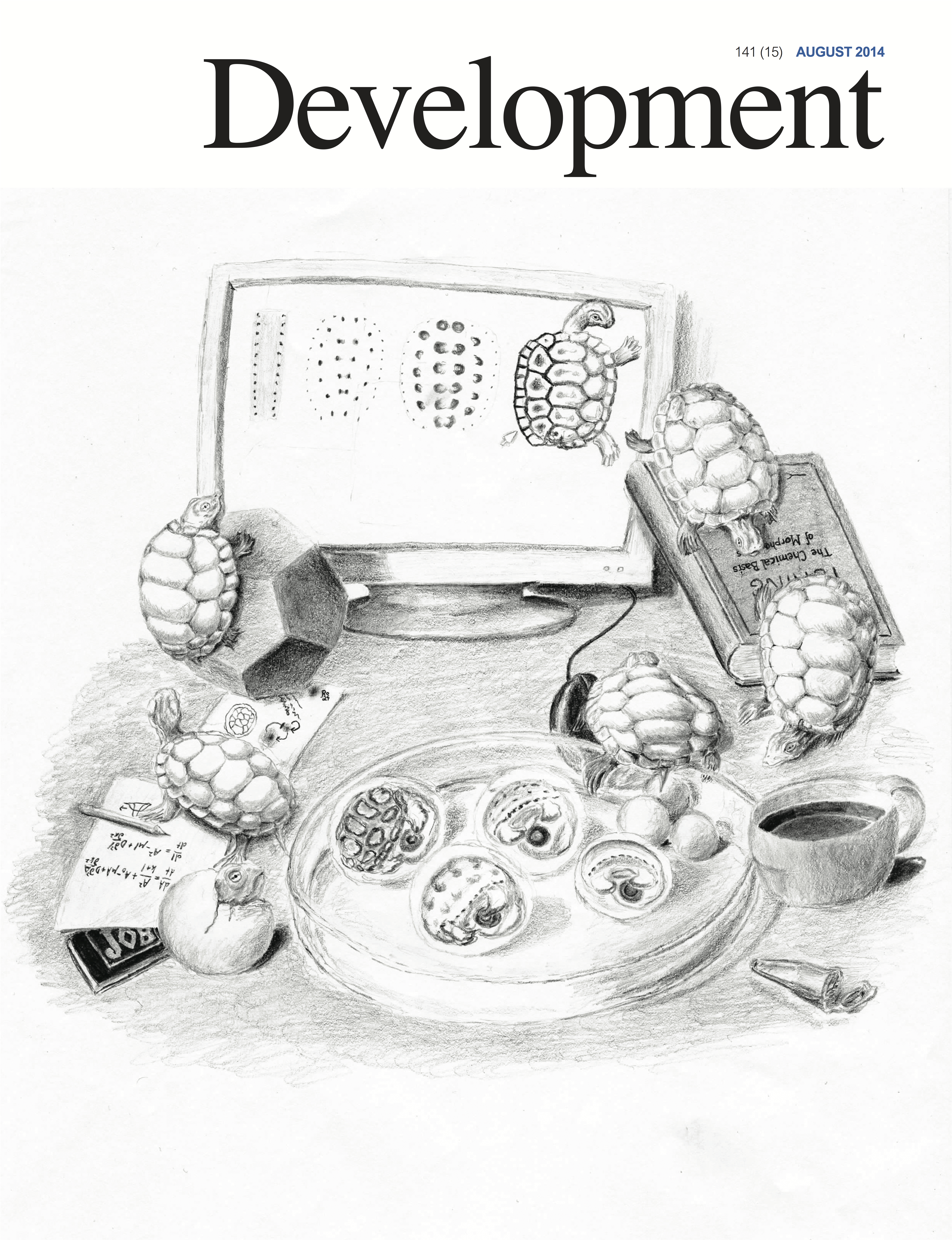
Here at Development we are very proud of our covers. Over the years we have featured many beautiful images, showcasing different model organisms and techniques and including a few unusual choices! Most of these images were submitted by our authors, but some of the most recent ones were voted by you here on the Node. Over the years, the style and content of the covers has evolved, and so they mark an interesting perspective on changes to the field and the journal. We wanted to share this collection with you, so during the last few months we have gone through our archive and collated some of our covers in the short movie below. What do you think of our selection? Did we include your favourite cover? Let us know what you think by leaving a comment below! You can also browse our full cover archive by visiting our website.


You can watch this and other movies on our YouTube channel.
 

Thumbs up (7 votes)
Loading...

Tags: ,
Categories: Images, Video

A day in the life of a butterfly lab

Posted by , on 21 February 2014

Hello! I’m Leila, a finishing PhD student in Patrícia Beldade’s lab at the Instituto Gulbenkian de Ciência, Portugal. We work on different topics within Evolutionary Developmental Biology, Evo-Devo, with the common interest on how development contributes to intra- and inter-specific variation and can influence evolutionary processes: developmental hierarchies (me), developmental plasticity, and the origin of novelty. The lab does not focus on a particular organism. Rather, we have flies, butterflies and ants. Because I’m interested in a highly diverse group – both taxonomically and morphologically – that can be manipulated experimentally, I chose butterflies.

Most of my work uses the African species Bicyclus anynana, established in the 80’s and living happily ever after in the lab. Butterflies are holometabolous insects, which means they go through four metamorphic stages: embryo (egg), larva (caterpillar), pupa (chrysalis), and adult (butterfly, Fig. 1A and 2A). For B. anynana, this cycle takes about 4 weeks at 27°C, and twice as much at 19°C. These are the temperatures that, in the lab, induce what are the natural wet- and dry-season phenotypes, respectively. Wet and dry morphs have different wing color patterns and life histories. The lab stock population has much genetic variation, which allows for artificial selection of distinct wing pattern and life history traits; they respond to artificial selection with particular ease. Many of our studies concentrate on a particular wing pattern element called the eyespot, which develops at the end of the larval stage and throughout the pupal stage. Eyespots are serially repeated structures, which are ideal for studies of modularity, and eyespots are also evolutionary novelties, that is, they only evolved within this group. And, of course, they are experimentally tractable: you can do many surgical manipulations and the prospective butterfly comes out just fine. I mean, fine – for us. This system is particularly interesting for understanding cell-fate determination because the wing is a 2D structure composed of parallel arrays of cells where each cell corresponds to one fate (or color, Fig. 2B). That added to the possibility of dissection of (much larger than flies) tissues, of tissue transplants, of pharmacological approaches by injection or tissue culture, of gene expression assays of any kind; genetic/genomic resources; and growing transgenic tools shines butterflies in the spotlight of Evo and Devo studies.


Fig1 Bicyclus anynana taller

(A) Life cycle of butterflies, with time corresponding to Bicyclus anynana (Satyrinae, Nymphalidae) development at 27°C. At the end of the pupal stage, pigmentation takes place, here illustrated by the orderly pigment deposition on wings to form patterns elements called eyespots. Scale bar: 1cm. (B) Day-night cycles are associated to many life-history transitions including when pupation (left panel), the onset of wing pigmentation, and eclosion (right panel) occur. [click in the image to make it bigger]


Studying comparative development relies on having as many species in captivity as possible. Butterflies can be bred or purchased online (‘normal’ people do that for teaching life cycles in schools, or releasing them at weddings). Among species available for lab studies, we count on B. anynana (Fig. 1), buck-eye Junonia coenia (Fig. 2), Heliconius (beautiful example of mimmicry), speckled-wood Pararge aegeria, the cabbage butterfly Pieris rapae and P. brassicae, the migratory monarch Danaus plexippus, and Vanessa cardui (feeds on nettle – really painful working with these). You cannot, however, have butterflies at any time because many species hibernate or are univoltine, i.e., one generation per year. But whenever spring comes, it is time to get your net (and camera; it is memorable) or set up your trap. If you try doing this yourself, you will probably catch a species that needs real sunlight to grow, or doesn’t like to be stared at while doing, you know, reproduction, or or or. Even though many species can be bred, it’s not easy. Better going to butterfly houses, where all the laborious work is done with a smile in their faces.


(A)     Life cycle of Junonia coenia (Nymphalinae, Nymphalidae): Movie 1 shows the transition from pre-pupa to pupa. Scale bar: 1cm. (B) Butterfly wings are 2D structures composed by juxtaposition of cells in parallel rows as tiles in a roof, and each cell bears a single color. The image shows an eyespot that, within this arrangement, forms concentric rings of different colors. Scale bar: 1mm.


Real-time recording of pupation in Junonia coenia. The prospective pupa strips the black larval epidermis by whole-body contractions (refer to Fig. 2 for before-and-after stages). At the end of this movie, the location of eyes, proboscis, antennae, and wings can be seen given the cuticle is much thinner in the boundaries between organs. These “pre-cuts” help the eclosing butterfly to break the pupal cage.

 
Food and hygiene are critical aspects of animal breeding, as any developmental biologist knows. Since a lab usually needs food in almost-industrial scale, one either has to use artificial diets or cultivate crops. Our species is a grass-eater and we feed larvae on maize, such that the lab weekly rotates in agricultural, maize sowing, tasks. We can get seeds from popcorn or from local cooperatives, but never transgenic, engineered to resist “pests,” like caterpillars. In fact, our maize greenhouse, a warm and moist environment with endless food and no predators, is a dreamland for butterflies and many other arthropods. We often need to release spiders, aphids, other Lepidopterans (moths love our greenhouse), the vast world of Dipterans et al.; but also charming vertebrates such as our resident gecko. For hygiene, we constantly bleach eggs and the butterfly incubators (a controlled environment, with authorized Metazoans only), daily clean and spray cages with hospital sterilizing agents, change gloves between cages, have all materials washed and bleached, and so on. Even with all this care, diseases can spread quickly and once, no matter what we did, the poor fellows were getting sicker and sicker. The entire lab mobilized for a day of master cleaning. Picture this: dozens of butterfly cages stacked like apartments, a group of very mature scientists with labcoats, masks, gloves; sweeping, layering the butterfly facility with detergent-water-bleach-water, UV lamps, under the misty atmosphere of dust dancing along “I will survive” for so long it made us dizzy. There is no way that wouldn’t form a deep bond between us, so we repeat the ritual every semester.

A typical day in a butterfly lab involves feeding larvae – they walk to the new leaves so we only need disposing old “deciduous” maize pots; cleaning their cages; giving adults their banana; freezing eclosed adults from an experiment, and all trash to make sure nothing stays alive; collecting, bleaching, and counting eggs to establish a new generation; and finding green pre-pupae camouflaged in green leaves for experiments of the next day or week. This usually takes about a third of a day, so with the remaining time we do wet-lab and office duties. Similar to what Andrew Mathewson said in “A day in the life of a zebrafish lab,” butterflies are somehow in the middle of the frenetic rhythm of yeast, worms, and flies but not so long as mice and Arabidopsis. So usually we run a couple experiments in parallel and it’s not uncommon to start the day in the tropical 27°C incubator, get timed pupae and start running gene expression protocols, proceed to the dark and cold microscopy room for immunohistochemistries that finished, perform wing transplants or DNA/RNA extraction or set up assays in tissue culture, return to the incubator and turn on the camera to record pupation time.

As I follow the sequential, hierarchical stages of development, I keep close track of (their) time, which often compromises the notion of weekday and weekend. We take time-lapsed photographs during the night to know very exactly when pupation occurred. The pupal stage follows circadian cues (Fig. 1B). When final instar larvae are done eating, they crawl into a hidden place during the night, curl up and get immobilized in the pre-pupal stage, when they reorganize their innards. One day later, shortly after lights go off, they pupate (Movie 1). Five days pass and pigmentation begins in their eyes (Movie 2), wings, antennae, legs, and whole body. To characterize the progression of pigmentation, I dissect late pupae for every single of the last 48h of their development. It is great to, as a job, study how butterfly wings develop and get their colors.


Pigmentation in the eyes is already visible through the pupal cage in fifth-day pupae of B. anynana. Movie assembled from time-lapse images taken every 5min during 7h.


The same individual of Movie 2 in its last day of pupal life, when all organs are ready and final sclerotization takes place; sclerotization is the process by which cuticular cells harden, rendering them impermeable. Movie assembled from time-lapse images taken every 5min during 6h.


Pigmentation starts in the afternoon of the 5th day and colors on eyes and wings are already visible through the pupal cage in the morning of the 6th day. Next day, 2h after light goes on, the eclosing butterfly breaks the softened pupal cage (Movie 3). Wings go first, then head, then abdomen; their long tongue, or proboscis, curls (a synapomorphy!); their wings stretch and pump haemolymph so they expand to become 2x, 3x, 4x larger and finally, an hour later, they attempt their first flight – and usually fall. Many important steps happen in the dark, probably a protective strategy for sessile pupae to move when no bird sees. Also, as butterflies are sensitive to temperature, it makes sense to be ready to fly with the rising sun, find nectar, find mates, and fill the world with joy.


Node day in the life new doodle squareThis post is part of a series on a day in the life of developmental biology labs working on different model organisms. You can read the introduction to the series here and read other posts in this series here.


Thumbs up (10 votes)
Loading...

Tags: ,
Categories: Lab Life

5-year Post-doctoral Fellowship to study planar polarity and tissue morphogenesis at the Crick.

Posted by , on 20 February 2014

Closing Date: 15 March 2021

A 5-year Wellcome Trust funded position is available in the lab of Dr Barry Thompson to study planar polarity and tissue morphogenesis.

The lab will move to the new Francis Crick Institute in summer next year, which will be an exciting environment for multidisciplinary science.

For more information:

www.crick.ac.uk

http://www.london-research-institute.org.uk/research/barry-thompson

email: barry.thompson@cancer.org.uk

Thumbs up (No Ratings Yet)
Loading...

Categories: Jobs

The Company of Biologists Workshop: From Stem Cells to Human Development

Posted by , on 18 February 2014

As you may know, the Company of Biologists runs a fantastic series of small Workshops on diverse topics across the life sciences (more details on our website).

I’m excited to announce that the latest in this series, “From Stem Cells to Human Development”, is now open for registration. The Workshop is being organised by our Editor in Chief, Olivier Pourquié, and two of our Academic Editors, Benoit Bruneau and Austin Smith. It runs from September 21st-24th 2014, and will be held at the beautiful venue of Wotton House, Surrey. This meeting brings together a great line-up of speakers with a common interest in understanding human development using stem cell systems – including  the establishment of pluripotency, development of the major lineages and tissue morphogenesis, as well as translational and ethical aspects of human stem cell research.
The current invited speaker list includes:

Clare Blackburn (University of Edinburgh, UK)
Elaine Dzierzak (Erasmus MC, Rotterdam, Netherlands)
Susan Fisher (University of California San Francisco, USA)
Göran Hermerén (University of Lund, Sweden)
Danwei Huangfu (Memorial Sloan Kettering Cancer Center, New York, USA)
Insoo Hyun (Case Western Reserve University, Cleveland, USA)
Gordon Keller (University Health Network, Toronto, Canada)
Jürgen Knoblich (Institute of Molecular Biotechnology, Vienna, Austria)
Arnold Kriegstein (University of California San Francisco, USA)
Rick Livesey (University of Cambridge, UK)
Alexander Medvinsky (University of Edinburgh, UK)
Hiromitsu Nakauchi (University of Tokyo, Japan)
Jenny Nichols (University of Cambridge, UK)
Janet Rossant (Hospital for Sick Children, University of Toronto, Canada)
Yoshiki Sasai (RIKEN Centre for Developmental Biology, Kobe, Japan)
Henrik Semb (University of Copenhagen, Denmark)
Hans Snoeck (Columbia University Medical Center, New York, USA)
James Wells (Cincinnati Children’s Hospital Medical Center, USA)
Joanna Wysocka (Stanford University, USA)

Unlike our previous workshops, this meeting will be open to a larger number of participants, with space for around 80 applicants. Application is open until May 13th, but given the limited number of places available, I would encourage anyone who is interested in attending to register early to secure their place. More details on this exciting event can be found here, as can the registration pages.

We hope to see some of you there in September!
 

September 2014 Workshop A4_2

(click on the poster to see the full size version)

Thumbs up (2 votes)
Loading...

Tags: , , ,
Categories: Events

Instituto Gulbenkian de Ciência’s PhD Programme: applications for the 2015 class are now open!

Posted by , on 18 February 2014

Closing Date: 15 March 2021

Applications for the 2015 class of the Instituto Gulbenkian de Ciência (IGC) PhD programme in Integrative Biology and Biomedicine are open until March 30th. This Programme is run in collaboration with ITQB (Instituto de Tecnologia Química e Biológica) and the Champalimaud Foundation.

The IGC PhD programme exposes students to a wide spectrum of different topics in the biological sciences. Unlike traditional programmes, students in our PhD programme are not required (or even encouraged) to choose a laboratory or topic until they have had a semester to discover the Institute’s scientific opportunities, and discuss them with their peers, postdocs and PIs.

The program normally accepts 9 to 12 students each year. Selected students receive full tuition and stipend support for 48 months, funded by Fundação para a Ciência e a Tecnologia (FCT; Portugal). The degree will be granted by Universidade Nova de Lisboa and Instituto Superior de Psicologia Aplicada.

Candidates of any nationality may apply, and there are no age restrictions. We do requireMaster’s degree from candidates applying from countries within the Bologna agreement region, or those with similar undergraduate programs (3-year programs). Candidates from countries with a 4 or 5-year university degree program, are also eligible.

We seek highly motivated students, with total commitment to the pursuit of answers to original questions in a multidisciplinary environment. The IGC PhD Programme welcomes applications from candidates with university degrees in any field, including those outside the life sciences.

Research at the IGC revolves around four main axes: Evolutionary Biology, Quantitative Biology, Integrative Cell and Developmental Biology, and Immunobiology. The broad-scoped nature of the IGC research programme favours original approaches to outstanding biological questions that promote bridges across different disciplines and methodologies.

Click here to access further details about the IGC PhD Programme and how to apply.


Learn more about the IGC PhD programmes in a video featuring PhD students, the Director of the PhD programme and the Director of the IGC:


For further information about the IGC consult: www.igc.gulbenkian.pt


Thumbs up (2 votes)
Loading...

Tags: ,
Categories: Jobs

In Development this week (Vol. 141, Issue 5):

Posted by , on 18 February 2014

Here are the highlights from the current issue of Development:


Time to update the mammalian CV

141_5 RemakeThe dorsal aorta (DA) and the cardinal vein (CV) are the first vascular pair to form during development. Although it is generally accepted that the DA derives from angioblasts, the cellular origin of the mammalian CV is currently unknown. Now, on p. 1120, Rong Wang and colleagues reveal that the mammalian CV is formed, at least in part, from endothelial cells that originate from within the DA. Using markers specific to either aortic or venous-fated endothelial cells, the authors show that the DA contains a mixed population of endothelial cells with either venous or arterial identity, but that the number of venous-fated endothelial cells decreases over time. The authors use time-lapse microscopy to visualise the migration of endothelial cells away from the DA and to the CV, and further show that this process requires ephrin B2/EphB4 signalling. The authors propose a mechanism whereby ephrin B2/EphB4-mediated cell repulsion drives the segregation of venous-fated endothelial cells away from the DA, facilitating their movement to the CV.


Mammary stratification caught on camera

13_1121 Fig1During development, select epithelial cells must undergo asymmetric division in order to generate a stratified epithelium. Previous studies have shown that basally positioned epithelial stem cells orchestrate this process, but the mammary epithelium has two distinct cell layers inside the basement membrane, so it remains unclear which cell type is responsible for stratification. Using elegant time-lapse imaging of three-dimensional mouse mammary epithelial cultures, Andrew Ewald and colleagues now reveal (p. 1085) that it is the apical luminal epithelial cells that divide vertically to generate the stratified mammary epithelium. The authors show that this is accompanied by the loss of tight junctions, as marked by ZO-1, as well as loss of apicobasal polarity in the new daughter cells. Importantly, the authors also demonstrate that this mechanism of stratification and loss of polarity operates during early oncogenesis in the mouse mammary epithelium. These data uncover a common cellular mechanism that underpins the developmental and oncogenic stratification of mammary tissue.


An epigenetic bag of TRX

ITIP1129The Drosophila Trithorax (TRX) protein plays a key role in maintaining active transcription of many master cell fate regulatory genes. Previously, TRX was thought to function mainly at the promoter by depositing the histone H3K4 trimethylation mark (H3K4me3) via its catalytic SET domain. However, recent findings have challenged this view, prompting new investigations into the function of TRX. Now, on p. 1129, Peter Harte, Feng Tie and colleagues reveal that TRX, along with TRX-related (TRR), is responsible for histone H3K4 monomethylation (H3K4me1), and not trimethylation, suggesting a role for TRX in stimulating enhancer-dependent transcription. In vivo studies support these findings, as a catalytically inactive form of TRX results in reduced H3K4me1, but no change in H3K4me3, in Drosophila embryos. The authors also show that TRX collaborates directly with CREB-binding protein (CBP) to promote robust H3K27 acetylation, which antagonises Polycomb silencing and may also stimulate enhancer-dependent transcription. These data provide exciting new insights into the mechanisms of epigenetic regulation in developing organisms.


Oct4: the plot thickens

ITIP1001The transcription factor Oct4 is well known for its role in maintaining pluripotency in vitro and for preventing ectopic differentiation of early embryos in vivo. Recent evidence also suggests a role for Oct4 in lineage specification; however, little is known about how this occurs and the manner in which Oct4 is required. Now, on p. 1001, Jennifer Nichols and colleagues report a non-cell-autonomous requirement for sustained Oct4 expression during primitive endoderm (PrE) specification. The authors confirm that maternal and zygotic Oct4 are not required for development to the blastocyst stage, but that conditional inactivation of Oct4 in the early blastocyst results in reduced expression of PrE markers Sox17 and Gata4, and a failure to generate PrE-derived tissue in chimeric assays. Surprisingly, the formation of PrE can be rescued if the conditionally inactivated Oct4 mutants are injected at the pre-blastocyst stage with wild-type embryonic stem cells, suggesting that Oct4 is not required to operate cell-autonomously in order to specify the PrE.


RAd migration in the cortex

ITIP1151The generation of layer-specific cortical neurons is fundamental to the circuitry of the developing and adult brain. Although the intrinsic drivers of neuronal specification are becoming increasingly understood, the extrinsic signals that guide migration and consolidate post-mitotic neuronal identity are less clear. In this issue (p. 1151), Shanthini Sockanathan and colleagues investigate the role of endogenous retinoic acid (RA) signalling in regulating the radial migration and laminar fate of post-mitotic cortical neurons. Using a dominant-negative RA receptor construct, the authors show that ablation of RA signalling in mouse embryos in utero not only delays migration of subsets of cortical neurons, but also results in a failure to maintain their correct regional identity following migration. This phenotype can be partially rescued by stabilised β-catenin, which the authors show is normally maintained by RA signalling. This work sheds light on the extrinsic mechanisms that control cortical neuronal development and has important implications for disorders in which cortical neuronal circuitry is de-regulated.


Maternal undernutrition stimulates embryo endocytosis

Figure2_1.cdrThe production of healthy offspring depends on many factors spanning from intrinsic genetic elements to variations in the in uteroenvironment. Poor maternal diet is associated with increased risk of cardiovascular, metabolic and behavioural disorders during later life of the offspring, but how the developing embryo copes with maternal dietary stress has not been well characterised. Now, on p. 1140, Tom Fleming and colleagues investigate the compensatory mechanisms that are activated when the early embryo is challenged by poor maternal nutrition. Using quantitative imaging techniques and extensive marker analyses, the authors show that a restricted-protein maternal diet results in stimulated endocytosis within both the trophectoderm and the primitive endoderm of the early mouse embryo to overcome the shortfall in nutrient supply. The authors show that enhanced trophectoderm endocytosis occurs in response to reduced branched-chain amino acids and is mediated via RhoA GTPase signalling. This exciting finding is an important step in uncovering the cellular mechanisms that underpin disorders caused by poor maternal nutrition.


PLUS…


Cell competition: how to eliminate your neighbours

CompetitionIt has been known for years, based on studies of Drosophila,  that viable cells can be eliminated by their neighbours through a process termed cell competition. New studies in mammals have revealed that this process is universal and that many factors and mechanisms are conserved.  Here, Amoyel and Bach provide an overview of the mechanistic steps involved in cell competition and discuss recent advances in the field, which have shed light on how and why cell competition exists in developing and adult organisms. See the Review on p. 988


The Mediator complex: a master coordinator of transcription and cell lineage development

MediatorMediator is a multiprotein complex that is required for gene transcription by RNA polymerase II. Here, Yin and Wang describe the most recent advances in understanding the mechanisms of Mediator function, with an emphasis on its role during development and disease. See the Primer on p. 977


Thumbs up (No Ratings Yet)
Loading...

Categories: Research

The first Xenopus imaging course

Posted by , on 17 February 2014

The first Xenopus imaging workshop was held at the Xenopus Resource Centre at the Marine Biological Laboratory in Wood’s Hole, MA from November 17th – 22nd


Kymograph of a beating heart in a Xenopus tadpole expressing GFP under a cardiac actin promoter.  By Kyle Jewhurst, Tufts University.
Kymograph of a beating heart in a Xenopus tadpole expressing GFP under a cardiac actin promoter. By Kyle Jewhurst, Tufts University.

Kymograph of a beating heart in a Xenopus tadpole expressing GFP under a cardiac actin promoter. By Kyle Jewhurst, Tufts University.


20 international frog researchers, from Japan to the UK to the US, all looking to use microscopy for many different applications, attended the workshop.  Stages of frog egg and embryo development, from late-stage oocytes to late tadpole stages, were of interest to the students attending; there were those wishing to dissect tissues for high resolution imaging compared to those wishing to image whole embryos; those wanting to do live imaging and those wanting to figure out how to get pictures of fixed samples used for in situ hybridisation. Although there were a wide variety of research goals, everyone was looking to use a similar set of techniques geared towards their own individual needs. I have included some examples of images taken at the course throughout this blog, so you can see what we got up to.


The course was very free in structure, which meant people with their own projects had time to prepare samples, whilst those with no agenda could try to learn how to use the microscopes and just play around with things.   mRNAs for microinjection, and embryos from both wild type and various transgenic frog lines were provided, to give people an opportunity to try out experiments at the course.  Microscopes from Zeiss, Nikon and Leica were made available by those companies, with reps from Nikon and Zeiss in attendance to help out and provide guidance on microscope use.


We were lucky to have a number of experts at the course: from the University of Southern California, Scott Fraser; from the University of Texas at Austin, John Wallingford, his graduate student Eric Brooks and postdoc Asako Shindo; and from the University of Pittsburgh, Lance Davidson and his graduate student Jo Shawky.  With such a high teacher:student ratio, there was a lot of opportunity to get time with each of the instructors.  Each instructor also gave talks about theory (for example, Scott Fraser gave an excellent overview of the theory of microscopy; whilst John Wallingford went through the do’s and don’ts of Photoshopping) and their own work, highlighting examples of the use of imaging in their own science.  Some excellent stories were told – we got a sneak preview to the work of Asako Shindo, published just recently in Science (Science 2014, 343, 649-652).


In addition to the showcasing of developed scientific stories, we also had the opportunity to showcase our own work each day, with an evening show-and-tell of everyone’s best images.  Not only did we get the opportunity to see what everyone else was working on – we also were able to critique the work of others and ourselves, to figure out how to improve the imaging technique for future use.


There was also the opportunity to make tools for frog manipulations – I now have my very own hair loops and eyebrow dissecting knife – staples for any frog researcher!


At the end of the day, after downing tools and critiquing images, we would get together for a quiet drink and a chat, an opportunity to talk to other researchers we might not necessarily meet at the conference. In addition to what you would normally talk about or see at conferences, because we were spending the day doing practical things, we were often able to look directly at someone else’s research as it happened and have discussions about what they were doing, looking at actual samples.


X.laevis

 Video of Stage 8 Xenopus laevis embryo with EB3-GFP labeling microtubule + ends and H2B-RFP labelling histones magenta.  By Romain Gibeaux, University of California, Berkeley.

Image2AnimationB

 Confocal z-stack image of sectioned Stage 45 tadpole, injected with tomato-Sert, showing nerve bundle connecting to otic placode.  By Gary McDowell, Tufts University.

Heartbeat medium zoom

Real-time beating heart in transgenic Xenopus tadpole expressing GFP under cardiac actin promoter.  By Kyle Jewhurst, Tufts University.

 
The course was the first of its kind that the frog community has held but it was very well received and all of us students had a great time.  In particular there was a lot of help provided by the National Xenopus Resource staff, Esther Pearl and Christy Lewis, who provided frog and microinjection support for the students, as well as ensuring mRNAs were ready and available for people to try.  Hopefully the  course will be run again as we all appreciated what a great resource this could be for our fellow researchers in the frog community.  Also, where else are you going to be able to generate the images for your own t-shirt!


T-shirt designed by Esther Pearl of National Xenopus Resource; images by Gary McDowell, Kyle Jewhurst and Emily Pitcairn, all of Tufts University.

 T-shirt designed by Esther Pearl of National Xenopus Resource; images by Gary McDowell, Kyle Jewhurst and Emily Pitcairn, all of Tufts University.


Gary McDowell, Center for Regenerative and Developmental Biology, Tufts University


You can follow the National Xenopus Resource on twitter at:

@XenopusNXR

You can also follow more frog posts at:

@BiophysicalFrog

bewareofthefrog.tumblr.com


Thumbs up (5 votes)
Loading...

Tags: , , ,
Categories: Events, Images