Microscopes 4 Schools: hands-on microscopy for children
Posted by SimonBullock11, on 16 July 2014
The moment I really got fascinated by biology was when, aged 16, I saw a water flea’s heart beating in a school lesson. Up until that point I liked the subject but had never been really excited. Labelling the parts of a flower or an eye was fine, but not thrilling!
I was discussing my experience with Daphnia with Isabel Torres, a like-minded post-doc in the same department, and we decided to establish an outreach project introducing the microscopic world to even younger children. The idea was to try to spark an interest in science that would persist for as a long as possible. It was an experiment; we did not know how much the children would take in or what they would like to see the most. We were very lucky to receive generous funding for educational microscopes and cameras from the Lister Institute and the MRC, who have supported primary research in my lab. We spent a fun few days trying different samples. One afternoon we had about a quarter of the LMB’s Cell Biology department along to marvel at Tetrahymena ingesting carmine particles!
Once reasonably confident in our “Microscopes4Schools” program we persuaded a local school to act as our guinea pigs. We found that the 10 and 11-year olds were thrilled by the microscopic world and were engaged by all the activities. With the help of many great volunteers from our institute, particularly Monica Brenni and Marwah Hassan, we’ve since run the workshop at several local schools, at three Cambridge Science Festivals and for hundreds of children at our institute’s open day.
The children use cheap, robust stereomicroscopes to identify mutant fruit flies and to match up samples (ladybirds, butterflies, seeds, flowers) with high magnification images that we provide. Activities with compound microscopes include looking at live Daphnia (!), Tetrahymena, Volvox and the children’s cheek cells.
We’ve learnt that it does not take a lot of money to run such an outreach activity. The stereomicroscopes cost approximately £40 (€50/$70) and the compound microscopes cost just under £200 (€250/$350), yet producing images of great quality (see diatom image below). Although we’ve typically taken several microscopes to schools I’ve recently run a scaled down version with a single educational compound scope and a CCTV camera, which I connected to a data projector. This was for five to seven year olds and they too were thrilled to see the samples.
Another successful initiative has been to lend a user-friendly digital microscope to local schools for two weeks so that they can take images of samples collected by the children. This has evolved into a “Science Image Award” (an example of an entry is shown below) with the best image winning an educational microscope, which is generously provided by a sponsor. This activity brings the children in contact with the natural world, which does not happen regularly for many of them.
We put together our experiences in a website, www.microscopes4schools.co.uk, which includes advice on sourcing suitable microscopes and supplies, as well as suggestions for experiments. Judging by Google Analytics this site has had a far-reaching impact (67,000 visitors from 186 countries in the two years it has been active).
As many of us engaged in outreach will attest, the whole process is a lot of fun and highly rewarding. Seeing children enthused by science is a great joy. Plus I still get to marvel at all kinds of things down the microscope, which is what got me into this game in the first place!
Simon Bullock
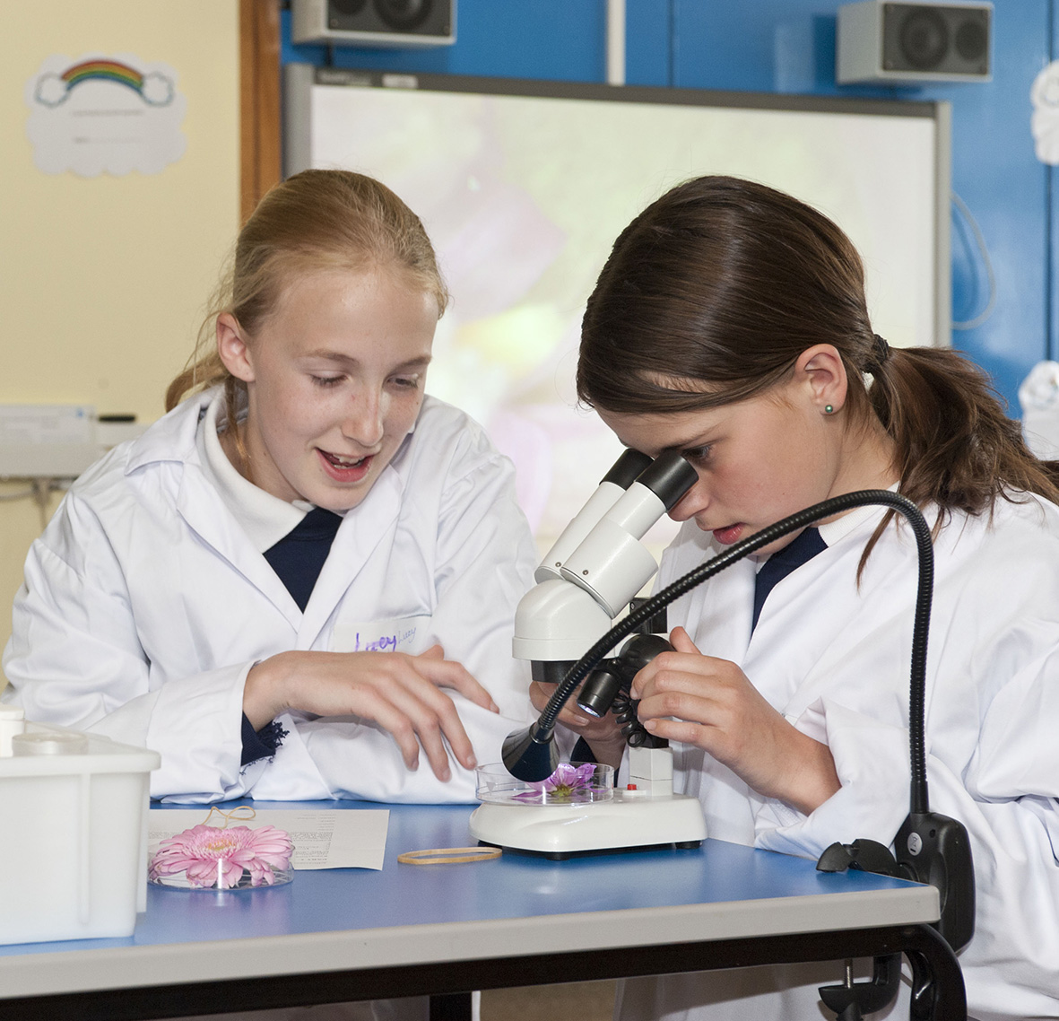
The “Stereomicroscope Challenge” in the classroom
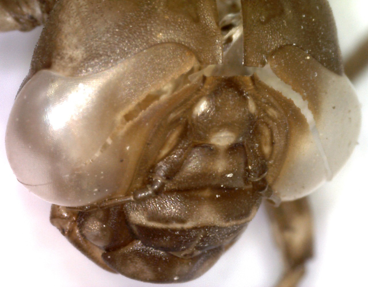
Entry for Science Image Award: Dragonfly pupal case
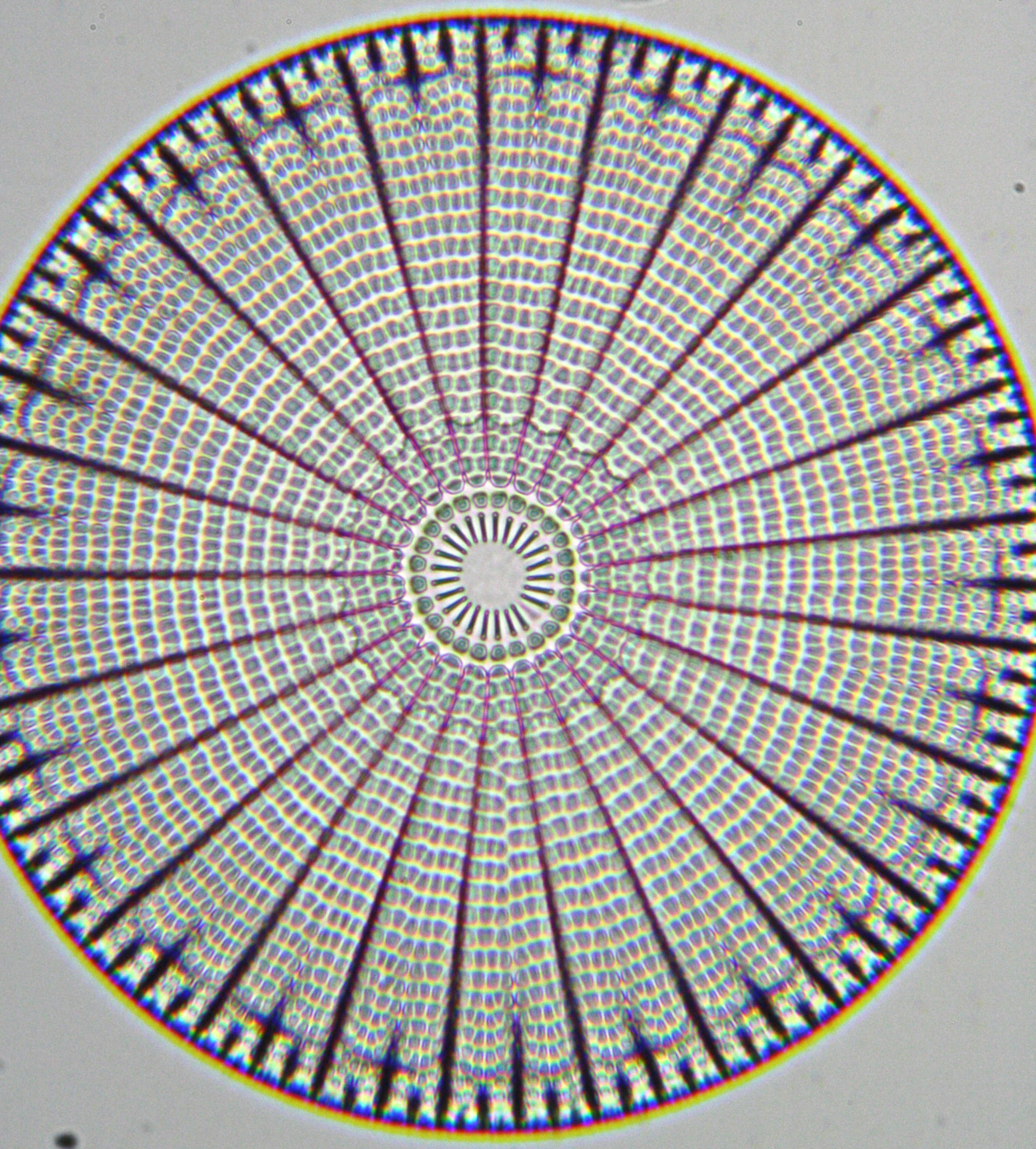
Image of diatom from £200 educational microscope
![]() This post is part of a series on science outreach. You can read the introduction to the series here and read other posts in this series here.
This post is part of a series on science outreach. You can read the introduction to the series here and read other posts in this series here.
 (1 votes)

 (No Ratings Yet)


 Embryonic stem cell (ESC) cultures display a marked heterogeneity in the expression of Nanog, one of several core pluripotency factors required for proper development in vivo. In addition, Nanog levels have also been shown to fluctuate in individual ESCs in vitro; however, the extent and functional consequences of these fluctuations in different pluripotency states has not been fully established. Now, on p.
  The balance between excitatory versus inhibitory neuron specification during development is crucial for sensory information processing in later life. The basic helix-loop-helix (bHLH) transcription factors Ascl1 and Ptf1a are crucial for establishing this specificity in the dorsal spinal cord, but how these two factors, which recognise a similiar DNA motif, can have opposite downstream effects is unclear. Now, on p.
  During blastocyst development, asymmetric cell divisions generate polar and apolar daughter cells, which organise into outer and inner positions, respectively, to form the trophectoderm (TE) and inner cell mass (ICM) lineages. The Hippo signaling pathway is crucial for setting up this early lineage specification, but how Hippo signaling relates to cell position and polarity remains unclear. In this issue (p.
  Cell polarity is fundamental for biological activity across many varied cell types within different animal species. Intracellular trafficking regulates the differential distribution of proteins that is fundamental to establishing cell polarity, but how cell polarity regulators exert their effects on trafficking machinery is largely unknown. Now, on p.
 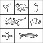 In March 2014, the RIKEN Center for Developmental Biology in Kobe, Japan, hosted a meeting entitled ‘Regeneration of Organs: Programming and Self-Organization’. Scientists from across the globe met to discuss current research on regeneration, organ morphogenesis and self-organization – and the links between these fields. As discussed by Daniel Goldman, a diverse range of experimental models and organ systems was presented at the meeting, and the speakers aptly illustrated the unique power of each. See the Meeting Review on p
 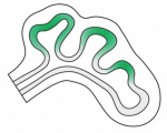 Branching morphogenesis is the developmental program that builds the epithelial trees of various organs, including the airways of the lung, the collecting ducts of the kidney, and the ducts of the mammary and salivary glands. R
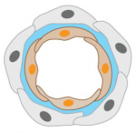 The formation of the vasculature is essential for tissue maintenance and regeneration, and understanding how vascular formation is coordinated in vivo can offer valuable insights into engineering approaches for therapeutic vascularization and angiogenesis. Here, Kyung Min Park and Sharon Gerecht discuss how the process of vascular development can be used to guide approaches to engineering vasculature. See the Review on p.