Applications for the 2015 class of the Instituto Gulbenkian de Ciência (IGC) PhD programme in Integrative Biology and Biomedicine are open until March 30th. This Programme is run in collaboration with ITQB (Instituto de Tecnologia Química e Biológica) and the Champalimaud Foundation.
The IGC PhD programme exposes students to a wide spectrum of different topics in the biological sciences. Unlike traditional programmes, students in our PhD programme are not required (or even encouraged) to choose a laboratory or topic until they have had a semester to discover the Institute’s scientific opportunities, and discuss them with their peers, postdocs and PIs.
The program normally accepts 9 to 12 students each year. Selected students receive full tuition and stipend support for 48 months, funded by Fundação para a Ciência e a Tecnologia (FCT; Portugal). The degree will be granted by Universidade Nova de Lisboa and Instituto Superior de Psicologia Aplicada.
Candidates of any nationality may apply, and there are no age restrictions. We do require a Master’s degree from candidates applying from countries within the Bologna agreement region, or those with similar undergraduate programs (3-year programs). Candidates from countries with a 4 or 5-year university degree program, are also eligible.
We seek highly motivated students, with total commitment to the pursuit of answers to original questions in a multidisciplinary environment. TheIGC PhD Programmewelcomes applications from candidates with university degrees in any field, including those outside the life sciences.
Research at the IGCrevolves around four main axes: Evolutionary Biology, Quantitative Biology, Integrative Cell and Developmental Biology, and Immunobiology. The broad-scoped nature of the IGC research programme favours original approaches to outstanding biological questions that promote bridges across different disciplines and methodologies.
Clickhere to access further details about the IGC PhD Programme and how to apply.
Learn more about the IGC PhD programmes in a video featuring PhD students, the Director of the PhD programme and the Director of the IGC:
Here are the highlights from the current issue of Development:
Time to update the mammalian CV
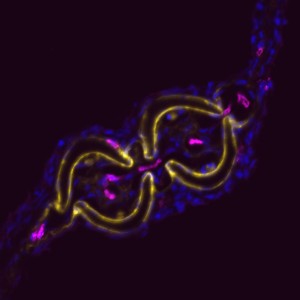
The dorsal aorta (DA) and the cardinal vein (CV) are the first vascular pair to form during development. Although it is generally accepted that the DA derives from angioblasts, the cellular origin of the mammalian CV is currently unknown. Now, on p. 1120, Rong Wang and colleagues reveal that the mammalian CV is formed, at least in part, from endothelial cells that originate from within the DA. Using markers specific to either aortic or venous-fated endothelial cells, the authors show that the DA contains a mixed population of endothelial cells with either venous or arterial identity, but that the number of venous-fated endothelial cells decreases over time. The authors use time-lapse microscopy to visualise the migration of endothelial cells away from the DA and to the CV, and further show that this process requires ephrin B2/EphB4 signalling. The authors propose a mechanism whereby ephrin B2/EphB4-mediated cell repulsion drives the segregation of venous-fated endothelial cells away from the DA, facilitating their movement to the CV.
Mammary stratification caught on camera
During development, select epithelial cells must undergo asymmetric division in order to generate a stratified epithelium. Previous studies have shown that basally positioned epithelial stem cells orchestrate this process, but the mammary epithelium has two distinct cell layers inside the basement membrane, so it remains unclear which cell type is responsible for stratification. Using elegant time-lapse imaging of three-dimensional mouse mammary epithelial cultures, Andrew Ewald and colleagues now reveal (p. 1085) that it is the apical luminal epithelial cells that divide vertically to generate the stratified mammary epithelium. The authors show that this is accompanied by the loss of tight junctions, as marked by ZO-1, as well as loss of apicobasal polarity in the new daughter cells. Importantly, the authors also demonstrate that this mechanism of stratification and loss of polarity operates during early oncogenesis in the mouse mammary epithelium. These data uncover a common cellular mechanism that underpins the developmental and oncogenic stratification of mammary tissue.
An epigenetic bag of TRX
The Drosophila Trithorax (TRX) protein plays a key role in maintaining active transcription of many master cell fate regulatory genes. Previously, TRX was thought to function mainly at the promoter by depositing the histone H3K4 trimethylation mark (H3K4me3) via its catalytic SET domain. However, recent findings have challenged this view, prompting new investigations into the function of TRX. Now, on p. 1129, Peter Harte, Feng Tie and colleagues reveal that TRX, along with TRX-related (TRR), is responsible for histone H3K4 monomethylation (H3K4me1), and not trimethylation, suggesting a role for TRX in stimulating enhancer-dependent transcription. In vivo studies support these findings, as a catalytically inactive form of TRX results in reduced H3K4me1, but no change in H3K4me3, in Drosophila embryos. The authors also show that TRX collaborates directly with CREB-binding protein (CBP) to promote robust H3K27 acetylation, which antagonises Polycomb silencing and may also stimulate enhancer-dependent transcription. These data provide exciting new insights into the mechanisms of epigenetic regulation in developing organisms.
Oct4: the plot thickens
The transcription factor Oct4 is well known for its role in maintaining pluripotency in vitro and for preventing ectopic differentiation of early embryos in vivo. Recent evidence also suggests a role for Oct4 in lineage specification; however, little is known about how this occurs and the manner in which Oct4 is required. Now, on p. 1001, Jennifer Nichols and colleagues report a non-cell-autonomous requirement for sustained Oct4 expression during primitive endoderm (PrE) specification. The authors confirm that maternal and zygotic Oct4 are not required for development to the blastocyst stage, but that conditional inactivation of Oct4 in the early blastocyst results in reduced expression of PrE markers Sox17 and Gata4, and a failure to generate PrE-derived tissue in chimeric assays. Surprisingly, the formation of PrE can be rescued if the conditionally inactivated Oct4 mutants are injected at the pre-blastocyst stage with wild-type embryonic stem cells, suggesting that Oct4 is not required to operate cell-autonomously in order to specify the PrE.
RAd migration in the cortex
The generation of layer-specific cortical neurons is fundamental to the circuitry of the developing and adult brain. Although the intrinsic drivers of neuronal specification are becoming increasingly understood, the extrinsic signals that guide migration and consolidate post-mitotic neuronal identity are less clear. In this issue (p. 1151), Shanthini Sockanathan and colleagues investigate the role of endogenous retinoic acid (RA) signalling in regulating the radial migration and laminar fate of post-mitotic cortical neurons. Using a dominant-negative RA receptor construct, the authors show that ablation of RA signalling in mouse embryos in utero not only delays migration of subsets of cortical neurons, but also results in a failure to maintain their correct regional identity following migration. This phenotype can be partially rescued by stabilised β-catenin, which the authors show is normally maintained by RA signalling. This work sheds light on the extrinsic mechanisms that control cortical neuronal development and has important implications for disorders in which cortical neuronal circuitry is de-regulated.
The production of healthy offspring depends on many factors spanning from intrinsic genetic elements to variations in the in uteroenvironment. Poor maternal diet is associated with increased risk of cardiovascular, metabolic and behavioural disorders during later life of the offspring, but how the developing embryo copes with maternal dietary stress has not been well characterised. Now, on p. 1140, Tom Fleming and colleagues investigate the compensatory mechanisms that are activated when the early embryo is challenged by poor maternal nutrition. Using quantitative imaging techniques and extensive marker analyses, the authors show that a restricted-protein maternal diet results in stimulated endocytosis within both the trophectoderm and the primitive endoderm of the early mouse embryo to overcome the shortfall in nutrient supply. The authors show that enhanced trophectoderm endocytosis occurs in response to reduced branched-chain amino acids and is mediated via RhoA GTPase signalling. This exciting finding is an important step in uncovering the cellular mechanisms that underpin disorders caused by poor maternal nutrition.
PLUS…
Cell competition: how to eliminate your neighbours
It has been known for years, based on studies of Drosophila, that viable cells can be eliminated by their neighbours through a process termed cell competition. New studies in mammals have revealed that this process is universal and that many factors and mechanisms are conserved. Here, Amoyel and Bach provide an overview of the mechanistic steps involved in cell competition and discuss recent advances in the field, which have shed light on how and why cell competition exists in developing and adult organisms. See the Review on p. 988
The Mediator complex: a master coordinator of transcription and cell lineage development
Mediator is a multiprotein complex that is required for gene transcription by RNA polymerase II. Here, Yin and Wang describe the most recent advances in understanding the mechanisms of Mediator function, with an emphasis on its role during development and disease. See the Primer on p. 977
The first Xenopus imaging workshop was held at the Xenopus Resource Centre at the Marine Biological Laboratory in Wood’s Hole, MA from November 17th – 22nd.
Kymograph of a beating heart in a Xenopus tadpole expressing GFP under a cardiac actin promoter. By Kyle Jewhurst, Tufts University.
20 international frog researchers, from Japan to the UK to the US, all looking to use microscopy for many different applications, attended the workshop. Stages of frog egg and embryo development, from late-stage oocytes to late tadpole stages, were of interest to the students attending; there were those wishing to dissect tissues for high resolution imaging compared to those wishing to image whole embryos; those wanting to do live imaging and those wanting to figure out how to get pictures of fixed samples used for in situ hybridisation. Although there were a wide variety of research goals, everyone was looking to use a similar set of techniques geared towards their own individual needs. I have included some examples of images taken at the course throughout this blog, so you can see what we got up to.
The course was very free in structure, which meant people with their own projects had time to prepare samples, whilst those with no agenda could try to learn how to use the microscopes and just play around with things. mRNAs for microinjection, and embryos from both wild type and various transgenic frog lines were provided, to give people an opportunity to try out experiments at the course. Microscopes from Zeiss, Nikon and Leica were made available by those companies, with reps from Nikon and Zeiss in attendance to help out and provide guidance on microscope use.
We were lucky to have a number of experts at the course: from the University of Southern California, Scott Fraser; from the University of Texas at Austin, John Wallingford, his graduate student Eric Brooks and postdoc Asako Shindo; and from the University of Pittsburgh, Lance Davidson and his graduate student Jo Shawky. With such a high teacher:student ratio, there was a lot of opportunity to get time with each of the instructors. Each instructor also gave talks about theory (for example, Scott Fraser gave an excellent overview of the theory of microscopy; whilst John Wallingford went through the do’s and don’ts of Photoshopping) and their own work, highlighting examples of the use of imaging in their own science. Some excellent stories were told – we got a sneak preview to the work of Asako Shindo, published just recently in Science (Science 2014, 343, 649-652).
In addition to the showcasing of developed scientific stories, we also had the opportunity to showcase our own work each day, with an evening show-and-tell of everyone’s best images. Not only did we get the opportunity to see what everyone else was working on – we also were able to critique the work of others and ourselves, to figure out how to improve the imaging technique for future use.
There was also the opportunity to make tools for frog manipulations – I now have my very own hair loops and eyebrow dissecting knife – staples for any frog researcher!
At the end of the day, after downing tools and critiquing images, we would get together for a quiet drink and a chat, an opportunity to talk to other researchers we might not necessarily meet at the conference. In addition to what you would normally talk about or see at conferences, because we were spending the day doing practical things, we were often able to look directly at someone else’s research as it happened and have discussions about what they were doing, looking at actual samples.
Video of Stage 8 Xenopus laevis embryo with EB3-GFP labeling microtubule + ends and H2B-RFP labelling histones magenta. By Romain Gibeaux, University of California, Berkeley.
Confocal z-stack image of sectioned Stage 45 tadpole, injected with tomato-Sert, showing nerve bundle connecting to otic placode. By Gary McDowell, Tufts University.
Real-time beating heart in transgenic Xenopus tadpole expressing GFP under cardiac actin promoter. By Kyle Jewhurst, Tufts University.
The course was the first of its kind that the frog community has held but it was very well received and all of us students had a great time. In particular there was a lot of help provided by the National Xenopus Resource staff, Esther Pearl and Christy Lewis, who provided frog and microinjection support for the students, as well as ensuring mRNAs were ready and available for people to try. Hopefully the course will be run again as we all appreciated what a great resource this could be for our fellow researchers in the frog community. Also, where else are you going to be able to generate the images for your own t-shirt!
T-shirt designed by Esther Pearl of National Xenopus Resource; images by Gary McDowell, Kyle Jewhurst and Emily Pitcairn, all of Tufts University.
Gary McDowell, Center for Regenerative and Developmental Biology, Tufts University
You can follow the National Xenopus Resource on twitter at:
Hi! I’m Tanvi, a third year PhD student in the Ettensohn Lab at Carnegie Mellon University in Pittsburgh, Pennsylvania, USA. The big question our lab is concerned with is how the genome encodes morphogenesis. We use transcriptional gene regulatory networks as a tool to study how transcription factors and signaling pathways regulate the expression of downstream genes involved in developmental anatomy. We use the sea urchin embryo as a model system as it is remarkably well-suited for regulatory network analysis and is a wonderful system for studying cell behaviors during development. The sea urchin embryo has a relatively simple morphology and is composed of only 10-12 different cell types. Large numbers of synchronously developing embryos can be obtained easily, and several specific cell types can be isolated relatively easily in large quantities. Just as importantly, these embryos are transparent and detailed cell behaviors can be observed in vivo, either during normal development or after different kinds of molecular manipulations, such as gene knockdowns. Detailed gene regulatory networks have already been established for various cell types of the embryo. The Strongylocentrotus purpuratus, or purple sea urchin, genome has been sequenced and a high quality transcriptome profile is available for different developmental stages. The genomes of several other echinoderms are at different stages of assembly and provide a terrific resource for comparative studies.
Our lab works with two different sea urchin species – the purple sea urchin, which is maintained at 15˚C, and the green sea urchin, which requires a warmer 23˚C. Both species are kept in temperature-controlled seawater tanks that are well-aerated and clean. The graduate students of the lab do most of the routine maintenance such as handling incoming urchin shipments, cleaning tanks, checking salinity and ammonia levels, feeding the urchins, and fishing out dead animals from time to time. Our extremely handy advisor deals with occasional tank breakdowns.
Purple sea urchins (S. pupuratus). Photo courtesy: ALAMY
Green sea urchins (L. variegatus). Photo courtesy: R. Wallocombe
We receive our adult sea urchins from California or Florida, depending on the species. When we need urchins, we simply send an email to a commercial supplier and a few days later we receive FedEx boxes containing fertile sea urchins cooled on ice! The urchins tolerate overnight shipping fairly well, but some release their gametes (or “spawn”) along the way. This could be a mild annoyance or a nightmare depending on how many animals spawn. Once the urchins arrive, we place them in separate cups (usually old yogurt containers) with seawater and float them on the tanks. We change the seawater every few hours. This acclimates the urchins to the tank temperature and water and minimizes stress. Any urchins that may have spawned require additional water changes and are kept isolated until they stop spawning.
Purple sea urchins recently transferred into their tank
After 6-8 hours, when all the urchins are stable, they are transferred into the tank. It is absolutely essential that urchins placed in the tank are not actively spawning, since seawater containing gametes (even a small number) will induce mass spawning in all the urchins! The next morning, I check on the tanks with fingers crossed, hoping that the urchins have not all spawned and died in a cloudy smelly mess at the bottom of the tank (this happens only rarely but is even more unpleasant than it sounds). The task of transferring newly arrived urchins to the tank is shared within the lab, which greatly reduces the stress and anxiety associated with it!
A day or two later, the urchins are delightfully low-maintenance. They cling to the walls of the tank and stay more-or-less stationary until we pluck them out to collect eggs or sperm. They can survive without feeding for many weeks, but we feed them sea kelp to keep them happy and full of gametes.
Spawning urchins shedding eggs (left) and sperm (right)
I work with embryos of the purple sea urchin. I usually work on embryos at the early gastrula stage, and set up embryo cultures about 24 hours before my experiment. Collecting gametes is a simple matter of shaking the urchins vigorously. This stresses them and causes them to release a fraction of their gametes, which I collect “dry” (sperm) or “wet” (eggs). I then plop the urchins into floating cups and later transfer them back into the tank. If I need more gametes than they provide willingly, I inject them with a 0.5M KCl solution, which induces them to shed all their gametes. Unfortunately, they usually do not survive this treatment. It is impossible to tell male purple sea urchins from females from external morphology alone, so picking the right urchin out of the tank is a matter of chance. I usually don’t need to go through more than 3-4 urchins before getting both eggs and sperm. The yield of gametes can be as high as a whopping 10-15 ml of eggs and 1 ml of sperm, if the animals are ripe. Sperm stays fresh for one week at 4˚C, which is convenient. Eggs are usually used the same day but can be stored overnight at 4˚C.
To set up an embryo culture, I simply mix eggs and sperm in a glass bowl filled with seawater! A small drop of sperm is more than enough to fertilize ~1 ml of eggs. This would, theoretically, give me about 1 million synchronously developing embryos! I check the culture under the microscope to ensure that almost all the eggs are successfully fertilized and then cover the bowl and keep it at 15˚C for 24 hours. The culture needs no maintenance and the embryos do not need to be fed unless a developmental stage later than the 72-hour larva is required.
The next day, I start my experiment by checking on the embryos. A good culture will have embryos that have developed synchronously with a morphology characteristic of their developmental stage. The culture may not develop well if the embryos are too concentrated or the quality of the eggs was poor. When the embryos reach the desired stage, I collect them by spinning in a clinical centrifuge.
My current experiments involve isolating a specific cell type of the embryo, the primary mesenchyme cells (PMCs), which build the embryonic endoskeleton. For this, I collect large amounts of 24-hour embryos and subject them to various washes using embryo-dissociation solutions. The embryos can withstand the mechanical forces of repeated centrifugation, which makes my life very easy! I separate the primary mesenchyme cells from other embryo cell types using a simple sucrose gradient. I am usually able to quickly isolate large amounts of PMCs. It is exceptionally rare to be able to isolate a specific population of early embryonic cells in such large quantities! The PMCs remain viable in culture for days, and carry out their developmental program autonomously. Needless to say, isolated PMCs have been an extremely useful model for me to study the development of a specific cell lineage. I am interested in understanding how genes involved in the morphogenesis of PMCs are regulated at the transcriptional level. I have been able to identify hundreds of genes differentially expressed in PMCs at the early gastrula stage using isolated PMCs. I am now working towards locating cis-regulatory elements mediating the expression of these PMC-enriched genes. My ultimate goal is to construct a detailed and comprehensive gene regulatory network that will explain how PMC morphogenesis is encoded in the genome.
Other lab members routinely set up and use embryo cultures for many interesting experiments involving injection of morpholinos or DNA constructs, transplantation or removal of various cell types, drug treatments to block certain developmental pathways, in situ hybridizations to analyze spatial gene expression patterns, and other kinds of experiments. The ease of working on sea urchins enables us to spend less time worrying about obtaining embryos and more time focusing on uncovering the mysteries of development!
This post is part of a series on a day in the life of developmental biology labs working on different model organisms. You can read the introduction to the series here and read other posts in this series here.
We are Alicia (1st year PhD student) and Marion (3rd year PhD student) and we work in an ascidian lab at CNRS, Montpellier in France. Alicia’s aim is to compare gene regulatory networks of Ciona intestinalis and Phallusia mammillata during early embryogenesis. Ciona is much better characterised, therefore, Alicia has so far only worked with Phallusia. Marion works on enhancers and aims to better understand the sequence determinants of their activity, using Ciona intestinalis as a model organism.
Phallusia mammillata in a jazzy setting: they are a brilliant model organism to study developmental biology. Unfortunately, we no longer have the blue backdrop for our tanks but we do have a starfish!
Working with ascidians
In the world, more than 70 labs work with ascidians[1]. Ascidians belong to the tunicate group; these are marine invertebrates that belong to the phylum Chordata. Tunicates are the sister group of vertebrates with whom they share some features of chordate embryonic developmental programme such as the formation of tadpole larvae. Due to their small and rapidly evolving genome, they are powerful organisms to study chordate evolution. Ascidians are simple genetically because they have not undergone whole genome duplication events and exhibit limited gene redundancy and compact regulatory regions.
The sea squirt Ciona intestinalis is a good model to study developmental biology; they have rapid development and the embryos have invariant lineages. Specialised databases such as Aniseed[2], which was created by the Lemaire lab, have catalogued an atlas of expression profiles, cell lineages and single cell morphological properties and also gene, transcript and protein data. One can retrieve information about each cell in the embryo through different identification markers (name, position), which facilitates the analysis and interpretation of results.
Phallusia mammillata and Ciona intestinalis are thought to have diverged about 300 million years ago but they still share a common invariant embryonic cell lineage. Phallusia arrived in our lab ~3 years ago because of its experimental advantages over Ciona: they have more eggs, transparent embryos and show much less seasonality in embryo production. After a few optimisations, we can now perform all of Ciona’s routine experiments on Phallusia. Comparative genomics approaches can be undertaken to study ascidian developmental programs as many ascidian genomes have now been sequenced.
Ascidians are simple organisms making them easy to analyse. They have a much lower cell number during embryogenesis when compared to vertebrates: 110 to 112 at the onset of gastrulation and about 2600 cells at the tadpole stage. Due to their invariant cell lineage, a phenotype can be characterised with single cell resolution. Electroporation provides a strong tool to easily and quickly make hundreds of transgenic embryos making ascidians a powerful model to study gene regulation.
No Ciona were harmed in the making of this video – our colleague Mathieu playing around with their siphons.
The routine in the lab
One of our favourite practicals is collecting the eggs and sperm from our dear Phallusia; this is done first thing in the morning in our 17°C embryo room. It is always exciting to see the volume of eggs that we manage to collect; it is cause for celebration when the Phallusia has 1.5ml or more. Sperm is not quite as exciting as we only require 10µl; nevertheless, it is still much appreciated.
Dechorionation is performed to remove the chorion and follicular cells which allows a better visualization of the embryonic development and facilitates any further experiments. Phallusia dechorionation lasts a couple of hours. In Ciona, this process is very fast and it is performed after fertilisation. Eggs are now very fragile and should be gently manipulated. Fertilisation is rather fast: sperm is activated (under the stereoscope, we can see the sperm become agitated), and then mixed with eggs. Most of the eggs are fertilized at the same time and thousands of embryos will develop in a synchronous way. From this point on, 20 minutes remain before the first cleavage. During this short time frame, electroporations can be performed.
Collecting our embryos at each stage is a day-long process; it takes the embryos up to 5 hours to reach early gastrula. We try to disperse the embryos as much as possible in the plates as they develop to avoid that they touch and fuse together to create what we call little monsters. Once they have reached the stage of interest, we fix the embryos and colour them if needed. From here on, our embryos can be kept for many months.
Microinjection is a very common technique in ascidian laboratories. It is used for 2 main goals: exogenous expression (microinjection of mRNA) and gene expression knock down (microinjection of morpholinos). Ascidian oocytes are particularly sensitive to microinjection when compared to other species and easily explode or get stuck to the material during microinjection until you get the technique right (we are lucky to have our post-doc Ulla). It is a rather slow technique demanding a high degree of patience and precision but its potential is fantastic. Unlike electroporation, microinjection allows for ubiquitous expression without mosaicism. You can also use interesting control situations with a single embryo. Microinjection of mRNA in a single cell at the 2 cell-stage allows to have half of an embryo expressing an exogenous mRNA whilst the other half remains wild type. Ascidians have bilateral symmetry until late embryonic stages. Although in terms of analysis microinjection techniques are very appealing they provide a much lower number of transgenics to work with than electroporation techniques.
Our ascidian tanks – a main attraction within our building where many come to admire our animals.
Shipment and animal care
We get a weekly shipment of Phallusia and Ciona. Before they are transferred to the tanks, the tanks are thoroughly cleaned during which time the animals are kept in the water in which they were transported. This waiting period means that their water temperature can gradually warm up to match the temperature of the tank water. We do not want to disturb the animals by transferring them before they are ready. We have to order each shipment a week in advance because a special boat and diving expeditions are organised to collect the animals by Roscoff Marine station. We thus need to plan ahead how many animals we need and order only what is required.
Our tanks are in the lab where we work so we regularly check how our animals are doing. To assess if the animals are healthy, we give the tanks a little tap to see if the animals respond by closing their siphons. The colour (and the smell) of the animals can also be an indicator of their health as they turn darker when they die. We have to remove dead animals as soon as possible because they raise nitrite levels in the tanks, which is bad for the other animals. Our sea squirts are kept in natural sea water which is delivered by an oyster farmer.
Working with wild animals
We have not yet started to culture ascidians in our lab; therefore, we work with wild animals delivered from Brittany. Ciona’s fertility depends greatly on sea water temperatures; the period during which they produce eggs spans from April to November in France. So we have to take this into account when planning our experimental work. Funnily enough, our experiments greatly depend on the climatic conditions 1000km from our lab! At each delivery, we could be in for a surprise: did our animals survive the trip? Do they have eggs? Will they develop properly? One pleasant surprise we occasionally get is clandestine animals attached to the Phallusia such as starfish, brittle stars, urchins, worms, annelids or porcelains and the list goes on. At the moment, we have a beautiful purple starfish as a lab pet and we feed her mussels.
Acknowledgments – We would like to thank our postdoc Ulla-Maj Fiuza for her time helping us write this article (and for her microinjection knowledge).
This post is part of a series on a day in the life of developmental biology labs working on different model organisms. You can read the introduction to the series here and read other posts in this series here.
This is a call for the registration to the EMBL Master Course Bioimage Data Analysis to be held from Monday, 12 May – Friday, 16 May 2014.
This course will focus on computational methods for analyzing images of proteins, cells and tissues, to boost the learning process of participants who have an immediate need to deploy image analysis in their own research. The course extends from the basic foundations of image processing and programming to the actual implementation of workflows using scripting in ImageJ macro and Matlab languages. The students will be guided to design such scripts to address some practical bioimage analysis projects. Among those course-projects, topics that are interesting for developmental biologists could be:
Quantitative Evaluation of Multi-cellular Movements in Drosophila Embryo
Tumor Blood Vessels: 3-D Tubular Network Analysis
For details on other topics, please visit the course website. We aim to gather expert knowledge to organize a world-leading course for image analysis in the fields of biophysics, cell biology and developmental biology.
The course will take place in Heidelberg, Germany at the EMBL Advanced Training Centre. Registration and motivation letter deadline is February 25, 2014. Please visit our course website for more details:
Why iodine deficiency during pregnancy may have disastrous consequences
Higher mammals, such as humans, have markedly larger brains than other mammals. Scientists from the Max Planck Institute of Molecular Cell Biology and Genetics (MPI-CBG) in Dresden recently discovered a new mechanism governing brain stem cell proliferation. It serves to boost the production of neurons during development, thus causing the enlargement of the cerebral cortex – the part of the brain that enables us humans to speak, think and dream. The surprising discovery made by the Dresden-based researchers: two components in the stem cell environment – the extracellular matrix and thyroid hormones – work together with a protein molecule found on the stem cell surface, a so-called integrin. This likely explains why iodine deficiency in pregnant women has disastrous consequences for the unborn child, affecting its brain development adversely – without iodine, no thyroid hormones are produced. “Our study highlights this relationship and provides a potential explanation for the condition neurologists refer to as cretinism”, says Wieland Huttner, Director at the Max Planck Institute in Dresden. This neurological disorder severely impairs the mental abilities of a person.
In the course of evolution, certain mammals, notably humans, have developed larger brains than others, and therefore more advanced cognitive abilities. Mice, for example, have brains that are around a thousand times smaller than the human one. In their study, which was conducted in cooperation with the Fritz Lipmann Institute in Jena, the researchers in Dresden wanted to identify factors that determine brain development, and understand how larger brains have evolved.
A cosy bed for brain stem cells
Brain neurons are generated from stem cells called basal progenitors that are able to proliferate in humans, but not in mice. In humans, basal progenitors are surrounded by a special environment, a so-called extracellular matrix (ECM), which is produced by the progenitors themselves. Like a cosy bed, it accommodates the proliferating cells. Mice lack such ECM, which means that they generate fewer neurons and have a smaller brain.
The scientists therefore conducted tests to see whether in mice, basal progenitors start to proliferate if a comparable cell environment is simulated. The result: “We simulated an extracellular matrix for the brain stem cells using a stimulating antibody. This antibody activates an integrin on the cell surface of basal progenitors and thus stimulates their proliferation”, explains Denise Stenzel, who headed the experiments.
Because a requirement of thyroid hormones for proper brain development was previously known, the researchers blocked the production of these hormones in pregnant rats to see if their absence would inhibit basal progenitor proliferation in the embryos. Indeed, fewer progenitors and, consequently, neurons were produced, likely explaining the abnormal brain development in the absence of thyroid hormones. When the action of these hormones on the integrin was blocked, the ECM-simulating antibody alone was no longer able to induce basal progenitor proliferation.
A combination of ECM and thyroid hormones thus appears necessary for basal progenitors to proliferate and produce enough neurons for brain development. Human brain stem cells produce the suitable environment naturally. “That is probably how, in the course of evolution, we humans developed larger brains”, says Wieland Huttner, summing up the study. The research produced another important finding: “We were able to explain the role of iodine in embryonic brain development at the cellular level”, says Denise Stenzel. Iodine is essential for the production of thyroid hormones, and an iodine deficiency in pregnant women is known to have adverse effects on the brain development of the unborn child.
– Researchers at IRB and IBMB-CSIC, in Barcelona, and at the University of Wageningen, in the Netherlands, reveal how auxin hormone-regulated proteins activate developmental genes in plants.
– Auxins are key components of plant growth and have many applications in agriculture. The biomedical application of these hormones are also being addressed.
– The study is published today in the scientific journal Cell.
A joint study published in Cell by the teams headed by Miquel Coll at the Institute for Research in Biomedicine (IRB Barcelona) and the Institute of Molecular Biology of CSIC, both in Barcelona, and Dolf Weijers at the University of Wageningen, in the Netherlands, unravels the mystery behind how the plant hormones called auxins activate multiple vital plant functions through various gene transcription factors.
Auxins are plant hormones that control growth and development, that is to say, they determine the size and structure of the plant. Among their many activities, auxins favor cell growth, root initiation, flowering, fruit setting and delay ripening. Auxins have practical applications and are used in agriculture to produce seedless fruit, to prevent fruit drop, and to promote rooting, in addition to being used as herbicides. The biomedical applications of these hormones as anti-tumor agents and to facilitate somatic cell reprogramming (the cells that form tissues) to stem cells are also being investigated.
The effects of auxins in plants was first observed by Darwin in 1881, and since then this hormone has been the focus of many studies. However, although it was known how and where auxin is synthesized in the plant, how it is transported, and the receptors on which it acts, it was unclear how a hormone could trigger such diverse processes.
At the molecular level, the hormone serves to unblock a transcription factor, a DNA-binding protein, which in turn activates or represses a specific group of genes. Some plants have more than 20 distinct auxin-regulated transcription factors. They are called ARFs (Auxin Response Factors) and control the expression of numerous plant genes in function of the task to be undertaken, that is to say, cell growth, flowering, root initiation, leaf growth etc.
Using the Synchrotron Alba, in Cerdanyola del Vallès (Barcelona), and the European Synchrotron, in Grenoble, structural biologist Dr. Miquel Coll and his team analyzed the DNA binding mode used by various ARFs.
For this purpose, the scientists prepared crystals of complexes of DNA and ARF proteins obtained by Dolf Weijers team in Wageningen, and then shot the crystals with high intensity X-rays in the synchrotron to resolve their atomic structure. The resolution of five 3D structures has revealed why a given transcription factor is capable of activating a single set of genes, while other ARFs that are very similar with only slight differences trigger a distinct set.
“Each ARF recognizes and adapts to a particular DNA sequence through two binding arms or motifs that are barrel-shaped, and this adaptation differs for each ARF,” explains Roeland Boer, postdoctoral researcher in Miquel Coll’s group at IRB Barcelona, and one of the first authors of the study.
The ARF binding mode to DNA has never been described in bacteria or animals. “It appears to be exclusive to plants, but we cannot rule out that it is present in other kingdoms. Our finding is highly relevant because we have revealed the ultimate effect of a hormone that controls plant development on DNA, that is to say, on genes.” says Miquel Coll.
Reference article:
Structural basis for DNA binding specificity by the auxin-dependent ARF transcription factors
D. Roeland Boer, Alejandra Freire-Rios, Willy van den Berg, Terrens Saaki, Iain W. Manfield, Stefan Kepinski, Irene López-Vidrieo, Jose Manuel Franco, Sacco C. de Vries, Roberto Solano, Dolf Weijers, and Miquel Coll
Cell (2014) 156, 577-589 http://dx.doi.org/10.1016/j.cell.2013.12.027
This article was first published on the 30th of January 2014 in the news section of the IRB Barcelona website