The community site for and by
developmental and stem cell biologists

Vote for a Development cover – Woods Hole – Round 3

Posted by , on 19 July 2012

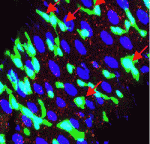
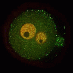
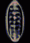
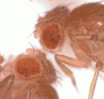
This week you don’t only get to decide which essay, from our competition, will appear in Development (see nominations, and the poll later today), but it’s also time to choose another cover from images from the 2011 Woods Hole embryology course. Vote in the poll below the images for the one you would like to see on the cover of Development. (Click any of the images to see a bigger version.) Poll closes on August 6, noon GMT.

1. 5th instar imaginal hindwing disk from the Painted Lady butterfly, Vanessa cardui. Immunostained for Engrailed in red. All nuclei are revealed by DAPI staining (blue), and trachae are shown in green. This image was taken by Alessandro Mongera, Maria Almuedo Castillo, and Jakub Sedzinski.

2. 3rd instar wing disk from Drosophila melanogaster. Triple flip-out clone system (courtesy of Melanie Worley and Iswar Hariharan) was used to reveal various cell lineage clones shown in yellow, blue, and purple. All nuclei shown in gray (DAPI). This image was taken by Lynn Kee.

3. Head of an adult C. elegans. DiI staining (red) reveals environmentally exposed neurons, while the JR797 GFP line allows visualization of all neurons (blue). This image was taken by Eric Brooks and John Young.

4. Ventral view of stage 16 Drosophila melanogaster embryo immunostained for Tropomyosin (green; muscle), Pax 3/7 (blue; segmentally repeated nuclei in CNS and ectoderm), and anti-HRP (red; cell bodies and axons of the nervous system). All nuclei shown in gray (DAPI). This image was taken by Julieta María Acevedo and Lucas Leclere.


While you’re here, why not also vote for the winner of our essay competition?

Thumbs up (5 votes)
Loading...

Tags: ,
Categories: Images

Navigate the archive

Use our Advanced Search tool to search and filter posts by date, category, tags and authors.

Essay nominee 2 – There’ll be dragons?

Posted by , on 18 July 2012

Below is the second of two essays nominated in our essay competition “Developments in development”. The other nominated essay appeared on the Node yesterday. Please read both essays, and come back on or after July 19 to cast your vote for the winner. The winning essay will appear in Development later this year. See the announcement for author bios, and read the first essay here.

There‘ll be dragons? – The coming era of artificially altered development

By Máté Varga

 As part of their yearly April’s Fool prank series, in 2006 The Economist ran a short article in their “Science and technology” section about a fictional company that planned to create mythical creatures in flesh and blood on demand. Scientists working for GeneDupe (as the company was called), would use their extensive knowledge about development, to introduce the genome of a lizard or horse into a computer, and with the help of evolutionary algorithms “mutate” it in a way that the new genome would be capable of giving rise to a dragon or unicorn, respectively. The DNA would be synthesized, introduced into an enucleated egg, and presto, in only a few months time the happy customer would walk away with a mythical pet.

Although the story contained several giveaways about the spoof, and anyone vaguely familiar with the status of the field could immediately see that the whole thing is nonsense, the article still had some aura of plausibility, as any good hoax should. More naive readers could have imagined it happening, and even science-types could have wondered, what if…?

Six years on, we are still far, far away from possessing the knowledge that would be required to recreate GeneDupe’s feat in the real world. But, arguably, we are inching closer. As more and more model and non-model organisms have their genomes unraveled, as we learn how genes are organized in developmental networks, as we decipher their enhancers and epigenetic codes, and find the exact mutations that can cause evolutionary changes, we do progress towards a future when GeneDupe-style simulations will become not only possible, but also common and, in certain situations, desirable.

This should not come as a surprise to anyone. After all, we have been tinkering with embryos for hundreds of years and even directly with genes for decades in order to get a comprehensive understanding of how a single fertilized egg can become a complex three-dimensional creature. A creature with intricate inner organ systems that can maintain it throughout its life and help it to react to its environment, adapt and procreate to have its genes passed on. Putting it more bluntly: just like Sylar, the negative protagonist of the TV series “Heroes”, we, developmental biologists, dissect things to see “what makes them tick”.

But once we have that knowledge, an obvious next step would be to try to use it, to control and alter developmental mechanisms in predictable ways – first in silico, then in vivo. This does not seem like a huge step from today’s transgenic and genome editing technologies, yet it would mean genome modification on a different level. Today’s transgenic approaches are, to a certain extent, still based on trial and error, as a comprehensive understanding of how gene regulation works is still missing. Acquiring that knowledge will enable us to introduce changes in the transcriptional program with surgical precision. And then, from mere tinkering-apprentices we would graduate to become Master Craftsmen, on par or not so far behind the Tinkerer-in-Chief, Natural Selection.

We have to be prepared that when we get the ability to do this sort of genetic engineering, the media coverage and reaction will be overwhelming. As Philip Ball describes in his wonderful book “Unnatural”, most human cultures possess a primordial fear of everything they consider to be against Nature’s will, and more often than not hold firmly to the irrational conviction that anything that does not exist in nature is against this will. This psychological reaction was on full display three decades ago, during the IVF debate, and can be seen these days in the gut-driven opposition against genetically modified plants (GMOs).

Therefore, it doesn’t take an oracle to foresee the debate that will ensue once a targeted approach of altering development will be available. The ghosts of Dr. Moreau and Frankenstein will be promptly summoned, scientists will be accused (yet again) of playing God. Outlandish claims will be made, biological equivalents of nanotech’s infamous “grey goo” meme will appear. Ordinary people might think that the end of the world is nigh, crazy biologists are just moments away of unleashing legions of deadly creatures on the unsuspecting inhabitants of the planet.

But amongst the brouhaha, valid arguments and well-grounded fears about the possible misuse of the technology will also be voiced. We should not only listen to these, but also anticipate them. As the recent public debate about the research on artificially modified influenza strains shows, even the scientific community can be caught off-guard with technological progress.

A blanket ban on the use of the novel technology would be useless and unworkable. Once the knowledge exists, people will try to use it. However, to state the obvious, what is doable, is not necessarily desirable, so we ought to seek a consensus on what should be ethically acceptable and what not. The rules should be straightforward and easily understandable even by a lay audience, as this is the only way not to repeat the PR-debacle of plant geneticists in the GMO-debate. For example, genetic modification of rare species threatened by uncurable diseases in order to avoid extinction could be favoured. Similarly, in order to protect whole ecosystems from collapse, altering key ecological species that can not adapt fast enough to the accelerating climate change could be considered.

In contrast, GeneDupe-style designer modification of pets or any species in a way that would make them commercially attractive, but would cause unnecessary harm and suffering for the animals, should be forbidden.

Ethically much murkier areas will be the ones that are directly related to developmental biology itself. Evolution does not plan in advance and, therefore, the phenotypes that we can observe today are often the result of historical contingencies (think of the inverted eye of vertebrates). A question that will be surely on several people’s mind is what if one removes these contingencies or introduces different ones. Will Stephen Jay Gould’s much quoted prediction about “rewinding the tape of life” be sustained, as it is widely agreed, and will different outcomes spring to life? Deciding whether this kind of research should be banned altogether, or allowed on a case by case basis, might have long lasting consequences.

And then there is the ethically most loaded question of them all, should the use of such technology on humans be even considered? Given the sorry history of eugenics in the past century, the answer seems like a forgone conclusion. Every single attempt to create the “perfect human” had calamitous consequences and it is hard to see currently any cold-headed rationale that could make a strong case for such programs. But we have to be aware that just as attempts to introduce gene therapy to cure particular diseases were shadowed by rumours of “gene doping”, the emergence of targeted development-altering methods might create the demand in some shadowy corners of the world to use it on humans.

All this, should make us wary. If the emergence of such new technology will catch us, biologists unprepared, in spite of achieving breathtaking things, like modern day Daedaluses we will pay a heavy price for our ineptitude to use our knowledge wisely.

——-

To vote for Máté’s essay, go to the poll

Thumbs up (20 votes)
Loading...

Tags: , ,
Categories: Discussion, Research

Essay nominee 1 – An Excitingly Predictable ‘Omic Future

Posted by , on 17 July 2012

Below is the first of two essays nominated in our essay competition “Developments in development”. The other nominated essay will appear on the Node tomorrow. Please read both essays, and come back on or after July 19 to cast your vote for the winner. The winning essay will appear in Development later this year. See the nominee announcement for author bios, and read the second essay here.

An Excitingly Predictable ‘Omic Future

By Joanna Asprer

 Developmental biology is an old field of study that finds its origins in the 18th century when Wilhelm Roux ablated cells from the two- or four-cell frog embryos and observed that the remaining cells could not give rise to an entire embryo. It is now more than a century after the publication of that seminal work, and we can attest to the saying that the more things change, the more they stay the same for here we are, still preoccupied with the same issue of stem cell pluripotency.

That matter aside, developmental biology has definitely come a long way. Where before, Roux and his contemporaries sought to study embryonic development through microsurgical manipulation of early embryos, we now alter model organisms on a genetic level, creating knockouts and transgenics, and we routinely analyze them on a molecular level by performing Q-RT-PCRs, ChIP assays, Co-IPs, immunohistochemical analyses and so on.

The most recent additions to our arsenal, the microarray and next gen sequencing, have been so pivotal to the progress of the field that they have ushered in an entirely new era, the ‘omics era. We are no longer limited to the analysis of one or several genes at a time; instead, we attempt to take in the full breadth of the changes that occur as development progresses, starting from the level of organisms, organs, tissues, cells, and molecules, to the level of genomes, epigenomes, and transcriptomes.

Interestingly, the field of developmental biology was slow to incorporate those technologies:

The pre-translational ‘omics era began in 1995 when the laboratory of Patrick Brown published a study about 45 Arabodopsis genes that were analyzed via microarray. The technology was quickly applied by those in the fields of cancer genetics, medical genetics, immunology and even drug development. It was only in 1999 that the first developmental papers studying Drosophila genes via microarray were published.

The lag seemed concordant with the general inaccessibility of the technology in its infancy. In fact, in the 1990s, scientists complained about restricted access to microarray technology, citing high cost, limited availability and patent disputes. Biotech companies responded by adding lower cost arrays to their portfolios, which prior to that mainly catered to industry R and D scientists. Meanwhile, institutions began to build core facilities to make the technology available to more scientists. Today, a Pubmed search using the keywords “microarray and embryonic development” yields close to 2000 results, and this is by no means an exhaustive search of all developmental studies that made use of array technology.

Next gen sequencing technology has also existed in some form as far back as the 1990s when it was only useful to developmental biologists for generating EST libraries. Soon after the technology was commercialized in 2005, it was used to catalog developmental miRNAs in several model organisms, an undertaking that has proven essential for the continued progress of developmental biology. However, medical genetics, cancer genetics, microbiology and virology made much bigger strides while using the same technology. This may be explained by the fact that the technology was primarily designed to sequence genomes and is understandably valuable in fields where there is great genetic variation within sample populations, and where there is a constant supply of new genomes to sequence.

For developmental biologists who make use of isogenic strains of organisms with fully-sequenced genomes, it was the subsequent application of next gen sequencing to the quantitative analysis of gene expression and gene regulation via RNA-seq and ChIP-seq that triggered its complete assimilation into the field. Still, even with the development of more relevant applications of the technique, there are currently under a hundred papers on Pubmed that can be located with the search words “RNA seq” or “ChIP seq” and “embryonic development“. This is again not an exhaustive list, but it gives us an idea that next gen sequencing is not as widespread or accessible as we would like it to be.

Both, next gen sequencing and microarrays are definitely not seen as routine tools on the level of even Q-RT-PCRs, at least not in the typical lab. Often, they are used as screens, performed with great cost and effort once in a blue moon; the intervening time is used to parse through the data and validate a multitude of individual results using more traditional techniques. This trend will continue for some time yet, but we are now at another turning point where the technological advancements with semiconductor chips are allowing us to sequence a human genome in one day for a mere thousand dollars, a far cry from the decade and billions of dollars that were needed to complete the Human Genome Project in the year 2000.

With the growing accessibility of next gen sequencing, more and more labs will do it more and more often. At some point, the affordability of new technologies will take high throughput experiments from the domain of core facilities and give direct, in-lab access to a wider variety of scientists; someday, hopefully soon, we will be able to do the high throughput experiments in our own labs, comparing what makes Cell A different from Cell B, answering questions about how Process C changes the behavior or identity of Cell A, and finding out what DNA or RNA sequences Protein D binds to in order to induce the development of Cell B from Cell A in response to Process C.

Based on what happened in response to the introduction of microarray cores, we can predict that this unparalleled access to high throughput technologies will accelerate progress in the field. True, the democratization of high throughput experiments is bound to bring with it a host of problems associated with inexperience, like misuse of equipment, faulty experimental design or misinterpretation of results. However, with time it will encourage a wider distribution of expertise which, combined with each lab having greater control over the equipment, will allow the execution of more experiments with possibly more complex experimental designs that will allow us to answer questions we could not tackle before.

So much data will be generated in this sequencing revolution that our search for the needle in a haystack will evolve into a search for the critical pieces of information hidden in terabytes of data. As the challenge shifts from data-gathering to data-mining, bioinformatics will be more important than ever before. There will be great demand to recruit bioinformatics experts in order to address developmental issues, and even classical developmental biologists will be compelled to get training if only to be able to design experiments properly and understand their collaborators sufficiently.

With teams of developmental biologists and bioinformatics experts working together, I envision a scenario where we would be able to speed up the process of mapping mutations generated by random mutagenesis using whole genome sequencing. It may be a long while yet before this becomes thinkable in the fly field, but it might already represent an economical alternative to maintaining large mouse colonies for long periods of time while doing segregation analysis.

Along those same lines, many mouse labs have experienced the tragedy of running across a mutant with an exciting phenotype only to observe that the phenotype has low penetrance, or worse, has disappeared with a transition from one genetic background to another. In this situation, it is quite likely that there is a modifier influencing the gene-of-interest. At the moment, we are given only two extreme options: throw in the towel and move on to something more tractable, or bull-headedly and blindly backcross into a defined background or two until the phenotype resurfaces or exhibits itself consistently.  Soon we may have the third, fast and productive option of sequencing genomes and finding the genetic change or polymorphism that segregates with the phenotype or the lack thereof. Thus, the lab tragedy can turn into a serendipitous discovery about the pathway the lab is working on.

Clearly, affordable next gen sequencing will be a great boon to basic scientists, but the ‘omics era comes at a time where we are experiencing a strong push for translational or disease-relevant science, courtesy of the grim economic outlook and the corresponding decrease in research funding and the demand for responsible use of public funds. The alignment of technological availability and the renewed thrust towards improving human health will work out very well for the field of medical genetics where the recent launch of whole exome sequencing for diagnostic purposes will reveal novel disease-causing mutations. This development promises to benefit other fields, including ours, as many of the discovered mutations will inevitably affect developmental genes and the human phenotype-genotype correlations will provide a nice complement to the studies done with model organisms.

Meanwhile, in developmental cell biology, great emphasis will continue to be placed on the study of different kinds of stem cells and their differentiation into cells that can be used for therapeutic purposes; this endeavor can benefit from high throughput analyses as well: At present, we rely on one or several markers to characterize pluripotent stem cells or the differentiated cells that we have turned them into, but we know that expression of one gene does not a cell type make. Even our markers tend to be expressed by multiple cell types, all of which represent possible end points of pluripotent stem cell differentiation. Moreover, expression of a certain marker doesn’t guarantee that an induced cell will behave and function exactly like the predicted cell type. Thus, there will be a point when it will be unacceptable to use only a few markers in identifying cells for therapeutic use, and instead transcriptome fingerprinting will be the norm.

I have no doubt that many completely new things will eventually revolutionize our field, but in my opinion, the future of developmental biology lies in the inevitable integration of the great trends that saw their beginnings in the last two decades: It will feature extensive use of next gen sequencing and bioinformatics to elucidate pre-translational ‘omics while finding the etiology of developmental diseases and refining the protocols for regenerative medicine. This may well be considered as a known and fixed future, but its predictability is not one that precedes boredom. For me, this is a future that is as exciting as any unknown development that anyone could imagine.

—–

To vote for Joanna’s essay, go to the poll.

Thumbs up (15 votes)
Loading...

Tags: , ,
Categories: Discussion, Research

Essay competition nominees

Posted by , on 17 July 2012

We’re pleased to announce the nominees of the first essay competition run by Development and the Node! We received a total of twelve eligible submissions – about epigenetics, data, model organisms, stem cells, and a range of other interpretations of the theme “Developments in development”. The submissions came from PhD students, recent graduates, postdocs, and lab heads on five continents.

Our two judges, Olivier Pourquié (Editor-in-Chief of Development) and Claire Ainsworth (science writer) read through the (anonymized) submissions to select the top essays based on content and writing.  They agreed on two essays both deserving to be in the running for the final prize.

And the nominees are….

Joanna Asprer – “An Excitingly Predictable ‘Omic Future”

Joanna has a doctorate in developmental biology from Baylor College of Medicine and has worked on brain, spinal cord, and inner ear development. She is currently a postdoc in Uma Lakshmipathy’s lab at Life Technologies where she studies embryonic and induced pluripotent stem cells.

and

Máté Varga “There’ll be dragons? – The coming era of artificially altered development”

Máté is a group leader at the Department of Genetics of Eötvös Loránd University in Hungary, where he works on tectal stem cells and early dorso-ventral patterning in zebrafish. He previously worked as a postdoc at University College London, and holds a PhD from the University of Pennsylvania.

The two nominated essays will appear in full on the Node today and tomorrow, and from Thursday onward you will be able to vote for your favourite in this poll. The final winner on August 15 will be published in Development. Both nominees will also receive an Amazon gift certificate worth £50.

Click the titles above to read the essays

Thumbs up (5 votes)
Loading...

Tags:
Categories: News

Postdoctoral Position on WNT signaling in craniofacial development, University of British Columbia, CANADA

Posted by , on 15 July 2012

Closing Date: 15 March 2021

A postdoctoral position is available immediately in the Richman lab to investigate the function of non-canonical Wnt signaling during craniofacial development in the avian embryo.  Our lab has developed new tools in which to visualize cell organization in post-migratory neural crest-derived mesenchyme (Geetha-Loganathan et al. 2011, Dev Dyn 240:2108–2119). Approaches used will include in vivo grafting to the face, static and time-lapse confocal microscopy,l micromass culture and expression profiling of cells with perturbations of the non-canonical signaling pathways. This project will lead to the discovery of the morphogenetic mechanisms underlying species specific form as well as the basis for human craniofacial abnormalities. UBC has new core facilities for 3D imaging including Optical Projection Tomography, tunable laser confocal microscopy and μCT. Applicants should have recently completed a PhD (2 years or less) and have related research experience in developmental biology and/or cell biology. Salary support is available from research grants but applicants will be encouraged to apply for independent support. Please email a CV, statement of research interests and contact information for three referees to:

Dr. Joy Richman,

Life Sciences Institute, UBC,

2350 Health Sciences Mall,

Vancouver, British Columbia, V6T 1Z3,

CANADA

richman@dentistry.ubc.ca

http://www.dentistry.ubc.ca/research/researchers/Richman/

Thumbs up (No Ratings Yet)
Loading...

Categories: Careers, Jobs

iPS cell culture scientist in the Scottish Centre for Regenerative Medicine

Posted by , on 13 July 2012

Closing Date: 15 March 2021

Job Summary:
Due to the set up of a new induced pluripotent stem (iPS) cell facility managed by Roslin Cells Ltd, we are looking to employ two Cell Culture Scientists to work within a small team engaged in the generation of new research grade and clinical grade cell lines for academic and commercial customers.
Job Description:
The post will also involve working with the development team to translate novel protocols for the manufacture of clinical grade cellular therapies for clinical trial.The posts will initially run for 1 year with the possibility of converting to a permanent position after a year.As a member of this team you will be involved in all aspects of reprogramming and maintenance of our cell lines and the further development of our iPS protocols working closely with Dr. Tilo Kunath.The position will also involve the day to day housekeeping of dedicated laboratories to ensure a high standard of organisation at all times with shared responsibility for out-of-hours cell culture maintenance.The successful candidates for this exciting role will be able to demonstrate:
  •  Excellent attention to detail with a desire to continually develop and improve our processes
  • The ability to work effectively within a small team
  • Ability to learn and share your knowledge with the development team and the wider team where appropriate
  • A determination to succeed with a “can do attitude”
  • Emotional resilience and an ability to work under pressure
  • Flexibility and enthusiastic, with a passion for lab work
  • Excellent communication and interpersonal skills
  • Exceptional organisational and time management skills
  • Customer focused with the ability to work to deadline and deliver results

Skills/Experience Required:

The successful candidates will have:

  •  Must have experience in culturing iPS cells and/or experience in culturing human pluripotent stem cells
  • A background in molecular biology and experience of cell characterisation assays
  • Computer literate with specific skills in the use of Microsoft Word, Excel and Power point
  • A working knowledge of GMP would be desirable

As well as being involved in an exciting and expanding company the company is offering an attractive salary and benefits package.

To apply, please send your CV and a covering letter to: enquiries@roslincells.com

Closing Date: 23 July 2012
http://roslincells.com/careers/2012/7/3/cell-culture-scientist.html

Salary: Attractive Salary & Benefits
Location: Edinburgh and Lothian

Thumbs up (No Ratings Yet)
Loading...

Tags: ,
Categories: Jobs

Evolutionary crossroads in development

Posted by , on 11 July 2012

The following editorial by Nipam Patel appears in Development issue 139(15). The corresponding Featured Topic on Evolutionary Crossroads in Developmental Biology includes all the primer articles mentioned and linked in the editorial.


Currently, most developmental biologists work on one or more of a relatively small number of experimental systems, such as Arabidopsis thaliana, Drosophila melanogaster (fruit fly), Xenopus laevis (frog), Caenorhabditis elegans (nematode), Danio rerio (zebrafish) and Mus musculus (mouse), and their research is largely focused on understanding developmental mechanisms at the genetic, biochemical and molecular levels. This bias toward certain species is easily understood – analyses in these organisms is greatly facilitated by the availability of an array of genetic, molecular and genomic resources that have been generated over the years by large communities of scientists. However, the field of developmental biology has a long and colourful history of experimentation with a remarkably varied assemblage of creatures, and many crucial discoveries were first made in species that are now relatively understudied. Furthermore, some species possess certain remarkable attributes that have generated interest for a very long time. For example, the axolotl (Ambystoma mexicanum, a Mexican salamander) is considered to be the champion of regeneration among vertebrates, and although the number of people working with axolotls is relatively small, they remain a species of great interest because of the potential breakthroughs that might come from them.

Another important reason that some developmental biologists have maintained interests in animals, plants and fungi outside of the main experimental systems comes from a desire to understand the evolution of development. Most of our model species are evolutionarily distant from one another, and in some cases certain aspects of their development are derived with respect to other closely related species. For example, the study of segmentation in Drosophila melanogaster has produced a multitude of remarkable insights into basic developmental mechanisms, but many of the well-understood steps in early patterning are atypical of the process of segmentation in arthropods as a whole. For this reason, developmental biologists have studied other arthropods, such as mosquitoes, beetles, crickets, grasshoppers, centipedes and spiders, to understand the diversity of developmental mechanisms at work in the process of segmentation. When placed into a phylogenetic framework, such comparative studies can also provide us with hypotheses as to how the process of segmentation has evolved within the arthropods and give us better insight into how segmentation is related between phyla.

The interest in such comparative studies and their implications stretches back through the entire history of developmental biology. The important evolutionary insight that they provide has long been recognised; Charles Darwin even devotes an entire chapter of The Origin of Species to a discussion of how development can help unravel the pattern and process of evolution. In more recent decades, genetics has proven to be a key bridge between developmental and evolutionary biologists. Although developmental and evolutionary geneticists often seem to speak very different languages, there is an increasing awareness of what one can contribute to the other, and a synthesis between the two has begun to yield remarkable insights in many cases. Furthermore, the ability to work with an increasing diversity of species has received a major boost owing to several technical breakthroughs. These include the genomic tools that allow us to quickly compare the genes that are shared and not shared between species, techniques such as in situ hybridisation, microarrays and transcriptome sequencing that facilitate comparative studies of the timing and pattern of gene expression and, finally, new tools for functional analyses that take advantage of breakthroughs in RNAi and transgenic technology.

For these reasons, research into the development of many species outside of the major experimental systems has flourished in recent years. Some, such as sea urchins, have indeed been studied for a very long time and can arguably now be placed in the pantheon of major ‘model’ systems. Their phylogenetic position as a sister group to the chordates provides insights into deuterostome origins, but at the same time they have made many direct contributions to our understanding of developmental mechanisms outside of any evolutionary context. Others, such as the cnidarian Nematostella, have risen to prominence relatively recently.

Starting about two years ago, Development began to publish a series of Primer articles under the banner ‘Evolutionary crossroads in developmental biology’, which aimed to review advances that have come from particular organisms, or closely related groups of organisms, that lie outside of the major experimental systems. Ten of these Primers have been published so far, and the organisms covered include Dictyostelium discoideum [slime mold (Schaap, 2011)], Cnidaria [including both Hydra and Nematostella (Technau and Steele, 2011)], cyclostomes [lamprey and hagfish (Shimeld and Donoghue, 2012)], tunicates (Lemaire, 2011), sea urchins (McClay, 2011), Physcomitrella patens [moss (Prigge and Bezanilla, 2010)], amphioxus (Bertrand and Escriva, 2011), annelids [including Platynereis, leech and Capitella (Ferrier, 2012)], spiders (Hilbrant et al., 2012) and hemichordates [including the acorn worm Saccoglossus kowalevskii (Röttinger and Lowe, 2012)]. Each Primer provides an overview of the phylogenetic position of the species, the experimental tools and techniques that are available for studying these organisms, and the evolutionary questions that can be addressed using this organism. It is important to remember that none of these are living ancestors, although some are thought to retain particularly striking ancestral features. For example, present day amphioxus is not the ancestor to all chordates, but it can be well argued that its genome has retained many ancestral features. Nevertheless, when placed into a phylogenetic context, each of the species discussed in these Primers can be used to make deductions about various common ancestors that did once exist. In so doing, this gives us insights into macroevolutionary processes that have shaped animal, plant and fungal diversity. Each article also highlights the usefulness of each species from a purely developmental perspective, and illustrates the impressive progress that can be made by relatively small communities of researchers applying modern tools.

Our world depends on maintaining biodiversity for its survival and, in a similar vein, the field of developmental biology is also strengthened by maintaining a wide diversity of experimental systems, each with its own unique and fascinating biology and place within the tree of life. This set of Primer articles is likely to expand as other non-model organisms are studied and developed, and will hopefully prove useful to those with a broad perspective on what it means to be a developmental biologist.

Thumbs up (2 votes)
Loading...

Categories: Discussion, News, Research

Moderation is key

Posted by , on 10 July 2012

Mae West was no biologist when she told us all that “Too much of a good thing can be wonderful.”  I shudder to think how little development would take place if any one cell type was produced in large amounts.  Thankfully, stem cells and those involved in tissue regeneration understand the importance of moderation. Today’s image is from a recent Development paper showing how Fox1/4 proteins restrict certain cell fates in lung epithelium, both during development and tissue regeneration.

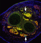
The epithelial layer in our lungs is made of several cell types, including secretory Clara cells, ciliated epithelial cells, neuroendocrine cells, and large goblet cells.  During irritation or injury, such as pollution or cigarette smoke, goblet cells increase the production of mucus.  Only a few goblet cells are typically found in healthy lungs, but asthma or COPD (chronic obstructive pulmonary disease) patients have an increased number of goblet cells.  A recent Development paper describes the role of the transcription factors Fox1 and Fox4 in regulating the cell fate decisions that produce goblet cells during development and tissue regeneration.  Specifically, Li and colleagues found that Fox1/4 restricted the goblet cell lineage program by repressing disulfide isomerase anterior gradient 2, Agr2.  In addition, Li and colleagues found that Fox1/4 deletion caused a catastrophic loss of tissue regeneration, by driving differentiation of all secretory cells into the goblet cell lineage program.  The images above show lung tissue after chemical injury by naphthalene, which causes a rapid loss of Clara secretory epithelial cells from the airways.  Control tissue (left column) regenerated by day 20.  Without Fox1/4 (right column), tissue regeneration was defective, as seen as the presence of too many goblet cells (top row, arrows) and the lack of ciliated epithelial cells (β tubulin IV in red, bottom row).

For a more general description of this image, see my imaging blog within EuroStemCell, the European stem cell portal.

ResearchBlogging.orgLi S, Wang Y, Zhang Y, Lu MM, Demayo FJ, Dekker JD, Tucker PW, & Morrisey EE (2012). Foxp1/4 control epithelial cell fate during lung development and regeneration through regulation of anterior gradient 2. Development (Cambridge, England), 139 (14), 2500-9 PMID: 22675208

Thumbs up (No Ratings Yet)
Loading...

Tags: , ,
Categories: Images, Research

In Development this week (Vol. 139, Issue 15)

Posted by , on 10 July 2012

Here are the research highlights from the current issue from Development:


Cxcr4a sets proliferative response to Hh

The Hedgehog (Hh) pathway controls both patterning and proliferation during development, but how do embryonic cells distinguish between these activities? On p. 2711, Pia Aanstad and colleagues provide data that indicates that proliferative responses to Hh signalling are context dependent. The researchers show that activation of Hh signalling promotes endodermal cell proliferation in zebrafish gastrula stage embryos but inhibits proliferation in neighbouring non-endodermal cells. Expression of the chemokine receptor Cxcr4a in gastrula stage endoderm determines the proliferative response to Hh signalling, they report, but does not affect the expression of Hh target genes involved in patterning. Finally, they show that Cxcr4a inhibits the activity of cAMP-dependent protein kinase A (a negative regulator of Hh signalling), and propose that Cxcr4a enhances Hh-dependent proliferation by promoting the activity of Gli. Together, these results indicate that Cxcr4a is required for Hh-dependent cell proliferation but not for Hh-dependent patterning. Thus, parallel activation of Cxcr4a might enable Hh signalling to control both patterning and proliferation during development.


Hh maintains testis somatic stem cells

Stem cells are specified and maintained by specific microenvironments called niches. In the Drosophila testis, somatic cyst stem cells (CySCs) give rise to cyst cells, which ensheath the differentiating germline stem cells (GSCs). Both stem cell pools are arranged around a group of somatic cells – the hub – that produce niche signals for both lineages. Now, Christian Bökel and co-workers report that CySC but not GSC maintenance requires Hedgehog (Hh) signalling in addition to Jak/Stat pathway activation (see p. 2663). The researchers report that CySCs unable to transduce the Hh signal are lost through differentiation, whereas Hh pathway overactivation in CySCs increases their proliferation. The additional cells generated by excessive Hh signalling remain confined to the testis tip and retain the ability to differentiate. Because Hh signalling also controls somatic cell populations in the fly ovary and the mammalian testis, these new observations reveal a greater organisational similarity between the somatic components of gonads across the sexes and phyla than previously appreciated.


ncRNAs keep genes silent

Noncoding RNAs (ncRNAs) help to establish transcriptional gene silencing during development by interacting with DNA and chromatin-modifying enzymes. But do they also help to maintain gene silencing? Here (p. 2792), Chandrasekhar Kanduri and colleagues explore the involvement of the Kcnq1ot1 ncRNA in the maintenance of gene silencing at the Kcnq1 imprinted domain in the mouse embryo. The Kcnq1 domain contains ubiquitously imprinted genes (UIGs), which show imprinted silencing in placental and embryonic tissues; placental-specific imprinted genes (PIGs), which are silenced on the paternal chromosome in the placenta only; and several non-imprinted genes (NIGs). By conditionally deleting the Kcnq1ot1 ncRNA at different stages of mouse development, the researchers show that this ncRNA is required to maintain UIG silencing throughout development, whereas silencing of some PIGs is maintained independently of Kcnq1ot1 ncRNA. Intriguingly, the researchers identify enhancer-specific histone modifications associated with actively transcribed NIGs. These, they propose, may limit the spread of ncRNA-mediated silencing. Together, these results suggest how ncRNAs might maintain transcriptional silencing in a spatiotemporal manner.


In vivo activities of miRNAs revealed

Hundreds of microRNAs (miRNAs) – short RNAs that mediate networks of post-transcriptional gene regulation – have been recorded in animals. Because cell-based assays and bioinformatics provide evidence for large numbers of functional targets for individual miRNAs, it is not obvious that manipulation of miRNAs will lead to interpretable phenotypes at the organismal level. However, on p. 2821, Eric Lai and co-workers describe a genome-wide transgenic resource for the conditional expression of Drosophila miRNAs and, surprisingly, report that the majority of the miRNA transgenes in their collection induce relatively specific mutant phenotypes when expressed in the developing wing. Many of these phenotypes resemble those produced by alterations in signalling and patterning genes and, notably, their specificities were not predictable from computational studies, thereby highlighting the usefulness of in vivo phenotypic assays of miRNA activity. Finally, the unexpectedly broad capacity of different miRNAs to generate specific dominant phenotypes in flies suggests that gain-of-function of diverse mammalian miRNAs may also generate an array of specific disease conditions.


A rib-tickling Hox10 motif

During the development of the vertebrate axial skeleton, Hox genes belonging to paralog group 10 play a role in blocking rib formation in the lumbar region of the vertebral column. Here (p. 2703), Moisés Mallo and colleagues investigate the molecular basis of the rib-repressing function of Hox10 proteins. The researchers identify two conserved motifs (M1 and M2) that flank the homeodomain of Hox10 proteins and show that M1, which is located next to the homeodomain’s N-terminal end, is required for Hox10 rib-repressing activity in mice. M1 contains two potential phosphorylation sites, they report, mutation of which to alanines results in a total loss of the rib-repressing properties of Hox10 proteins. Other experiments suggest that the activity of M1 requires interactions with more N-terminal parts of Hox10 and that M1 might also regulate Hox10 activity by altering the protein’s DNA-binding affinity through changes in the phosphorylation state of two conserved tyrosines in the homeodomain. Together, these results provide new insights into the regulation of rib development by Hox10 proteins.


Modelling EGFR patterning of fly epithelium

Epidermal growth factor receptor (EGFR) signalling regulates numerous processes throughout Drosophila development. For example, during oogenesis, an EGFR activation gradient induced by Gurken (a TGFα-like ligand secreted from the oocyte) patterns the follicular epithelium. On p. 2814, Stanislav Shvartsman and colleagues present a revised mathematical model for this important process, which initiates the formation of two dorsal eggshell appendages. Each of these appendages is derived from a primordium that comprises a patch of cells expressing the transcription factor gene broad (br) and an adjacent strip of cells expressing rhomboid (rho), which encodes a protease in the EGFR pathway. Previous models of eggshell patterning have not fully accounted for the coordinated expression of br and rho. The new model, however, proposes that the sequential action of feed-forward loops and Notch-mediated juxtacrine signals activated by the EGFR signalling gradient establishes rho expression, successfully describes the wild-type br and rho expression patterns, and accounts for changes in these patterns in response to genetic perturbations.


Plus…

Toward a blueprint for regeneration

Tissue regeneration has been studied for hundreds of years, yet remains one of the less understood topics in developmental biology. The recent Keystone Symposium on Mechanisms of Whole Organ Regeneration, reviewed by Gregory Nachtrab and Kenneth Poss, brought together biologists, clinicians and bioengineers representing an impressive breadth of model systems and perspectives. See the Meeting Review on p. 2639


Evolutionary crossroads in developmental biology: annelids

Annelids (the segmented worms) have a long history in studies of animal developmental biology, particularly with regards to their cleavage patterns during early development and their neurobiology. As reviewed by David Ferrier, Annelida are playing an important role in deducing the developmental biology of the last common ancestor of the protostomes and deuterostomes.

See the Primer on p. 2643


Evolutionary crossroads in developmental biology: the spider Parasteatoda tepidariorum

Spiders belong to the chelicerates, which is an arthropod group that branches basally from myriapods, crustaceans and insects. Hilbrant, Damen and McGregor describe how the growing number of experimental tools and resources available to study Parasteatoda development have provided novel insights into the evolution of developmental regulation and have furthered our understanding of metazoan body plan evolution.

See the Primer on p. 2655


Also see the related Editorial by Nipam Patel, Development‘s evo-devo Editor.


Thumbs up (1 votes)
Loading...

Categories: Research

Transcending boundaries: MBL Embryology 2012

Posted by , on 6 July 2012

The most striking realization I have had over the course of last four weeks spent at MBL, Woods Hole is how limitless is the scientific spirit . Pioneers of classical embryological manipulation techniques appreciating the importance of mathematical modeling, groups going to non-model organisms in search of an answer, groups identifying novel questions by merely observing or rather comparing differences between two organisms in one aspect of development, all are examples of the same. Working with a wide range of organisms we have been able to appreciate the diversity of body plan, and its molecular, cellular and behavioral attributes. It’s like learning is fun so, trying to create a five-limbed tetrapod and two-headed frog was fun but also every failed attempt made us realize the importance of temporal and spatial context in development. Not every experiment performed has to be hypothesis driven and so we see fish organiser grafted into frog and mouse organiser into chick, imagination and curiosity are the only drive here. We enjoy the freedom to explore and experiment. Faculties and TAs are available around the clock, eager to help better define the question and design experiments. This encourages to think and ask questions without bothering about practical limitations. A glimpse of the wonderful scientific outcome of this can be seen in the “Fish-bowl” previously known as “Sweat-box”, the one hour post-talk discussion session with the speaker. The speaker is bombarded with questions by students, not all of whom are working in the same field. Many of the questions, including the naive ones, provide novel directions or lead towards yet unexplored possibilities. It’s the most fulfilling one-hour of the day for me and I hope all my course-mates and faculties share the feeling.

Though I am part of a vertebrate developmental biology group, organiser grafts, tissue transplants, gastrulation, chimeras etc. have mostly been text book concepts for me. Learning these classical techniques and concepts from the experts in the field was overwhelming. As they went down memory lane, we learned the evolution of the field. Sitting through the talks we were introduced to the discovery aspects of many molecular and cellular phenomena that so far we have been reading as facts. How the field started from inquisitive observation and systematic documentation followed by attempts to interpret the same. Hypotheses were generated and tools to validate the hypothesis were created. Need based emergence and evolution of the fields of molecular biology, imaging, biomechanics, bioinformatics and so on took place. You are introduced to different model organisms, their advantages as well as limitations. Also to the most recent techniques available for different kind of expression and functional analysis for different organisms, along with feedback on the performance . You get to hear about the questions that led to adoption of non-model organisms in labs and that make groups run more than one lab spread across globe on seasonal basis. In the lab session, you find faculties and TAs happy to help you try any and every experiment you can think of, always ready with tips from their experience and demonstrations. This deroots almost any hesitation one has in working with new model organisms or trying different techniques. It’s all about daring to try something new and different combined with patience and perseverance. In the last two weeks I have had the privilege of working with five different vertebrate species with experiments ranging from classical grafting, skeletal preps., bead implantation to assess the morphogenetic potential of proteins and drugs, to laser ablation, mouse embryo culture, mouse in utero electroporation and TALEN injection in transgenic fish lines. Imagine !!!

You set up a time-lapse last night to capture cellular movements along with lineage tracing by injecting dye in a two-cell zebrafish embryo. Today morning you found that due to improper sealing , and subsequent evaporation of embedding media, you could not capture anything. You are very upset and sitting quiet and calm at the dining table. “What happened?”, comes from your friends. And then comes a long list of strange disasters, time-lapse of an unfertilized egg, time-lapse of a dead embryo, embryo crawling out of the field just few minutes into time-lapse and so on. You just can’t help laughing all your worries away and happily start the next attempt for the same experiment. The group has participants from places spread all over the world. Strangely, you don’t feel the diversity unless, one of your group members suddenly in the middle of the night in the confocal room, tired after a long day, starts speaking Pourtuguese. It’s only after looking at your expression-less face with wide open eyes that she recalls you being an Indian . You enjoy a refreshing laugh together and move on with your experiment trying to find the best possible orientation of the mouse embryo for the time lapse. That small element lying there somewhere deep within us which wants us to read “the mind of nature” is what connects us all beyond our differences.

Thumbs up (14 votes)
Loading...

Tags: , , ,
Categories: Education, Events