The community site for and by
developmental and stem cell biologists

Postdoctoral Position in Skeletal Patterning

Posted by , on 10 April 2012

Closing Date: 15 March 2021

A postdoctoral position studying skeletal development is available in the laboratory of Dr. Amy Merrill at the University of Southern California’s Center for Craniofacial Molecular Biology (CCMB).  CCMB offers a highly supportive and interactive environment with a strong research profile in craniofacial development and repair.  Work conducted in the Merrill laboratory integrates human genetics and developmental biology to study normal and abnormal craniofacial development.  We are looking for an enthusiastic candidate who will carry out an independent research project aimed at understanding spatiotemporal signals that pattern bone and cartilage.  Ideal applicants will be highly motivated and have recently completed doctoral training in Developmental Biology.  

Please provide a cover letter, CV, and contact information for three references by email to: amerrill@usc.edu

Thumbs up (1 votes)
Loading...

Tags: , ,
Categories: Jobs

Navigate the archive

Use our Advanced Search tool to search and filter posts by date, category, tags and authors.

The 2012 UK National Xenopus Conference

Posted by , on 10 April 2012

The UK national Xenopus conference is an annual event held to discuss the exciting and extremely varied work carried out across the UK using the African clawed frog (Xenopus) as the model organism of choice. The event provides opportunity for PhD students and Postdocs from Xenopus labs round the UK to present their data to experts within the Xenopus community, a community which can easily be described as passionate and welcoming to all new members. This year’s meeting was held on the 19th of March at the Wellcome Trust Sanger Institute (Cambridge, UK). This institute, opened in 1992, is famous for its substantial participation in the sequencing of the human genome. As a single institute it alone contributed the most sequencing data for what is now considered the gold standard human genome sequence. The meeting was held in a conference room lined with stands for some of the many sponsors of the event. These included companies such as sequencing giants Illumina, morpholino pioneers Gene Tools and many others including Techniplast, Sigma-Aldrich, Agilent Technologies, Biostatus and Cellectis Bioresearch.


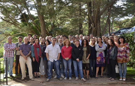
A busy room full of keen Xenopus researchers


The talks kicked off with a welcome from Derek Stemple, a senior investigator at the Sanger Institute who is currently using Zebrafish and Xenopus tropicalis as human disease models as well as being involved in the Zebrafish genome sequencing project. His welcome outlined the history of the Sanger Institute and gave a strong message of the importance it has played in revolutionising genome sequencing over the last 20 years and its continuing research into genomics and human disease models. This posed as a great introduction for the first talk by Amanda Hall who is currently working on the Xenopus tropicalis mutation resource project at the Sanger Institute. This project aims to create Xenopus knockouts which can be used to study various disease models. Invitro sperm ENU mutagenesis has been used to generate 6000 F2 mutant individuals the progeny of which can be maintained as genomic DNA libraries and frozen sperm for verification once positive phenotypic analysis is carried out on an F3 carrier population. The genomic library will undergo a reverse genetic screen known as TILLING to indentify 175 preselected mutations. This project can greatly benefit the Xenopus community as all mutations will be made available to view on an online database on the Sanger Institute website. Also all mutant sperm and living animals can be made available for use in further research.

The second talk of the day was by Anita Abu-Daya who is currently working in Lyle Zimmerman’s lab at the National Institute of Medical Research (NIMR) in London. Lyle’s lab has collected a variety of weird and wonderful Xenopus tropicalis mutations over the last few years by the means of gynogenetic screens. In her talk, Anita spoke of a mutant called Whitehart, which after phenotypic analysis was identified to have no circulating blood. Genetic analysis of Whitehart revealed it to have a mutation resulting in a premature stop codon in the Smad 4.1 gene, the mediator Smad of both TGFβ and BMP signalling. But if the embryos lack such an important Smad then why is the phenotype not more extreme? This was deduced to be down to redundancy by the similarly expressed Smad 4.2. This work in summation gave a potential role for BMP signalling for the maintenance of blood. The third talk was given by Nick Owens from Mike Gilchrists lab also at the NIMR. His talk outlined the importance of biological variability when using techniques such as RNAseq. To prove this importance he uses two inbred mothers whose eggs are fertilised with the same father. 8 pools of 20 embryos were collected from both parent combinations to undergo sequencing. His data generally shows the more replicates the better to avoid biological variability. Three replicates will always give more reliable results than two.

The next set of talks included one from Tom Bates of Esther Bell’s lab (Kings College London). His work showed a novel TGFβ inhibitor, Coco, to be expressed in the animal pole of gastrula stage embryos. Knock down of Coco by host transfer techniques and morpholino injection showed a loss of dorsal tissues. His work could be summarised by a model suggesting Coco to be regulating germ layer specification via its inhibition of TGFβ in the animal pole. Siwei Zhang of Enrique Amaya’s lab (University of Manchester) spoke about the role of Fezf2/TLE4 in diencephalon development. He showed Fefz2 to be expressed during Xenopus neurulation and in the forebrain of tailbud and tadpole stage embryos. mRNA injection of Fefz2 resulted in dorsal anteriorized embryos leading to a hypothesis of Fefz2 regulating Wnt signalling in the diencephalon area. Next up was Natalie Gibb from Stefan Hoppler’s lab (University of Aberdeen). She demonstrated a role for Sfrp1 in promoting myocardial differentiation. Her talk contained some stunning images and videos demonstrating the ability of Sfrp1 to modulate the size of the developing heart. Coinjection of Sfrp1 and Wnt6 rescued the dual axis phenotype usually associated with Wnt overexpression. This indicates a role for Sfrp1 in modulating Wnt signalling during myocardial development. The last talk of this set was from Neil Roberts (University of Manchester). His work used Xenopus as a disease model to characterise mutations which may result in the unusual disease, Urofacial syndrome a human disease with symptoms including bladder dysfunction and abnormal facial expressions. His work, funded by Kidney Research UK, indicates potential for mutations in the Heparanase-2 gene to be causative of the disease.

Between talks a delicious buffet lunch was supplied which allowed all conference attendees to sit outside and enjoy the outrageously beautiful day upon which the conference took place. With the sun shining everyone moved outside to continue their discussions and meet new people within the community. Also at this time a tour was provided of the Sanger Institutes very own Illumina HiSeq equipment allowing certain attendees to see these powerful machines in action. This also gave the opportunity for us to talk to some of the sponsors. One which I found particularly interesting was Biostatus whom have developed what they are calling CyGEL, a thermoreversible gel which can be used to immobilise living organisims such as the Xenopus allowing live cell imaging.


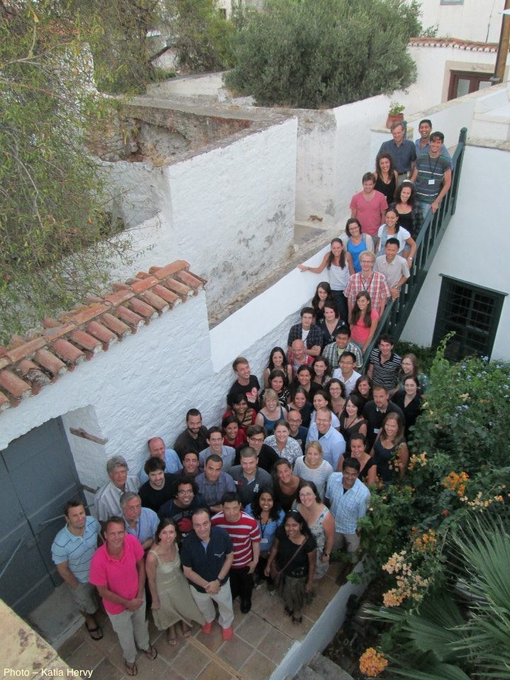
Xenopus researchers enjoying the beautiful weather


The next round of talks started with Jerome Jullien of John Gurdon’s lab. He demonstrated genome wide reprogramming of somatic nuclei by the Xenopus oocyte transcription machinery. Nuclear transfer was shown to increase Serine 2 phosphorylation found on RNA polymerase II, a phosphorylation state associated with transcription elongation. Clara Collart of Jim Smith’s lab (NIMR) gave the next talk on the role of YRNAs during midblastula transition (MBT). YRNAs are expressed maternally and their expression increases after MBT. Mopholino knockdown of specific YRNAs results in normal looking embryos until MBT which is known to be the onset of transcription. All previous transcription occurring in the embryo is maternal. Clara demonstrates that YRNAs are capable of interacting with chromatin after MBT to promote the onset of DNA replication in Xenopus. Hugh Woodland (University of Warwick) finished this set of talks with a rather puzzling and insightful presentation demonstrating the relationship between oocyte and follicle cell RNA localisation. RNAs are known to exchange via transport pathways between the oocyte and macrovilli/nanotubules extending into the surrounding follicle cells. Conversely, Hugh’s data provides evidence for particles in the oocyte nanotubules to represent an RNA transport pathway from follicle cells to the oocyte. This suggests that follicle cells may contribute to stored egg RNAs. To solve this conundrum he has transplanted primordial follicle mesoderm from Xenopus borealis into Xenopus laevis to see which species RNA will be present in the follicle cells and oocytes. The problem is he still has a long time to wait for these frogs to fully mature.

The last set of the day started off with Eric Theveneau from Roberto Mayor’s lab (UCL). He presented the importance of placodes, found in the gaps between branchial arches, in modulating neural crest migration. He uses Sdf1 as a marker for the placodes to demonstrate an intrinsic attraction of neural crest cells towards Sdf1 positive cells leading to efficient co-ordination of these cell types during cranial morphogenesis. Roberto Paredes (University of Manchester) next spoke of his research into the behaviour of myeloid cells after injury. He has developed an effective method to visualise myeloid cells in vivo demonstrating a fast response by neutrophils in response to injury and a comparatively slower response by macrophages. The last talk of the day was given by Matt Guille (University of Portsmouth) who when not conducting his own research is an indispensible member of the Xenopus community for his work in managing the European Xenopus Resource Centre in Portsmouth. Today however, he was not talking about the centre but his own research on histone function in early Xenopus development. The specific histones H2A.Z1 and H2A.Z2 were the primary focus of the talk. These are implicated to be involved in transcription activation and differ by only 3 amino acids. They give similar expression patterns found at several stages of Xenopus development. Knock down of H2A.Z1 shows a loss of Lmo2, Tbx, Hex and Brachyury shown by insitu hybridisation and animal cap experiments.

One of the most important parts of the UK national Xenopus conference is the community discussion at the end. This gives anyone the chance to voice concerns over all manner of topics including the Portsmouth resource centre, funding and even problems arising with scientific techniques. The key points made in this discussion included the announcement of the opening of the American Xenopus resource centre which can be found at Woods Hole, Massachusetts. A general update of the resources available at the Portsmouth centre. Finally, a suggestion to all Xenopus researchers to keep a careful log of antibodies they have previously used and found to be successful. This kind of information could save Xenopus researches a lot of valuable time and money by producing an accessible database of these antibodies. The day ended with the usual drinks and nibbles and for me a short drive home to Norwich. I had a fantastic day at the conference and would strongly recommend attending it next year if you are working in the Xenopus field. I personally will be eagerly anticipating what new and exciting research next year’s meeting will bring.

Thumbs up (10 votes)
Loading...

Tags: ,
Categories: Events

Let the collections commence!

Posted by , on 6 April 2012

All is well. The Kazakh family is unbelievable. They have
been catching more than 20 females each night although almost every
one of them died the first two nights. We think it’s because they overheated during the day before they were able to bring them to me. So now they’re bringing the
animals each night after they finish the collection which was
about 1 am the first night and 2 am last night. I am becoming one with
my nocturnal animals. The unfortunate thing (besides my lack of sleep)
is that these guys are much more agressive than the animals in my
colony back home, and especially pissed off and active at night after
being trapped in a small cage. Thank goodness for my gloves with leather
fingertips. They try desperately to take their vengeance on my hand and
instead get a mouthful of leather. Perhaps it still makes them feel
good to think they’re punishing me.

Many of the embryos are too old for my analyses, but the pregnancy
rate is so high this year that I’m still getting a lot of embryos of
the stages I want. 25 litters so far, to be precise. Which is more
than 90 embryos for two night’s work. At this rate I’ll have a
bumper crop in no time and perhaps be finished a bit early. Definitely
in time to switch gears for phase 2 of the biomechanics work when the
grad student working with me arrives from Boston in a couple of weeks.

Not sure what happened this year though. The climate is no different
from when I’ve been here before.The temperatures are tracking the
same. There’s hardly any vegetation yet, and the trees haven’t budded.
But the jerboas have all clearly fed well and are breeding at least
1-2 weeks earlier than before. Maybe there are annual cycles I’m not
aware of. A professor at the Academy of Sciences told me that about
once in 5-7 years there are almost no jerboas. Maybe this is the
opposing peak of that valley.

In any case, I can’t complain. Given the difficulty of this work and
all of the many many factors that are out of my control, I have to say
I am pretty pleased. Fingers crossed this run of good luck continues.

Thumbs up (2 votes)
Loading...

Categories: Lab Life, Research

Origins of Tissue Stem Cells – June 28-29, 2012 in Edinburgh

Posted by , on 5 April 2012

Upcoming deadlines: oral abstract, poster abstract and early bird registration – 23 April 2012

Meeting chairs: Dr Clare Blackburn and Dr Val Wilson (University of Edinburgh)

Topics:
•  Gut and associated organs: when, where and how do stem  cells arise?
•  Specifying the hematopoietic lineage
•  Stem cell development
•  Specifying the neural lineage
•  PGCs

Invited speakers: check out the list of invited speaker on the meeting website  (www.abcam.com/edinburgh)

Meeting deadlines – 23 April 2012:
•  Oral abstracts
•  Poster abstracts
•  Early bird registration

Meeting website: www.abcam.com/edinburgh

Thumbs up (No Ratings Yet)
Loading...

Categories: Events, News

I was so hungry…

Posted by , on 5 April 2012

I ate a horse. Seriously. I ate horse meat tonight. I wouldn’t have ordered it, and I don’t feel so good about myself for doing it, but I believe in adapting and respecting a culture which includes eating pretty much anything (that’s not obviously unsafe) that’s put in front of me (with the exception of bugs). I had been invited to join another banquet in honor of an English couple and their son who are visiting to studying some of the chinese herbal plants. We went to a big multistory fancy Uyghur restaurant that’s more in the Chinese style with Uyghur influence. The first restaurant I’ve been to here with security guards out front and a bag check before you walk in the front door. We were taken up an elevator to the third floor and ushered to a small banquet room where each seat had a hotpot burner with broth and the waitstaff passed out aprons. What a scene. My good friend “Alice” (her English name) was instructed by the host to order, so a whole bunch plates of raw meat and vegetables came out onto the table. She seemed to take great pleasure in ordering some of the most expensive things on the menu knowing the host (a retired scientist and regional party official) wouldn’t refuse. The meat and vegetables get dunked into the pot of boiling spicy broth until they’re cooked and then fished out and eaten. A small plate of what looked like sliced salami rolled around in front of me, and Alice gave a sly smile and said “horse”. Really. Horse. So I took a piece to try it – in part so I can now say I’ve eaten horse. It was already cooked – dry roasted and lightly smoked and tasted different from any other kind of meat I’ve had. Interesting.

Other than that I’ve just been spending my weekend in Urumqi arranging things to return to Fukang on Monday and dive into the dissections. I had to make a few solutions, so now all of that is done. I needed to find a freezer and fortunately a professor here decided to buy a new small deep freezer for the lab and will loan it to me first for a few weeks. So we spent 2 hours yesterday and another 4 hours today running about between three different stores trying to find a freezer in stock that would be easy to get paid for by government money. As we were leaving the institute this morning, a throng of about 200 middle school students carrying pails of water rushed passed us led by kids holding red flags. I asked what in the world was going on, and my friends said it’s international environment day. I thought they meant earth day, but that’s not for a few more weeks. I have no idea what was the holiday, but I saw so many ordinary people (not city workers) out cleaning things – street lamps, fences, bus stops. There was a whole line of about a dozen people with rags squatted down busy wiping the black grime off of a fence that runs between the street and sidewalk. All in business suits. Common attire here. Years ago I once saw a guy laying paving stones wearing a business suit and dress shoes.

And the first part of this week is the Tomb Sweeping (Qingming) festival in honor of the ancestors, so there are carts all over the city selling fake paper money, incense, plastic fruit, and small plastic trees. The custom is to visit the cemeteries and give these offerings (burning the money) in honor of the ancestors. And since Monday and Tuesday are a holiday, everyone has had to work saturday and sunday to make up for the time. Fascinating…

Since I started this post 4 days ago, I have made it back to the field station and have collected 25 litters of embryos in 2 days. Success! However, the internet is down, so we had to find an internet cafe in the town. The smoke is getting to be a bit more than I can handle for now, so stay tuned for the next update…

Thumbs up (4 votes)
Loading...

Categories: Lab Life

2012 Canadian Developmental Biology Conference Overview

Posted by , on 4 April 2012

A few weeks ago, over 150 Canadian and international graduate students, post-docs and scientists met for the 6th biennial Canadian Developmental Biology Conference. This year’s conference was held in the middle of the world-renowned Rocky Mountains, in the beautiful town of Banff, Alberta. Although the blue skies and snow-covered mountaintops beckoned, a program filled with captivating speakers, 100 great poster presentations and even a dancing lesson from Albertan cowboys proved to be a worthy competitor for our attention.


Based on conversations with conference participants, one of the best features of this conference was its diversity. The talks focused on five research themes, covering a variety of model systems including worm, fly, frog, mouse, fish, Arabidopsis and planaria. The first theme was embryogenesis and cell polarity. It included a talk from Helen McNeil (University of Toronto) on the mechanisms by which fat family cadherins signal through the mitochondria to effect planar cell polarity in Drosophila and a talk from Janet Rossant (University of Toronto) on the signaling pathways involved in early lineage decisions in mouse embryos. The second theme, epigenetics and development, included among others, Sabine Cordes’ (University of Toronto) talk about the polycomb-dependent epigenetic mechanisms in neural development. The next two sessions focused on cell signaling and cell fate specification in development.  Some of the great talks in these sessions included a talk by Andrew Waskiewicz (University of Alberta) about the use of zebrafish to study ocular birth defects such as coloboma and a talk by Brent Bobick (University of Calgary) on how the transcription factor Shox2 is necessary in mice to repress cartilage formation. Lastly, the conference finished off with a session on cell size and proliferation. This session encompassed talks from Nam-Sung Moon (McGill Univeristy) on factors that synergize with Rb in Drosophila as well as a talk from Bret Pearson (University of Toronto) about how planaria control the proliferation of their adult stem cells.

On top of all the great sessions, one of the highlights of the conference was the keynote lecture by Utpal Banerjee (UCLA). He focused on recent work in his lab which uses hemaopoetic cells in Drosophila to study how stress signals can interact with and influence cell metabolism. The chance to see such a distinguished researcher speak was truly inspirational for all of us graduate students in the early stages of our careers. As well as wowing the crowd with his science, Utpal Banerjee also took time to participate in a special session on teaching methods in undergraduate developmental biology. In this session, he focused on methods such as “Research Deconstruction” which have been implemented at UCLA with great success thus far.  Also of note was the second special lecture by Pierre Chambon (IGBMC) on transcriptional control by nuclear receptors.

After all the science, we had a chance to experience some Albertan culture at the “Cowboy and Western” banquet offsite at a huge tent in the mountains.  While keeping warm by the gigantic bonfire, we ate barbeque and learned how to do traditional line dancing from a duo of cowboy singers who were the entertainment for the night.  It was a great chance to let loose, catch up with old acquaintances and meet some new friends!

Overall this was a great conference filled with interesting talks, engaging poster sessions and lots of time to interact with the best scientists and researchers from around the country.  Congratulations to all of the following winners of the poster competition, each of whom won $100: Namal Abeysundara, Corey Arnold, Ben Chan, Devon Germain, Felix Gunawan, Xue Han, Yoichi Kawabe, Lauren Killip, Kate Krivy, Karen Lange, Saiqun Li, Stephanie McMillan, Stephen Nemec, Stanley Neufeld, Jeremy Saban, Tara Stach, Simone Superina, and Chris Wang. Also congratulations to the winners of a Society of Developmental Biology sponsored $1000 travel credit to go to the 71st International SDB meeting in Montreal in July.  The graduate student winner was Steffen Biechele and the postdoc winner was Elizabeth RideoutOn behalf of us, and all the conference participants, we’d like to say a big thanks to the conference organizers Sarah Childs and Carol Schuurmans (University of Calgary) for such a stimulating meeting.  Hope to see everyone at the 7th Canadian Developmental Biology Conference in Montreal in 2014!

By: Lauren Killip and Corey Arnold

Thumbs up (7 votes)
Loading...

Tags:
Categories: Events

In Development this week (Vol. 139, Issue 9)

Posted by , on 3 April 2012

Here are the highlights from the current issue of Development:


Haematopoiesis to knock your SOX7 off

During vertebrate development, haematopoietic and endothelial cells emerge from a common mesodermal progenitor, the haemangioblast. Upon haematopoietic commitment, the haemangioblast generates blood precursors through populations of endothelial cells with haemogenic properties, but what regulates the generation of haemogenic endothelium? Here (p. 1587), Valerie Kouskoff and colleagues use mouse embryonic stem cells to investigate the role of the transcription factor SOX7 in haemangioblast differentiation. The researchers report that, within blast colonies (which recapitulate early haematopoietic development in vitro), SOX7 is expressed in haemogenic endothelium cells and is downregulated in nascent blood precursors. Enforced expression of Sox7 in blast colonies, they report, blocks haematopoietic differentiation and sustains the expression of endothelial markers. They also show that SOX7 binds and activates the promoter of VE-cadherin, an adhesion molecule that is expressed in haemogenic endothelial cells but downregulated during blood cell formation. Together, these results identify SOX7 as a transcriptional regulator of genes expressed in the haemogenic endothelium and provide new insights into early embryonic haematopoiesis.

A novel photo-morpholino on/off switch for genes

To investigate the molecular mechanisms of development, it is useful to be able to turn genes both on and off in a spatially restricted manner. Although the development of photo-activated morpholino oligonucleotides (photo-MOs) has made it possible to turn genes off at specific times and places, finding a way to deactivate MOs and restart gene expression has proved more elusive. Here (p.1691), Alexandra Tallafuss, Philip Washbourne and colleagues describe an approach in which they can turn genes off and on using sense photo-MOs (S-photo-MOs) and complementary antisense photo-MOs (AS-photo-MOs), respectively. S-photo-MOs bind to and block the activity of regular morpholinos, and exposure of the S-photo-MOs to UV light allows the morpholinos to become active and block gene expression; AS-photo-MOs block gene function like regular morpholinos, but normal gene function can be restored by light inactivation. Importantly, the researchers demonstrate the feasibility of their new approach in whole zebrafish embryos by studying notochord induction and neural crest development, and in single cells by temporally manipulating Gal4 transgene expression.

See also the authors’ post about this paper on the Node.

Endoderm tugs at heart strings

During vertebrate heart development, cardiac progenitors form bilateral fields within the lateral plate mesoderm that move towards the embryonic midline and fuse to form the heart tube. It is generally accepted that, during this process, in addition to its inductive signalling role, the endoderm serves as a mechanically passive substrate for the migrating cardiogenic mesoderm. Now, Victor Varner and Larry Taber suggest that the endoderm also has an active mechanical role during heart tube assembly (see p. 1680). Using label-tracking experiments in combination with actomyosin inhibitors, the researchers show that, in chick embryos, the endoderm actively shortens around the anterior intestinal portal, and that myosin-II-dependent contraction drives this shortening. Inhibition of contractility using actomyosin inhibitors, they report, produces cardia bifida and foregut defects. Moreover, computational modelling suggests that the endoderm (not the mesoderm) is the dominant contractile tissue layer during heart field migration. Thus, the researchers suggest, during avian cardiogenesis, active contraction of the endoderm pulls the heart fields towards the embryonic midline.

Notch tunes pancreatic endocrine progenitor behaviour

During pancreatic development in mammals and zebrafish, Notch-responsive cells (NRCs) give rise to endocrine cells, but how does Notch signalling regulate the proliferation and differentiation of pancreatic progenitors at the single-cell level? On p. 1557, Nikolay Ninov and co-workers use live imaging and a novel transgenic reporter system that allows the dynamic assessment of Notch signalling to answer this question in zebrafish larvae. The researchers show that zebrafish NRCs exhibit different levels of Notch signalling that, in turn, regulate distinct cellular outcomes. Specifically, high levels of Notch signalling induce quiescence, lower levels promote progenitor amplification, and sustained downregulation of Notch signalling triggers a multistep process that includes cell-cycle entry and amplification followed by endocrine differentiation. Importantly, the researchers show that progenitor amplification and differentiation can be uncoupled by modulating the duration and/or level of Notch signalling downregulation. Thus, different levels of Notch signalling drive distinct behaviours in pancreatic endocrine precursors, a finding that could inform the development of regenerative therapies for type 1 diabetes.

Apical domain size matters for neurogenesis

Early in retinal development, neuroepithelial progenitor cells begin to divide in a neurogenic mode in which at least one daughter cell exits the cell cycle and differentiates into a neuron. Now, on p. 1599, Brian Link and colleagues investigate the cell biological mechanisms that influence neurogenesis by analysing zebrafish and mouse retinal neuroepithelia deficient for Llgl1 (lethal giant larval protein homologue 1), a protein that is implicated in apicobasal polarity, asymmetric division, cell shape and cell-cycle exit. The researchers show that Llgl1-deficient neuroepithelia retain apicobasal polarity but have expanded apical domains. Importantly, Llgl1-deficient neuroepithelia display increased Notch activity and reduced neurogenesis. Expansion of the apical domain through inhibition of Shroom3 also increases Notch activity and reduces neurogenesis. These results, together with studies of interkinetic nuclear migration (the apical-to-basal movement of neuroepithelial nuclei that is linked with cell-cycle exit) in Llgl1-depleted retinal precursors, suggest that the size of the apical domain of retinal neuroepithelia modulates the strength of the polarised signals that influence neurogenesis.

Mitotic-meiotic switch awry in testicular teratoma

The formation of testicular teratomas and other testicular germ cell tumours (TGCTs) is thought to involve defects in male germ cell development. On p. 1577, Jason Heaney and co-workers provide new insights into TGCT development by studying strains of mice with different teratoma incidences, including the 129 strain, which has a high spontaneous rate of TGCTs. In 129 mice, TGCTs are visible at embryonic day (E) 15.5 as microscopic foci of pluripotent stem cells (embryonal carcinoma cells). The researchers report that germ cell proliferation, the expression of the pluripotency factor Nanog and the premature expression of genes normally expressed in pre-meiotic adult male germ cells, such as cyclin D1 and Stra8, at E15.5 are all directly related to increased tumour risk. Notably, embryonal carcinoma cells co-express genes involved in germ cell pluripotency and differentiation, and deletion of Stra8 in 129 mice reduces their teratoma incidence. Together, these results indicate that deregulation of the mitotic-meiotic switch in male germ cells contributes to teratoma initiation.

Thumbs up (No Ratings Yet)
Loading...

Categories: Research

Sub-Nuclear Structures and Disease

Posted by , on 3 April 2012

Sub-Nuclear Structures and Disease
28 June-1 July 2012
The Møller Centre, Cambridge, UK

Now in its third year, this Wellcome Trust meeting will focus on the biology of sub-nuclear structures including the nucleolus, cajal and PML bodies. These structures have key roles in normal and diseased cells and they interact in a dynamic way. A fundamental understanding of these sub-nuclear structures can lead to advances in our understanding of infectious disease and cancer.

The conference will bring together cell biologists, microbiologists and virologists working on normal and tumor cells and researchers interested in how these structures are affected by infectious and acquired disease across all eukaryotic systems.

Please note: Due to work to expand the Wellcome Trust Conference Centre at Hinxton, Sub-Nuclear Structures and Disease 2012 will take place at The Møller Centre, Churchill College – part of the historic University of Cambridge

Scientific Organisers:
Susan Baserga Yale University, USA
Julian Hiscox University of Leeds, UK
David Matthews University of Bristol, UK
Brian McStay NUI Galway, Ireland

Invited Speakers include:
Susan Baserga Yale University, USA
Richard Gardner University of Washington, USA
Ingrid Grummt Deutsches Krebsforschungszentrum, Germany
Ross Hannan Peter MacCallum Cancer Centre, Australia
Valerie Lallemand-Breitenbach Hopital Saint-Louis, France
Angus Lamond University of Dundee, UK
Greg Matera University of North Carolina, USA
Brian McStay NUIG, Ireland
Karla Neugebauer Max Planck Institute, Germany
Craig S. Pikaard Indiana University, USA
Michael Taliansky The James Hutton Institute, UK
David Tollervey University of Edinburgh, UK
Adrian Whitehouse University of Leeds, UK

Abstract submission is strongly encouraged as several talks will be selected from abstracts.

For more information and to register, please see: https://registration.hinxton.wellcome.ac.uk/display_info.asp?id=294

Thumbs up (No Ratings Yet)
Loading...

Categories: Events

Photo-Morpholinos

Posted by , on 3 April 2012

In the study of the roles of genes during development, one problem that is often faced by researchers examining ‘late’ roles of genes is one of obscuration by temporal pleiotropy. That is: gene mutations and gene knock-down obscure late roles in development, because early phenotypes, such as cell death or malformed embryos, make it impossible to examine later functions. A common way to achieve gene knock-down in developing zebrafish and Xenopus tadpoles, is by using Morpholinos (MOs). These are antisense oligonucleotides composed of morpholine-modified nucleotide subunits, commonly used to block the initiation of translation or to block splicing. We were faced with just such a pleiotropy problem with a set of genes we are currently studying. We considered trying to cage MOs ourselves, rendering them temporally activatable, by linking two complementary MOs together using a photo-cleavable crosslinker, much as has already been described previously (e.g. Shestopalov et al. 2007). We called Gene Tools LLC, the company that synthesizes MOs, to ask them whether they had any suggestions with the chemistry. Paul Morcos, at Gene Tools LLC, mentioned that they had been developing such a product for a while and thought they were close to having something worth trying out.

A few months later, Paul called, asking if we would like to test their new photo-controllable MOs. They had designed a photo-cleavable subunit with the size and chemical properties so as to ‘blend in’ with the other MO subunits, but that could be cleaved by UV light. This made it possible to design (1) sense-photo-MOs (S-photo-MOs), which would base pair with ‘regular’ MOs and thus block their activity until UV exposure, and (2) antisense-photo-MOs (AS-photo-MOs), which would function just like ‘regular’ MOs until inactivation by UV exposure. These could then be used to turn gene function off at a given time point (using S-photo-MOs mixed with complimentary MOs), or, in a novel application, turn gene function on at a given time point (using AS-photo-MOs). These MOs would then allow researchers to study early or late gene function respectively, creating a more complete picture of gene function during development; and to find the critical time period in which gene function is necessary for the proper development of specific cell populations. Furthermore, we considered that by using a laser, we could generate cell-autonomous  knock-down of genes in specific cell populations, eliminating the need for cell transplant experiments from WT into mutant backgrounds, or vice versa.

We chose ntla, sox10 and gal4 genes as targets with which to beta-test the photo-MOs. Both ntla and sox10 knock-down have easily identifiable phenotypes. Gal4 was chosen as it would potentially give researchers the additional temporal and spatial control over transgene expression for a wide variety of applications. The results are published in the latest edition of Development, showing that these new reagents from Gene Tools work surprisingly well. As with regular MOs, the ideal working concentration needs to be determined, and one needs to find the best ratio of S-photo-MO to regular MO (1:1 is a good starting point). Once optimized these reagents perform well, almost completely blocking MO activity before UV and completely releasing MO activity after UV. Interestingly, our results confirmed that ntla is only necessary as late as tailbud stage to drive medial precursor cells towards a notochord fate, and that sox10 is not required for neural crest formation, but for maintenance. Experiments with Gal4 photo-MOs allowed us to restrict expression of a transgene (in this case GFP) to primary and not secondary motoneurons. This was not possible until now using conventional transgenic approaches. We hope these tools go on to help researchers circumnavigate problems of temporal pleiotropy during development and help enhance the expression patterns of transgenes in the future.

ResearchBlogging.orgAlexandra Tallafuss, Dan Gibson, Paul Morcos, Yongfu Li, Steve Seredick, Judith Eisen, & Philip Washbourne (2012). Turning gene function ON and OFF using sense and antisense photo-morpholinos in zebrafish Development, 139 (9), 1691-1699 : 10.1242/dev.075390

Thumbs up (8 votes)
Loading...

Categories: Research, Resources

Exhausted but satisfied

Posted by , on 31 March 2012

The breakfast bell rang early this morning, so I’ve only just gotten my shower this evening which is a good thing since I was filthy and covered in sand. We headed back to the spot where we set traps last night, and lo and behold I think we only lost one.  And I could be wrong on that.  I’m guessing because the spacing at one point was longer than it should have been, but I haven’t yet counted.  It’s too bad I have no interest in those stinking gerbils! Out of the 50 traps we placed yesterday, 12 had a gerbil in them but not a single jerboa. Some of that could have to do with the fact that we set them early in the day, and gerbils are diurnal.  So it’s hard for a jerboa to land itself in an already occupied trap.  But regardless, fail. We did, however, see good jerboa tracks this time since the sand was dry.  They’re pretty easy to tell since they are in pairs spaced about 10 inches between strides. The gerbils make scurrying tracks, so they’re quite distinguishable from each other.  So the animals are out and now to catch them!

After collecting the traps, we drove up to the forestry station we’d been sent to where we thought the people were located who collected for me in the past. We waited and waited but no one showed up.  Turns out they had a kid turn sick and went to Fukang city for the day.  So we made our way back to the field station to wait for some phone calls to be returned and in the meantime got hooked up with the man I will call “The Fixer”.  I remember him from before.  He’s good at arranging things.  And he remembered me and the people we were talking about.  However it wasn’t the family we had gone to visit today – it was another Kazakh family off along a different road.  So we loaded The Fixer into the car with us and went off to meet with the right family who did turn out to be the folks we’ve hired before.  A small leathery old man with green eyes and wearing a flat cap took us into a room to sit down for negotiations on the rug covered platform that doubles as the family bed. We came to an agreement for a set price per female jerboa they catch and bring to us for the hour drive to the field station.  Not a bad deal.  All were pleased, we provided them with nets and great headlamps, and we will see the outcome in a couple of days.
Thumbs up (1 votes)
Loading...

Categories: Lab Life