The community site for and by
developmental and stem cell biologists

Stem Cells versus Progenitors

Posted by , on 13 February 2011

With so much research focusing on stem cells, I’ve been wondering lately whether researchers are overlooking other important, multipotent cell groups, specifically what are called “progenitor” cells. But then another part of me wonders whether these two groups are so very different from each other. Technically, the main difference between stem cells and progenitors is their lifespan, with progenitors’ being much shorter, but the line here seems blurry; most adult stem cells cannot be cultured for extensive amounts of time before they differentiate or senesce.

I was reminded of the issue of stem cells versus progenitors by a paper that came out earlier this month in The Journal of Clinical Investigation that showed, surprisingly, that patients with androgenic alopecia (AGA), or male pattern baldness, had a normal number of hair stem cells in their scalps, but a depleted number of different hair progenitor cells. The progenitors now look like a likely culprit for AGA. It’s been well-studied how stem cells in hair follicles give rise to new hairs over time, and it’s known that progenitors derived from these stem cells play key roles in this process, but it had not been studied with relation to AGA previously. It’s possible that the stem cells in bald AGA scalps are somehow dysfunctional or inactivated, and this could cause the loss of progenitor cells, but it still needs to be looked into (If you’d like to read more detailed coverage of this paper, I wrote a technical blog post about it on my blog All Things Stem Cell and a layman article on it for my column Biology Bytes.)

I wonder what would have happened to this recent study if when the researchers had found out that the number of hair stem cells was the same in haired and bald scalps, they then moved on to investigating other, maybe non-cellular suspects, without looking at the progenitors. Perhaps they would have then discovered a molecular abnormality in the stem cells, and then suspected the downstream progenitor groups. I just can’t help but wonder how many other diseases and biological phenomena have been investigated with a primary focus on the stem cells involved, when in some cases the progenitors may be a better initial indicator for what’s changed in the system. Or maybe using the terms “stem cells” and “progenitors” is really splitting hairs; stem cells vary significantly in potency and proliferation capacity from group to group, so maybe we should just expand the already expansive term “stem cells” to encompass a broader range of cells. While I like to think that a cell type’s name doesn’t affect whether a researcher studies it, I’d imagine it’s easier to get funding for “stem cell” research than “progenitor cell” research (or, with some funding agencies it may be the other way around), and this may definitely affect a researcher’s focus with funding as tight as it is.

Thumbs up (6 votes)
Loading...

Tags: , , ,
Categories: Discussion, Funding, Research

Navigate the archive

Use our Advanced Search tool to search and filter posts by date, category, tags and authors.

What’s new at the Node?

Posted by , on 10 February 2011

We’ve been busy – both behind the scenes and in plain view – so it’s time for another update.

Contest
We’re currently running our very first contest, with a prize generously donated by TipArt. If you’d like to win a chance to commission some unique science-themed art, remember to send in your images before the end of this month.

Loading issue and other technical stuff
We’re aware of the slow loading front page, and are actively looking for someone to fix this, as well as other issues. Preferably a UK-based web developer with knowledge of PHP/Wordpress. If you know anyone fitting that description, send them our way!

Reporting comments
We’ve removed the link to “report comments” because it was attracting too much random and unnecessary clicking (making us have to re-approve every single comment on the site – no exceptions.). Have a look at the Help Page to see what to do if you ever come across any offensive/spam content on the Node.

Coming up
We’re currently working on getting author biographies with every post. It will use the text you have entered in your profiles, so have another look at that one of these days and make sure it’s up to date, and something you wouldn’t mind showing up under you posts. More on this later.

Thumbs up (No Ratings Yet)
Loading...

Categories: News

Embryonic stem cell origin — food for thought

Posted by , on 9 February 2011

What I love about developmental biology is the collaborative nature of the field.   The vast majority of biologists feel that by sharing ideas, data, and reagents, we can learn more than if we were all to work alone with blinders on our heads.  A recent paper in Development puts forth a hypothesis about embryonic stem cell origin.  Hopefully, the ideas presented will serve as food for thought for the stem cell field and lead to a new understanding about ES cells.

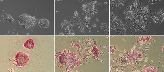
Embryonic stem (ES) cells are valuable in the study of human diseases and development due to their pluripotent nature, and have the potential for treating disease and replacing damaged tissue.  Because of their amazing potential, it is crucial to understand how and when pluripotency occurs, and how stem cells can be cultured.  Naïve pluripotency occurs twice during development—first in early epiblast tissue and again in the germ cell lineage—and this reflects the relationship between pluripotency and the mammalian germ line.  The recent Hypothesis paper by Nichols and Smith suggests that ES cells could be cultured via two routes to reach naïve pluripotency—directly from early epiblast tissue or during the specification of primordial germ cells in culture.

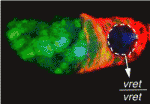
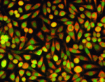
Images above show an early mouse embryo (left) and a colony of ES cells growing on a layer of fibroblast cells (right).  In the embryo, epiblast cells are red, hypoblast cells are green, and trophectoderm is blue.

For a more general description of this image, see my post on EuroStemCell, the European stem cell portal.

ResearchBlogging.orgNichols, J., & Smith, A. (2010). The origin and identity of embryonic stem cells Development, 138 (1), 3-8 DOI: 10.1242/dev.050831

Thumbs up (4 votes)
Loading...

Tags: , ,
Categories: Images, Research

In Development this week (Vol. 138, Issue 5)

Posted by , on 8 February 2011

Here are the highlights from the current issue of Development:

From pluripotent to pancreatic fates

A reliable method for generating insulin-producing β-cells from human pluripotent stem cells (hPSCs) would provide new therapeutic options for people with diabetes. So far, no-one has developed such a method but, on p. 861, Gordon Keller and colleagues provide new insights into the complex signalling networks that underlie β-cell differentiation. The generation of β-cells from hPSCs requires efficient endoderm induction followed by patterning and specification to a pancreatic fate. The researchers show that the duration of nodal/activin A signalling plays a pivotal role in endoderm induction and that WNT signalling enhances the subsequent development of pancreatic lineage cells. Inhibition of BMP signalling at specific stages is also essential for the generation of insulin-expressing cells. Importantly, report the researchers, optimal stage-specific manipulation of TGFβ and WNT signalling yields cell populations that produce insulin at levels similar to those made by the pancreas. However, because these cells also make other hormones, further studies are needed to discover how to convert these polyhormonal cells into functional β-cells.

Full Fat signalling in mammals: Dchs1 and Fat4 pair up

In Drosophila, Dachsous and Fat act as ligand and receptor, respectively, for a signalling pathway that regulates planar cell polarity (PCP) and transcription via the Hippo pathway. Mammals encode multiple Fat and Dachsous proteins but do they have an equivalent Fat signalling pathway? On p. 947, Yaopan Mao and colleagues report that murine Dchs1 and Fat4 function as a ligand-receptor pair during development. The researchers show that Dchs1 and Fat4 single mutants and Dchs1 Fat4 double mutants exhibit similar phenotypes. These phenotypes include the formation of kidney cysts and cochlear defects, suggesting that Dchs1-Fat4 signalling influences PCP in mice. However, the researchers also identify non-PCP-related requirements for Dchs1-Fat4 signalling in the development of other organs. In particular, they show that Dchs1 and Fat4 are needed for growth, branching and cell survival during early kidney development. Together, these results identify Dchs1 and Fat4 as a ligand-receptor pair for mammalian Fat signalling and identify new requirements for Fat signalling in multiple organs.

Capicua mediates response to RTK signalling

Receptor tyrosine kinase (RTK) signalling pathways regulate many developmental decisions, but how RTK signalling controls the expression of its target genes in different contexts is poorly understood. Here, Gerardo Jiménez and co-workers reveal that octameric DNA-binding sites for the transcriptional repressor Capicua (Cic) are critically involved in RTK signalling in Drosophila (see p. 915). They show that the regulation of terminal gap gene expression by the Drosophila RTK Torso in early embryos depends on octameric Cic-binding sites in the enhancer region of the gap gene huckebein. Moreover, these Cic-binding motifs are essential for recruitment of the Groucho co-repressor to the huckebein enhancer in vivo. Cic-binding sites also respond to EGFR RTK pathway activation in the embryonic neuroectoderm and in the developing wing. Finally, using synthetic enhancer constructs, the researchers show that Cic-binding motifs provide the regulatory information necessary to translate RTK signalling inputs into precise transcriptional responses in different tissues. Thus, they conclude, octameric Cic-binding motifs are general response elements for RTK signalling in Drosophila.

A new spin(dle) on stem cell division

Stem cells divide asymmetrically to balance self-renewal and differentiation, thereby maintaining tissue homeostasis. But what coordinates the divisions of multiple stem cell populations in complex tissues? To address this question, Yukiko Yamashita, Alan Hunt and colleagues (see p. 831) have been studying stem cell division in the Drosophila testis, which contains both germline stem cells (GSCs) and somatic cyst stem cells (CySCs). GSCs divide asymmetrically by maintaining a fixed cell polarity within the stem cell niche. Now, the researchers use time-lapse live imaging to show that CySC asymmetric division involves the repositioning of a randomly located mitotic spindle during or near anaphase onset. Spindle repositioning, they report, requires functional centrosomes, the motor protein Dynein and the actin-membrane linker Moesin, and is required to achieve the high-fidelity asymmetric CySC divisions that maintain both GSC and CySC numbers. The researchers speculate that the use of multiple mitotic schemes may be a general mechanism whereby divisions of different stem cell populations are coordinated in complex tissues.

Eyeing up neuronal circuits

The Drosophila optic lobe shares many characteristics with mammalian visual systems and might provide a powerful model for investigating the formation of visual processing circuits. Little is known, however, about the mechanisms that create neuronal diversity and organise neuronal circuits in the medulla, the optic lobe’s primary region. Now, on p. 983, Makoto Sato and colleagues describe the key features of the developing fly medulla. They show that, during larval development, the medulla is subdivided into concentric zones that are characterised by the expression of the transcription factors Drifter, Runt, Homothorax and Brain-specific homeobox. The birth order of the medulla neurons correlates with the expression pattern of these factors, they report, and each neuronal type exhibits an extensive but defined pattern of migration that disrupts the concentric zones during early pupal development. These results, and those of clonal analyses, lead the researchers to suggest that the concentric zone genes may form a genetic hierarchy that specifies neuronal identity and establishes neuronal circuits in the developing medulla.

Marking up germline imprints

Genomic imprinting – epigenetic modifications that ensure that certain genes are expressed from only one of the two inherited chromosomes – is crucial for normal development. Mammalian imprinted genes are associated with differentially methylated regions (DMRs) that are CpG methylated on one parental chromosome. At least 21 DMRs become methylated in the mouse germline and, on p. 811, Hiroyuki Sasaki and co-workers analyse a panel of these gametic DMRs. The extent of methylation of these DMRs differs significantly from that of embryonic DMRs, they report, suggesting that gametic DMRs should be used to identify the features that establish imprinting in the germline. They also show that maternal gametic DMRs appear as unmethylated islands in male germ cells, and unexpectedly identify widespread oocyte-specific non-CpG methylation. Finally, they report that DMR methylation changes dynamically during early development, indicating that DMRs are not fully protected from preimplantation epigenetic reprogramming. These results underscore the importance of using gametic DMR sequences for the study of imprint establishment.

Also…

PAR proteins are conserved regulators of cell polarity, and recent studies, reviewed here by Jeremy Nance and Jennifer Zallen, have identified elaborate links between PAR proteins and cytoskeletal proteins that help set-up molecular asymmetries and hence establish polarity within a cell. See the Review on p. 799

Thumbs up (No Ratings Yet)
Loading...

Tags: , , , , , , , , , , , , , , , , , , , , , ,
Categories: Research

MBL Embryology Course

Posted by , on 8 February 2011

Images taken by students in the 2010 MBL Embryology Course

Dear students, postdocs and mentors,

We write to share our enthusiasm about the MBL Embryology course and to encourage graduate students and postdoctoral fellows to apply for the 2011 summer course “Embryology: Concepts and Techniques in Modern Developmental Biology” at the Marine Biological Laboratory from June 5 – July 17, 2011.  The application deadline has now been extended, and the MBL will accept applications up to Feb. 15th (although the website will continue to give a Feb 1 deadline).  Further course description and application information is available at

http://www.mbl.edu/education/courses/summer/course_embryo.html

This course provides a unique intensive laboratory-lecture experience in contemporary developmental biology.  Students receive instruction from leaders in the field and also have the opportunity to conduct a series of laboratory exercises/investigations using state-of-the-art equipment and a wide range of model and non-model developmental organisms.  We believe that the unique educational experience provided by the Embryology Course is not available at any home institution.  In our experience, students leave this course with an increased breadth of understanding together with practical and novel laboratory experiences and a greatly expanded network of scientific colleagues.

The curriculum is divided into three areas: 1) modern comparative embryology and molecular phylogeny, cell lineage and cell specification; 2) pattern and organ formation; and 3) transcriptional regulation and the analysis of gene networks and developmental pathways.  Students will be exposed to a broad variety of marine and terrestrial invertebrates and vertebrates, and the increasingly sophisticated methods employed to analyze their development. Daily lectures, extended discussions and frequent informal talks provide an intense intellectual experience where students and faculty alike are immersed in sophisticated and continuously changing conceptual and experimental explorations.

We assure you that funds are available to provide substantial financial assistance to defray the cost of tuition.  In 2010 the MBL was able to provide up to 75% of these costs from NIH funding to the course as well as from endowed scholarships.  We expect to provide similar financial aid in 2011.

From our experience as faculty in the course and now as the co-directors, it is our belief that sending a student to this course is a proven, sound investment for the scientific future of the student, lab, and the developmental biology community, and requires only a relatively short absence from the home institution.

If you have any questions please contact Lee, Nipam or Carol Hamel, Admissions Coordinator, at admissions@mbl.edu

Sincerely,

Nipam Patel

University of California, Berkeley

nipam@uclink.berkeley.edu

Lee Niswander

University of Colorado Denver

Lee.Niswander@ucdenver.edu

Thumbs up (3 votes)
Loading...

Tags: , ,
Categories: Education, Events, News

JSDB meeting

Posted by , on 8 February 2011

There’s still time to register for this years JSDB meeting. The deadline for abstract submission has been extended until February 10th.

JSDB meetings have been held almost entirely in English in recent years and not only offer an exciting scientific program, but also a chance to discover Japan.  This year the conference will be held in Okinawa, the largest of the subtropical Ryukyu islands which form the southernmost tip of the Japanese archipelago. Okinawan culture is unique; even if you have visited Japan before, Okinawa offers something different and is well worth a trip.

Check out the JSDB homepage for more information.

Thumbs up (1 votes)
Loading...

Tags: ,
Categories: Events

Reflections on Natural History

Posted by , on 7 February 2011


One of the highlights of working at RIKEN CDB is undoubtedly the arrival upon my desk of “Library News”. Within this publication lies the eclectic column written by institute deputy director Shigeru Kuratani. These bimonthly articles are lively and often illuminating insights into the field of evolutionary morphology, frequently embracing topics such as art, literature, philosophy, and architecture.

There is plenty to entertain developmental biologists, including reviews of several classic anatomy texts such as De Beer’s “The Development of the Vertebrate Skull” and Ramón y Cajal’s “Histology of the Nervous System of Man and Vertebrates”. The pursuit of rare texts and prints is a common theme in many issues and a real sense of enthusiasm for science, history, and adventure is evident as Dr. Kuratani details his quests for classic anatomical text books throughout his career, for treasured natural history plates from the antiquarian markets on the banks of the Seine, and for particular rare specimens of moths resident in the mountains of Japan.

An enlightening and entertaining way to pass time over a cup of coffee, these library columns are now available from the Kuratani lab website (http://www.cdb.riken.jp/emo/clm.html).

Thumbs up (4 votes)
Loading...

Categories: Resources

Natural Disasters and University Disruptions

Posted by , on 6 February 2011

Natural disasters can be powerfully destructive forces. At the very least, they have a habit of interrupting our lives and work. Damage varies depending on the intensity of nature’s fury and how prepared a city (or institution) is for a particular emergency. This can also influence how challenging recovery will be, in the aftermath.

The University Queensland was fortunate in many ways after the Southern Queensland floods, during which 3/4 of the state was submerged. Its St. Lucia campus lay next to the Brisbane River, but it only experienced low-level flooding, and so a handful of buildings were affected. Most escaped damaged. Partly this came from a lesson learned in 1974, the last time the university had experienced intense flooding (levels of up to 6.7 meters. this time water levels rose to about 4-5 meters). Since then, new buildings were predominantly established on higher ground. Post flooding, clean up crews were able to clear the debris and the university re-opened within days.

About 3 weeks later, Cyclone Yasi, category 5 (highest level there is, with gale force winds of up to 285 km/hr) hit Northern Queensland. Major cities in that region include Cairns and Townsville. The damage has been extensive according to recent news reports, with uproot trees, property damage, beached boats etc. Now, thousands are currently without power.  However, as cities were well prepared and properly evacuated, the death toll remains at one.

(Images from Flickr CC, by robandstephaustralia)

James Cook University (JCU), world renown for environmental research, closed its campuses at Townsville and Cairns on the morning of Feb. 2 in anticipation of the cyclone. Over 12 hours later, Yasi hit in the wee hours of Feb. 3. Clean up has been taking place, with the university set to re-open its campuses on Monday, Feb. 7. Many universities, like JCU, has a list of emergency procedures to take, in the event of a disaster or other threats to campus and students. It has a website informing staff and students on how the university responds.

This includes appointing an emergency controller who will  relay messages to staff, arrange any special measures for protecting equipment etc. Another page lists off how to ensure personal safety. As cyclones affect Queensland every year, it’s recommended steps can be quite detailed. i.e. During a storm, it may be necessary to shut off power and wear hardy clothing.

In a previous post on UQ and the Flood, Pablo Astudillo commented that Chile’s universities experienced an Earthquake, 8.8 on the richter scale (last year in Feb. 2010), followed by a tsunami and possibly some fires. Perhaps the only thing invariably worse than one natural disaster, is two or several in a row.

So, for any of you reading this, what kind of natural disruptions have you experienced? And how has this affected your research? Click here for a short survey.

Thumbs up (2 votes)
Loading...

Categories: Lab Life, News

EuroStemCell: connecting scientists with European citizens

Posted by , on 3 February 2011

I’m one of a team of science communicators, scientists, clinicians and social scientists involved in a project called EuroStemCell. It’s an EU-funded project that unites more than 90 European stem cell and regenerative medicine research laboratories in a coordinated effort to engage with the public about our science, and support others to do the same. We’ve got lots going on – this post gives you an introduction to what we’re all about, and we’ll keep you updated as the project unfolds. (more…)

Thumbs up (3 votes)
Loading...

Tags: ,
Categories: Outreach, Resources

the Node image competition

Posted by , on 3 February 2011

[updated 24/2] – New deadline: March 15

To celebrate the successful first half year of the Node, we’re running a competition.

A Node
Latin, Nodus – a knot
In general – a point of intersection
In developmental biology – an embryonic tissue that
patterns a developing embryo

All you have to do is send us an image (it can be, for example, a photo, drawing or microscopic image) related to:
(a) developmental biology; and
(b) some sort of intersection.
Make sure to include a brief description of the image, and only send in your own work.

Think about it carefully, and think out of the box.

Here are a few ideas to get you started….
Two developmental biologists in a knot (well – it’s an intersection of sorts…)
The intersection of two points in development
The intersection of ideas of developmental significance

Email your image to thenode@biologists.com before February 28 March 15 (new deadline!), noon GMT

In March, we will post the top 3 images on the Node, and then everyone will be able to vote for their favourite. Don’t worry if you want to stay anonymous – we don’t have to put your name up on display – just the picture that you send us!

The winner will get to commission their own TipArt work, worth $50 (US dollars), to prominently display in their lab or at home. TipArt is a website celebrating creativity at the lab bench with artwork made of pipette tips.

Terms and Conditions are available here
Competition Rules are available here
Closing date is February 28th March 15 2011 (12:00GMT)

Thumbs up (8 votes)
Loading...

Tags: , , , ,
Categories: Images, News