I have long been interested in understanding how development emerges at the intersection of molecular and spatial organization. On one hand, decades of work have identified the genes and signaling pathways that control cell fate. On the other hand, classical embryology and biophysics have revealed how cells move, change shape, and assemble into tissues. Yet, directly connecting these two layers—linking gene expression to spatial patterns and morphogenesis at the scale of a whole embryo—has remained challenging.
When I joined the Schier lab in March 2020, single-cell RNA-seq approaches had already enabled the reconstruction of developmental trajectories with remarkable molecular detail [1]. For the first time, we could computationally line up cells along developmental paths and begin to understand how cell fates emerge at the whole-embryo level. But something always felt missing to me: these trajectories existed in abstract space, detached from the physical embryo. We could describe where cells were going, but not where they actually were.
At the same time, there was growing interest in spatial transcriptomics within the lab and through our membership in the Allen Discovery Center for Cell Lineage Tracing. This created an opportunity to bridge molecular and spatial information in developing systems. With my background in imaging and technology development, I was particularly drawn to the idea of building a method that could map gene expression across whole embryos, while preserving spatial organization at high resolution.
Technically, the initial setup went relatively smoothly. Ahilya Sawh (then in Susan Mango’s lab at the Biozentrum, now leading her own group at the University of Toronto, Canada) had previously established a FISH-based system for chromosome tracing in C. elegans [2, 3]. With support from the Biozentrum Imaging Core Facility, we adapted this system onto a Nikon microscope, making it more accessible for biological applications. Having this foundation in place was reassuring—it meant that the challenge ahead was not starting from zero, but rather pushing something promising to its limits.
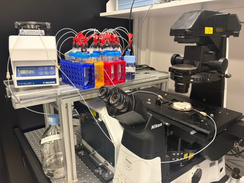
Fig.1 weMERFISH imaging platform for whole-embryo spatial transcriptomics. The weMERFISH system integrates a spinning disk confocal microscope with a microfluidic setup to enable automated, multiplexed imaging over extended periods.
Making it work in a whole embryo
Early on, we realized that if we wanted to understand gene expression in a whole embryo, measuring just a handful of genes would not be enough. We needed to look at hundreds of genes at the same time, but without an impractical number of imaging rounds. This led us to explore MERFISH, which uses a combinatorial barcode system to identify genes across multiple rounds of imaging and allows many genes to be read out efficiently [4, 5]. In practice, it felt like a good balance between scale and feasibility for what we wanted to do.
While the foundation was in place, adapting MERFISH to whole embryos still required several key innovations. An important part of this effort was our close collaboration with Bogdan Bintu at UCSD in the USA. During his time in Xiaowei Zhuang’s lab at Harvard University, Bogdan developed high-throughput imaging approaches that combine MERFISH with 3D chromatin organization [6], contributing deep experience in both experimental and computational aspects. He had already begun implementing important technical improvements and generously shared his strategies with us, as well as providing essential support for the instrumentation and computational pipeline.
In the end, making this work required combining several key improvements, each addressing a different limitation of the system:
• Stability during long-term imaging
Whole-embryo MERFISH imaging can take weeks, as the sample must be imaged plane by plane, region by region, and across multiple hybridization rounds. Early on, this long-term imaging became one of the most frustrating bottlenecks. Using conventional approaches, in which mRNAs are anchored to a gel and repeatedly probed, we saw signals gradually fading over time. Even under carefully controlled RNase-free conditions, degradation was unavoidable.
The solution came from rethinking the problem: instead of anchoring RNA, we anchored the primary DNA probes using acrydite modifications. This seemingly simple shift made a huge difference. The signal remained stable even after a month of continuous imaging, and we could use harsh stripping conditions (80–100% formamide) without worrying about losing the probes. It was one of those moments where a technical fix suddenly makes everything feel possible.
• Seeing deeper without losing signal
Imaging deep tissues introduced another challenge. Spinning disk confocal microscopy is essential for thick samples, but it comes with reduced signal compared to widefield imaging. We initially explored computational approaches to recover signal, but in practice, signal and noise often looked too similar to confidently separate.
Instead, we turned to a physical solution: branching amplification. By boosting the signal at the molecular level, we were able to image more reliably, reduce exposure time, and make the data much more interpretable, especially in deeper regions of the embryo.
• Designing for flexibility
Another challenge was making the method usable in practice. Traditional MERFISH requires encoding the combinatorial barcode directly into the primary probes, effectively locking the experiment into a fixed readout strategy.
We wanted something more flexible. By introducing a gene-specific linker system, we decoupled probe design from readout. This means that researchers can design large probe libraries first, and decide later how to read them out—sequentially for a few genes, or combinatorially for many. This flexibility turned out to be important not just technically, but psychologically: it lowers the barrier to trying the method in the first place.
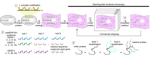
Fig. 2 Workflow and design principles of weMERFISH. Primary probes with acrydite modifications hybridize to mRNAs and are anchored in a gel, enabling stable imaging over many cycles. Gene-specific linker probes and sequential fluorescent readout identify transcripts through a combinatorial barcode. Signal is enhanced by branching amplification for imaging in whole embryos.
When the data started to speak
Once we had the data, the analysis moved quickly. I remember feeling a mix of excitement and disbelief: after spending three years building the system, suddenly everything was there at once. The work was tremendously accelerated by the incredible team behind it: Jakob El Kholtei and I co-developed the weMERFISH method and the data processing pipeline. KJ Jenie, an exceptionally talented undergraduate at the time in Bogdan’s lab, built the MERFISHEYES website (https://schier.merfisheyes.com), making the dataset accessible and explorable. A key component of the project came from my postdoctoral colleague Jialin Liu, who had generated a comprehensive scMultiomics dataset of zebrafish development to study the regulatory logic of cell type specification [7], and very generously made it available to us. Integrating his scMultiomics data with the weMERFISH data allowed us to comprehensively map gene expression and chromatin accessibility in space, creating a multiomic atlas. Mariona Colomer-Rosell performed the analysis of these multiomic data and helped uncover principles of tissue-specific gene regulation at the whole-embryo level. Bringing together these different pieces was essential for turning the dataset into something we could truly interpret.
One of the most fascinating aspects for me was the concept of “time” in development. In single-cell data, we often reconstruct “pseudotime” trajectories, but seeing these trajectories mapped into real space was incredibly satisfying. Along the zebrafish tail, for example, we could directly observe the progression from progenitors to differentiated cells as a spatial gradient. It was one of the first moments where the abstract and the physical truly aligned.
We also applied a spatial version of RNA velocity [8, 9], using nuclear versus cytoplasmic transcripts as a proxy for transcriptional dynamics. What surprised us was that, especially in early development, the inferred transcriptional dynamics mirrored physical cell movements during morphogenesis. At first glance, this feels intuitive. But the underlying mechanisms are very different: transcriptional regulation and cell movement are controlled by distinct processes. The fact that they align so closely suggests a deeper coupling between gene expression dynamics and morphogenesis. This was one of those observations that stayed with me, because it hints at something fundamental that we don’t yet fully understand.
Another memorable part of the journey was the path from preprint to publication. When we first posted the work on bioRxiv and launched the MERFISHEYES website, the response was immediate and very encouraging. People started exploring the data, reaching out with questions, and even visiting the lab to learn how to set up the method. Seeing the dataset being used so quickly made us realize that it could become a resource for the community much earlier than we had expected.
At the same time, the peer review process pushed the work in important ways. The reviewers appreciated the technology and the dataset, but also challenged us to go further, especially to better connect the method to biological questions and to take fuller advantage of the multimodal data. Addressing these comments led to substantial additions and improvements throughout the paper. We expanded the analysis of subcellular transcript localization, strengthened the RNA velocity framework, benchmarked data integration methods more rigorously, and added new analyses such as cell–cell communication.
Perhaps most importantly, the revision motivated us to develop MERFISH-FATE in collaboration with Guoqiang Yu’s group (Tsinghua University, China), integrating spatial transcriptomics with live imaging to directly link gene expression changes to morphogenetic movements. Specifically, we mapped corresponding regions between a weMERFISH embryo and a live-imaged embryo at early gastrulation, where cells had been tracked throughout development. We then followed these trajectories forward and mapped the descendant cells back to their corresponding regions at mid-gastrulation, effectively connecting gene expression patterns across time.
This became a great extension of the story and shifted the paper from a largely static atlas into a more dynamic view of development. We then spent months simply looking at how patterns evolve—scrolling through images, comparing stages, trying to build intuition. Across many genes, we saw surprisingly complex dynamics. One example is tbxta, which is expressed at the embryonic margin at both early and mid-gastrulation. It would be natural to assume this reflects simple inheritance. But when we incorporated cell dynamics, we found that some cells activate tbxta while others turn it off. What looked like a static pattern was actually the result of dynamic and opposing processes. Moments like this made me appreciate how much information is lost when we only look at snapshots, and how powerful it is to connect gene expression with cell behavior. This is a direction we are now continuing to explore in more depth in a recent preprint describing fate mapping in zebrafish embryogenesis and beyond [10].
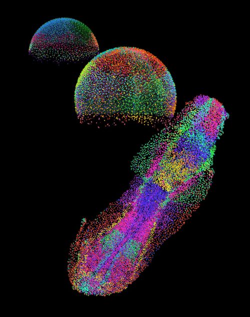
Fig. 3 An embryo in the making. Each color shows a distinct group of cells as they organize during early development of a zebrafish embryo.
Looking ahead: from description to understanding
weMERFISH provides a way to map gene expression and cellular states across intact, developing tissues in 3D. For me, a particularly exciting direction is to extend this approach to larger and more complex systems: whole organs, organoids, and beyond. Moving into these systems will allow us to study how spatial gene expression patterns scale with size and geometry, and how these patterns are adapted across evolution.
At the same time, a major direction in the field, in my view, is the integration of multiple modalities. In this work, we combined weMERFISH with chromatin accessibility and embryo morphogenesis, and this naturally raises broader questions: how does gene expression relate to 3D genome organization? To protein distribution? To lineage history and cell behavior? Each of these layers captures a different aspect of cellular identity, and I believe only by combining them can we begin to understand how cells make decisions in their native context.
This is also a direction I am eager to pursue in my own future work. These multimodal datasets are not just richer—they fundamentally change the type of questions we can ask. Instead of only describing patterns, we can begin to build models that explain how these patterns arise. We can start to ask causal questions: which molecular features predict a cell’s future behavior? Which spatial contexts bias fate decisions? And how conserved are these relationships across tissues and embryos?
Ultimately, I am particularly interested in understanding how variability between individual cells gives rise to robust and reproducible tissue structures. Development is remarkably reliable, despite underlying variability. Technologies like weMERFISH bring us closer to uncovering these principles, and to understanding how a single fertilized egg reliably gives rise to a complex organism.
Access the article:
Wan, Y., J. El Kholtei, I. Jenie, M. Colomer-Rosell, J. Liu, Q. Zhang, J. Navajas Acedo, L. Y. Du, M. Codina-Tobias, M. Wang, W. Zheng, E. Lin, T. H. Chuang, O. Mayseless, A. Sawh, S. E. Mango, G. Yu, B. Bintu, and A. F. Schier. 2026. “Whole-embryo spatial transcriptomics at subcellular resolution from gastrulation to organogenesis.” Science 391 (6790): eadt3439. https://doi.org/10.1126/science.adt3439.
References
Farrell, J.A., et al., Single-cell reconstruction of developmental trajectories during zebrafish embryogenesis. Science, 2018. 360(6392).
Sawh, A.N., et al., Lamina-Dependent Stretching and Unconventional Chromosome Compartments in Early C. elegans Embryos. Mol Cell, 2020. 78(1): p. 96–111 e6.
Sawh, A.N. and S.E. Mango, Multiplexed Sequential DNA FISH in Caenorhabditis elegans Embryos. STAR Protoc, 2020. 1(3): p. 100107.
Chen, K.H., et al., RNA imaging. Spatially resolved, highly multiplexed RNA profiling in single cells. Science, 2015. 348(6233): p. aaa6090.
Moffitt, J.R. and X. Zhuang, RNA Imaging with Multiplexed Error-Robust Fluorescence In Situ Hybridization (MERFISH). Methods Enzymol, 2016. 572: p. 1–49.
Su, J.H., et al., Genome-Scale Imaging of the 3D Organization and Transcriptional Activity of Chromatin. Cell, 2020. 182(6): p. 1641–1659 e26.
Liu, J., et al., Decoding the regulatory logic of specification and differentiation during vertebrate embryogenesis. bioRxiv, 2024.
La Manno, G., et al., RNA velocity of single cells. Nature, 2018. 560(7719): p. 494–498.
Bergen, V., et al., Generalizing RNA velocity to transient cell states through dynamical modeling. Nat Biotechnol, 2020. 38(12): p. 1408–1414.
Wang, M., et al., High-Fidelity Long-term Whole-embryo Lineage and Fate Reconstruction by Iterative Tracking with Error Correction. bioRxiv, 2026: p. 2026.03. 12.711203.
The laboratory of Assistant Professor Dr. Jette Lengefeld is inviting applications for a position as Laboratory Technician who is extensively experienced in experimental mouse work.
About the group The Lengefeld laboratory is working on understanding how the failure to regenerate tissue with age is a major health issue. A contributor to this decline is the loss of stem cell function. Despite the essential role of stem cells, it is still unclear how they fail to maintain their functions during aging and disease. We discovered a new aspect of stem cell aging in vivo: cellular enlargement. With age and damage, stem cells increase in size causing their functional decline. However, we are only beginning to understand how size impacts stem cell fitness and the physiological importance of this process remains unsolved.
The position This position includes several responsibilities. The main activities are listed below:
1. Supporting research projects of lab members, with a strong drive to intellectually contribute. Tasks will include mouse genotyping, mouse blood sampling, mouse monitoring and communication with mouse facility staff. We expect you to fully understand the experimental design and underlying reasons for performing the experiment.
2. Performing administrative tasks for group leader, research meetings and bills.
3. Active intellectual contribution to research discussions such as lab meetings, journal clubs, and project discussions.
Required qualifications and experience – Extensive experience in experimental mouse work and authorization to work with mice is required (please do not apply if not applicable).
– Minimum required education: Master’s degree.
– Experience in hematology will be prioritized.
– The candidate must be fluent in English and Finnish language skills are an advantage.
– The candidate is collaborative, communicative, and comfortable working in an international and multidisciplinary environment.
Salary and contract The position is offered for a fixed term of one year starting in May 2026 or as negotiated. Salary will depend upon the applicant’s level of skills, knowledge, and abilities and is based on the university salary system. A trial period of 6 months will be applied.
Interested? Application should include the following documents: cover letter (max 1 page), CV of max. 4 pages, full publication list, and names of three references.
The deadline for the applications is 30.04.2026, but the positions will be filled immediately once suitable candidates have been identified.
Please submit your application, together with the required attachments, through the University of Helsinki electronic recruitment system by clicking the “Apply for the position” – link below. Internal applicants (i.e., current employees of the University of Helsinki) must submit their applications by logging in to the SAP system: https://msap.helsinki.fi. For technical support regarding the recruitment system, please contact rekrytointi@helsinki.fi.
If you have any questions about the position, please do not hesitate to contact postdoc Dr. Emilie Cerezo emilie.cerezo(at)helsinki.fi.
Link to apply: https://jobs.helsinki.fi/job/Helsinki-Laboratory-Technician%2C-group-Lengefeld/1356106757/
A transcriptional code controlling fluid shear stress-induced gene expression Lucija Fleisinger, Susann Bruche, Hyewon Lim, Anna Rataj, Helena Rodriguez-Caro, Amaury Genovese, Vinesh Vinayachandran, Svanhild Nornes, Dorota Szumska, Dhruv S Gupta, Indrika Ratnayaka, Kira Chouliaras, Marek Giers, Simon J Conway, Alice Neal, Sophie Payne, Martin A Schwartz, Mukesh K Jain, Brian G Coon, Sarah De Val
Single-Cell Atlas of Transcription and Chromatin States Reveals Regulatory Programs in the Human Brain Yang Xie, Lei Chang, Guojie Zhong, Jonathan A. Rink, Tatiana Báez-Becerra, Ethan Armand, Wubin Ding, Kai Li, Eric Bonne, Audrey Lie, Hannah S Indralingam, Keyi Dong, Timothy Loe, Bohan Huang, Zhaoning Wang, Ariana S. Barcoma, Jackson K. Willier, Kyle W. Knutson, Jiayi Liu, Silvia Cho, Stella Cao, Kaitlyn G. Russo, Carissa K. Young, Jessica Arzavala, Yareli Sanchez, Aleksandra Bikkina, Natalie Schenker-Ahmed, Colin Kern, Zoey Zhao, Amit Klein, Jesus Flores, Chu-Yi Tai, Jacqueline Olness, Alexander Monell, Siavash Moghadami, Cesar Barragan, Chumo Chen, William Owens, Carolyn O’Connor, Michelle Liem, Mikayla V. Marrin, Cynthia Rose, Shane N. Alt, Nora Emerson, Julia Osteen, Jacinta Lucero, Daofeng Li, Rebecca D. Hodge, Ting Wang, C. Dirk Keene, Xiangming Xu, Quan Zhu, Joseph R. Ecker, M. Margarita Behrens, Bing Ren
Tracking morphological development in stony corals Garrett J. Fundakowski, Viviana Brambilla, Kyle J. A. Zawada, Cher F Y Chow, Emily Croasdale, Amelia J. F. Errington, Luisa Fontoura, Wilhelm J Marais, Rachael M. Woods, Pim Edelaar, Kevin Lala, Joshua S. Madin, Maria Dornelas
Early development of male germ cell clones shapes their reproductive success Tatsuro Ikeda, Maurice Langhinrichs, Tamar Nizharadze, Chieko Koike, Yuzuru Kato, Katsushi Yamaguchi, Shuji Shigenobu, Kana Yoshido, Shinnosuke Suzuki, Toshinori Nakagawa, Ayumi Maruyama, Seiya Mizuno, Satoru Takahashi, Nils B. Becker, Hans-Reimer Rodewald, Thomas Höfer, Shosei Yoshida
Observing concurrent subcellular dynamics in large living tissues Charles S Wright, Sanjeev Uthishtran, Laura Z Kreplin, Hetvi R Gandhi, Abhishek Patil, Harrison M York, Samyukta Sita, Samuel A Manning, Elliot Brooks, Guizhi Sun, In-won Lee, Wing Hei Chan, Sara Hlavca, Samuel Crossman, Helen E Abud, Jan Kaslin, Avnika A Ruparelia, Peter D Currie, Kieran F Harvey, Jose M Polo, John Carroll, Senthil Arumugam
bioRxiv: the preprint server for biology Richard Sever, Samantha Hindle, Ted Roeder, Sol Fereres, Olaya Fernández Gayol, Sanchari Ghosh, Martina Proietti Onori, Emma Croushore, Kevin-John Black, Linda Sussman, Janet Argentine, Wayne Manos, Marisol Muñoz, Josh Sinanan, Tracy K. Teal, John R. Inglis
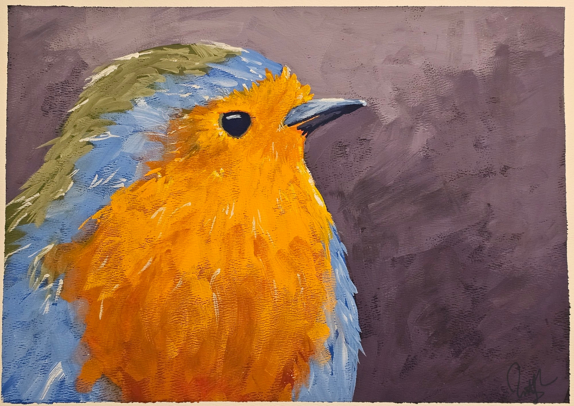
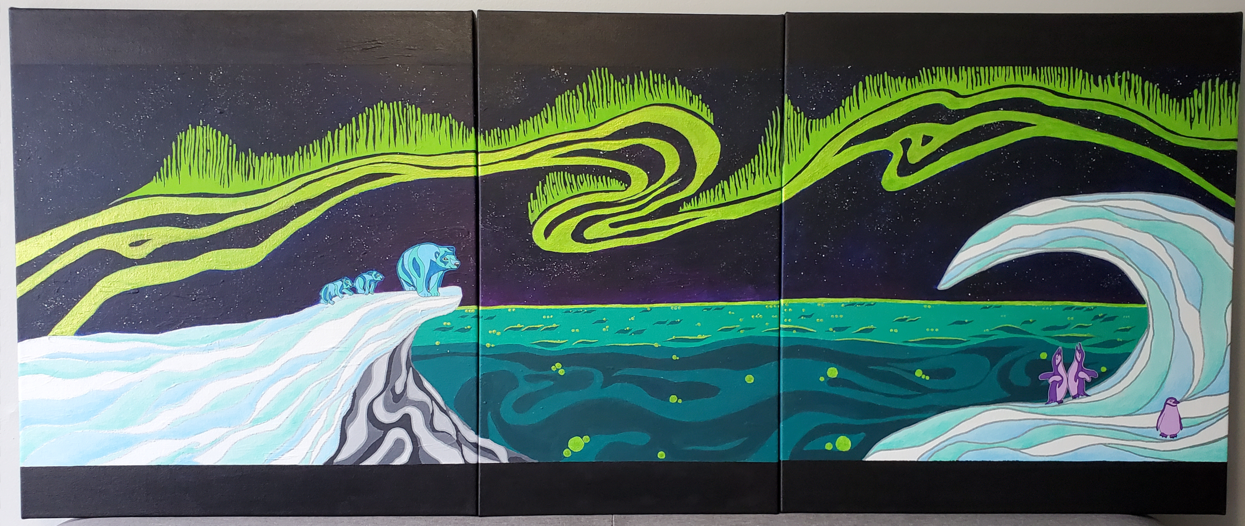
In this SciArt profile, we meet Brittany Carr, an Assistant Professor at the University of Alberta in the Faculty of Medicine and Dentistry, Department of Ophthalmology and Visual Sciences. In her artwork, Brittany uses acrylic, watercolour, gouache, and ink to create pictures of the natural world, while she is also a fan of using microscopy for ‘science’ art.
Can you tell us about your background and what you work on now?
During my PhD research, I was interested in pharmacological control of myopia, and investigating off-target drug effects of muscarinic antagonists in the chicken eye. I then switched to inherited retinal degeneration for my postdoctoral studies, where I learned to use frogs as a model organism. I studied the effects of loss of two genes: PROM1 and CDHR1 on photoreceptor outer segment morphogenesis and retinal degeneration. I started my independent research career in 2022. I am still interested in PROM1 and in using frogs to develop other models of inherited and age-related blindness. We have a few interesting projects in the lab that a new for me including looking at microglia and retinal inflammation, and retinal development.
Were you always going to be a scientist?
I was always interested in science as a kid and read every science book that I could get my hands on. I was the first person in my family to go to university, however, so I didn’t know how it was possible for me to actually become a ‘real’ scientist. I joined a pre-med undergraduate program with the intention to go to Optometry school. I was fortunate enough as a 3rd year undergraduate student to meet an incredible mentor, who invited me to join his lab and gave me free reign to do science. The first time I prepared an immunofluorescence slide of chicken retinal amacrine cells and looked at it under the microscope, I was absolutely hooked. From then on, I knew academia was the only place I wanted to be and I was lucky enough to have landed myself in a lab where there was a mentor who knew exactly how to help me make it happen.
And what about art – have you always enjoyed it?
Yes, I have always enjoyed art and drawing. I spent a lot of time in elementary, middle- and high-school drawing instead of taking notes or doing homework during my classes. There was a time where I was at a crossroads and had to make the decision to choose between art school and science. I chose (at the time) to pursue optometry. Then, later on, when I discovered microscopy I got to learn a technique where I could express myself artistically again in a scientific career. I am a self-taught artist, but I recently moved a few blocks away from an Atelier, where I can now take art classes. It’s been a lot of fun getting to spend time just painting and growing my art skills.
What or who are your most important artistic influences?
I have always been attracted to the ‘creepy’ or ‘weird’ artists, or loose, bright, and impressionistic landscape artists. People like H.R. Giger, Hieronymus Bosch, Francisco Goya, Francis Bacon. On the complete opposite end of the spectrum, Hester Berry, Julia Veenstra, and a whole bunch of local artists, including Di, Erinn Evans, Oksana Zhelisko, Jared Robinson, and Justina Smith.
How do you make your art?
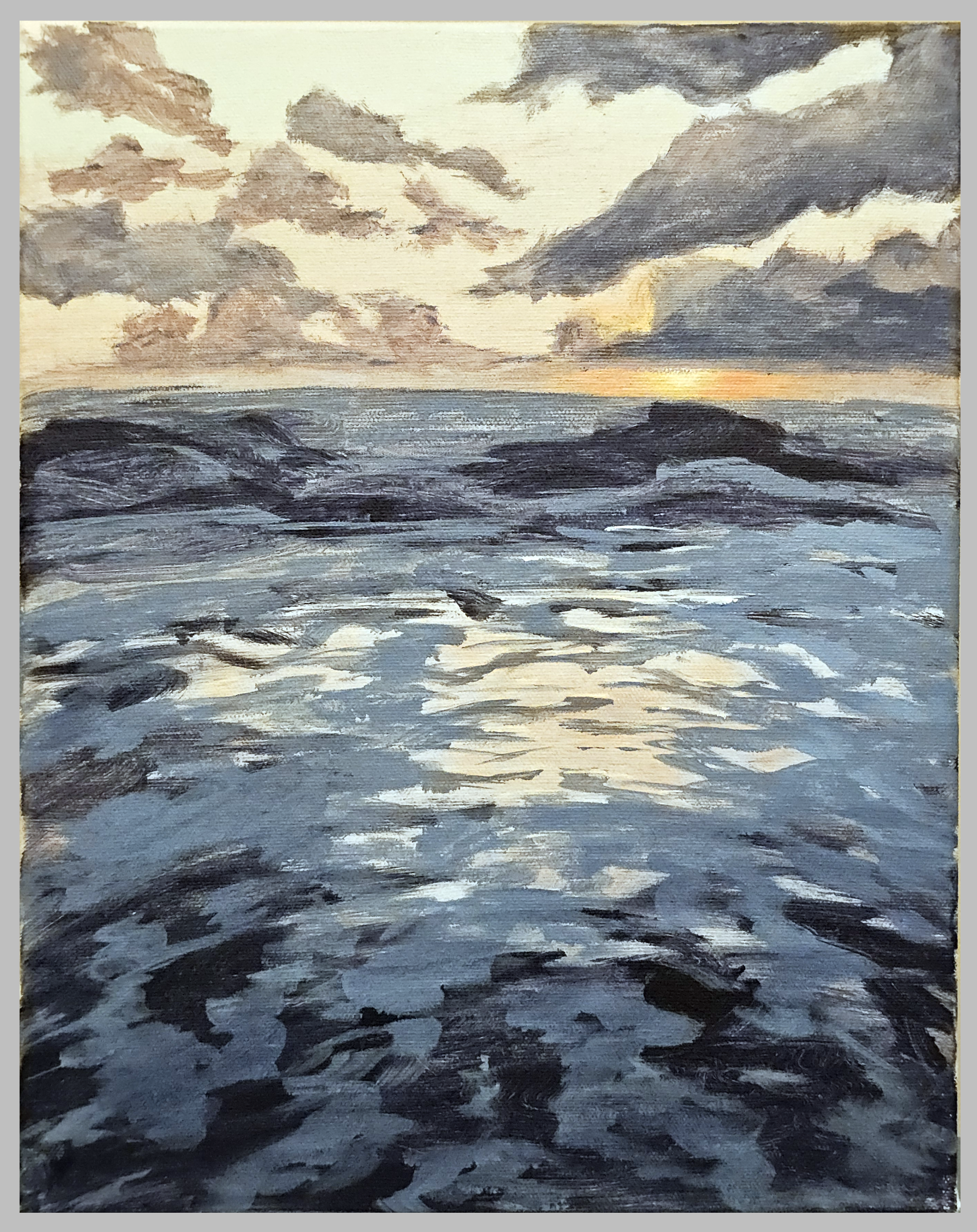
I dabble in a bunch of mediums, but primarily acrylic, watercolor, gouache, and ink. Practically speaking, I make most of my art in classes now, because running a lab and doing research is a lot of work, and nothing forces you to make art like paying money for a 4-6 hr block of time to do nothing else but make art. When the weather is nice, I like to take my sketchbook and work plein air. I am fortunate to live somewhere surrounded by nature, so I like to take advantage of that.
Does your science influence your art at all, or vice versa, or are they separate worlds?
I think that I like the same themes between my science and my art (nature, bright colours, high contrast), but they have two different goals. For science, you can make aesthetic things, but the goal is always to convey knowledge. Because of this, I approach my ‘science’ art differently than I do my traditional art, which for me, is just to make things that make me happy. I definitely take micrographs for aesthetic reasons, and most of these end up on social media and my webpage, not so often in actual papers or diagrams. The subjects that I draw in traditional art are not overtly science-themed, and instead are more focussed on landscapes and ‘macro’ nature, such as birds and animals, or silly things that I do just for the joy of it with no need to convey a message.
What are you thinking of working on next?
I’m the type of person that doesn’t paint until inspiration hits me, and then I can’t not paint until the idea in my head is realized. I live in a fairly quirky city now, and so I do want to do some small paintings of “Just Edmonton Things” that I have seen or experienced since moving here that I found funny or interesting.
How/ where can people find more about you?
I am most active on bluesky @drbjcarr.bsky.social, where I post mostly about science, but share my art and photography too.
Recently, I attended the biennial meeting of the German Society for Developmental Biology (GfE). This was my second time attending one of these meetings, and I was looking forward to it, having missed the last one a couple of years ago. Everyone from my scientific circle, here in Germany, thinks of these meetings as having a homely feeling with a close-knit familial atmosphere, and this meeting was no different. The meeting this time was even more special with members of the Dutch (DSDB) and Spanish (SEBD) societies joining in as well.
The meeting took place in Potsdam over the course of 4 days, on a university campus: a welcome trend in the GfE meetings, where the meetings are typically organised in an academic backdrop, instead of a commercial conference center. I believe this is a great strategy to keep registration fees low, making the meeting widely accessible to the community. With plenty of coffee and food to go around, the meeting struck a nice balance between great science and the time to digest it all.
These meetings are always a great opportunity to reconnect with your local scientific network. Having attended one of the previous iterations, I was looking forward to seeing some of my old colleagues. I am sure many others were also able to interact with colleagues from Spain and the Netherlands, creating new connections. With about 150 participants, the meeting was just the right size to not be overwhelming, with the international crowd finding ample opportunities to intermingle. The relatively small size naturally obviates the need for having parallel sessions, thus not forcing one to make the difficult choice of missing out on interesting talks. Despite its small size, the meeting had a significant presence on social media, with #GfE2026 trending on the feeds.
Covering topics from the basics of embryonic development to disease modeling, the conference showcased the latest and greatest in classical model systems, as well as emerging ones. As usual, the presence of in vitro embryo models was noteworthy, with a concerted drive towards increasing throughput and reproducibility in these systems. Surprisingly, -omics techniques (especially, single-cell RNA-Seq) were a bit underrepresented, giving the impression that perhaps the community has now gotten over the novelty-driven early excitement. Instead, there was an exciting abundance of talks and posters focusing on the role of mechanical regulation in biological systems (cell-, tissue- mechanics, mechanical manipulations and characterizations, etc.) at all stages of development.
Speaking of posters: while the quality of the posters was excellent, the duration of the poster sessions left something to be desired. Given how well organised this meeting was, commenting about shorter poster sessions feels nitpicky. However, there seems to be a broader emerging trend in conferences that needs to be addressed: more often than not, the space for poster sessions is limited, preventing the presenters from displaying the posters throughout the meeting. It is disheartening to have one’s poster propped up for a couple of days at best, not getting the attention that it deserves, after having spent hours preparing it. We, as a community, need to make a change and Make Poster-sessions Great Again: poster sessions should not feel like an afterthought. Participants should be allowed to display and celebrate their work throughout the meeting, with even- vs. odd-numbered posters being presented in different poster sessions. In any case, I particularly appreciated the novelty of many of the findings presented in talks and posters, with many unpublished results, whether completely new or freshly available as preprints.
One of the highlights of the conference was the PhD Award Lecture by Tatiana Lebedeva and the Hilde Mangold Award Lecture by Maik Bischoff. Tatiana walked us through her experiments with Nematostella vectensis embryos, where she focused on germ layer specification and gastrulation. It was great to see her grit and optimism despite the painstakingly difficult journey of trying to create transgenic animals to visualize β-catenin expression in embryos of this species. Maik talked about his work on the emergence of chirality in biological systems through tissue interactions. Although working with Drosophila melanogaster – a conventional model organism – he demonstrated how the field needs to use these experimental systems to ask increasingly challenging questions. Listening to these and other talks, I couldn’t help but wonder about the future of model organisms in developmental biology research. While research on non-model species is a necessary challenge and a welcome change for the field, work by Maik and others at the conference showed that model organisms such as Drosophila melanogaster still have their relevance. We are certainly in an age in which what was once frontier research in model organisms is now a territory being increasingly captured by non-model organisms. The only way to keep these conventional models relevant is to ask increasingly challenging questions and push the limits of what was once possible. (See this recent preprint, which talks about diminishing representation of model organisms in scientific literature over the past couple of decades, and what that might mean for the future of basic and applied research in biology.)
Thankfully, the weather was somewhat on our side, with some sunshine allowing us to sit outdoors during lunch times. The conference dinner on the waterfront was exceptional: I don’t remember having had better food at a conference in recent memory, and from what I hear, I missed out on a similarly excellent food during the last meeting. Keeping up with the tradition, the dance party followed, with great music till 1 am, when we were gently “forced” out of the restaurant. I suppose the next meeting (in 2028 at Heidelberg) has a lot to live up to.
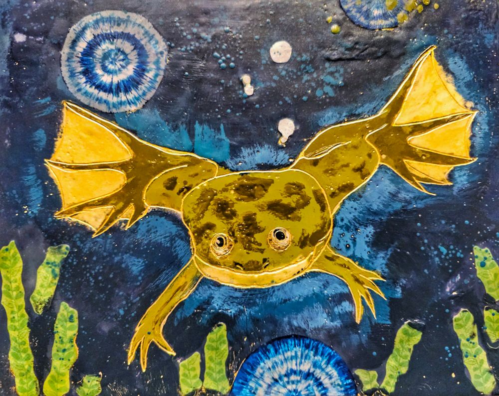
Acknowledgments: Thanks to Alex Eve and Verena Kaul for comments.Cover image, courtesy of meeting organisers and Ingrid Lohmann.
The School will be a 4-day (23-26 June 2026) Theory&Computation course in a splendid Swedish inland Resort (2 hours away from Stockholm – our bus will bring you there at no additional cost from a nearby train station).
The first editions of the school were in 2022 and 2024—both a success (click here or here if you are curious).
– The teachers will be prominent international scientists, but also former participants who are now invited as teachers – you will learn from your peers via hands-on computational sessions. By participating this year, you will also be able to present your candidacy to become a teacher in the next edition!
– The event is designed to favor frequent interactions between PhD students/Postdocs with successful scientists in the field, in multiple meet-the-speaker sessions, and during free outdoor time, as the invited speakers are asked to be around and available throughout the school.
– The school will focus on key relevant topics at the interface between technology, biology, and computation, including how to integrate computationally and conceptually all the analytical modalities such as gene expression in single cell, epigenetics and 3D genome.
– The school will also include a flash course of scientific writing tailored for the field (this is a regular, highly appreciated part of the school).
-The school is sponsored by PALS (a merge of the two prestigious Knut of Alice Wallenberg Foundation & SciLifeLab).
Among many other, you will benefit from hands-on-computational sessions on:
– Single-cell multiome Data Integration & workflow management and reproducibility – Combination of ATAC-seq and 3D-genomics – Non-coding variants Prioritization & advanced Transcription Factor motif analysis – Inferring Gene Regulatory Networks from large-scale epigenomics – Visium HD Spatial Transcriptomics
My name is Andrea Murillo, and I am delighted to share that I am the new Community Manager for the Node. I started my research journey as a physiologist and later found my way into endocrinology during my PhD, where I worked with my favourite worm and developmental model species, Capitella teleta. Throughout my PhD, I investigated components of the estrogen signalling pathway across life stages of C. teleta. That is how the wonder of developmental biology first wormed its way into my heart.
After finishing my PhD, I started working for The Company of Biologists as the Science Communications Officer. In that role, my passion for science communication grew, and my appreciation for biology deepened as I wrote about the Company’s fantastic journal content across many fields. But it was the science and the community surrounding the Node and Development that truly hooked me (I will stop with the worm puns now!).
As Community Manager, I am excited to build on the great work done by Eva, Cat, Aidan, Helen and Joyce and to continue some of our users’ favourite series and features. In my previous role, I worked closely with both my predecessor, Joyce, and the two Community Managers from our sister sites: Reinier from preLights and Helen from FocalPlane. I am thrilled to be a part of the team as the Node Community Manager, a transition which they have generously supported. I’m planning to bring some of my own fresh ideas to the Node, and I hope you will like them.
My first chance to meet some of you in person will be at the British Society for Developmental Biology 2026 Spring Meeting next week. If you are attending, please stop by and say hello. I am really excited to learn about your research and, most of all, to meet the people who make great developmental biology and stem cell research happen.
Preprints have become an indispensable part of our research ecosystem. Over the last 10 years, the biological community has witnessed an exponential growth in both submissions and readership of preprints. Arguably, the main drivers behind this growth are the ability of preprints to speed up the dissemination of research and broaden access to results long before formal publication.
Development and our not-for-profit publisher, The Company of Biologists, have a long history of actively supporting preprints (Prosée and Brown, 2025). In 2018, the Company launched preLights, a community-run platform that highlights noteworthy preprints across the biological sciences. Over time, preLights has evolved to provide support and training for early-career researchers to develop their writing skills for summarising and critiquing new work. In addition, Development’s own community site, the Node, posts monthly preprint lists from developmental biology and related fields. These lists are among the most-read posts on the Node, demonstrating the value of preprints within our community. Over time, preLights, the Node and Development have started working together more closely to highlight noteworthy preprints; a recent collaboration between preLights and the Node saw the introduction of curated preprint highlights in the form of ‘preLighters’ choice’ posts and a selection of preLights posts from the stem cell and developmental biology community feature in the journal as quarterly ‘Preprint Highlights’.
As part of this preprint ecosystem, Development launched its ‘In preprints’ series in early 2022 to bring curated, contextualised coverage of preprinted findings directly to our readership (Briscoe and Grewal, 2022). These articles are intended to complement other initiatives, such as preLights, in guiding readers to the preprints that matter the most in the field. We know that Development’s ‘In preprints’ articles receive, on average, over a thousand views within the first 12 months of publication and continue to be read in the years that follow. Development has now published around 60 ‘In preprints’ articles on topics ranging from single-cell lineage tracing techniques (Rodriguez-Fraticelli and Morris, 2022) to human stem cell-based embryo models (Moris and Sturmey, 2023) and Polycomb complexes (Iwasaki et al., 2023) to leaf-shape transitions (Byrne, 2024). You can browse all the ‘In preprints’ articles published to date in our dedicated subject collection.
Preprints featured in these articles have mainly been selected by Development’s in-house Reviews Editors. We are now expanding this initiative by appointing a small group of Preprint Editors – active researchers with their finger on the pulse of preprint literature – to commission and write ‘In preprints’ articles. This is an exciting opportunity to co-curate the preprint literature directly with members of the Development community, bringing specialist expertise and diverse perspectives to bear on an ever-growing body of work. We intend to appoint Preprint Editors who represent specialist topics within the broader scope of Development, and we hope that, as expert academics, they will be able to identify and highlight the best preprints from their fields as part of their routine exploration of the research literature.
We are currently accepting applications for Preprint Editors, and our call will close on Monday 30 March 2026. Applicants with at least 3 years of postdoctoral experience or principal investigators from any career stage are welcome to apply. In exchange for their contributions to the project, Preprint Editors will receive formal recognition and financial remuneration, including the option to receive support to attend relevant conferences. To find out more about this initiative, please visit our webpage: https://journals.biologists.com/dev/pages/preprint-editors.
We look forward to working with our first Preprint Editors following their appointment later this year to further strengthen our coverage of important preprinted work. We also anticipate that this programme will continue to evolve in response to community needs. This next step marks our commitment to supporting the reach of preprinted work and bringing curated, quality research to the attention of our community.
Biology Open is proud to partner with the European Zebrafish Society (EZS) to offer travel grants for the 13th European Zebrafish Meeting (EZM2026) to be held 7-11 July 2026 in Vienna, Austria. This travel grant is designed to provide financial support for early-career researchers based in the Global South.
The aim is to strengthen links for future collaboration and enhance the researcher’s career opportunities, which is in line with The Company of Biologists’ core value of supporting biologists.
More information and an application form can be found on the EZS website.
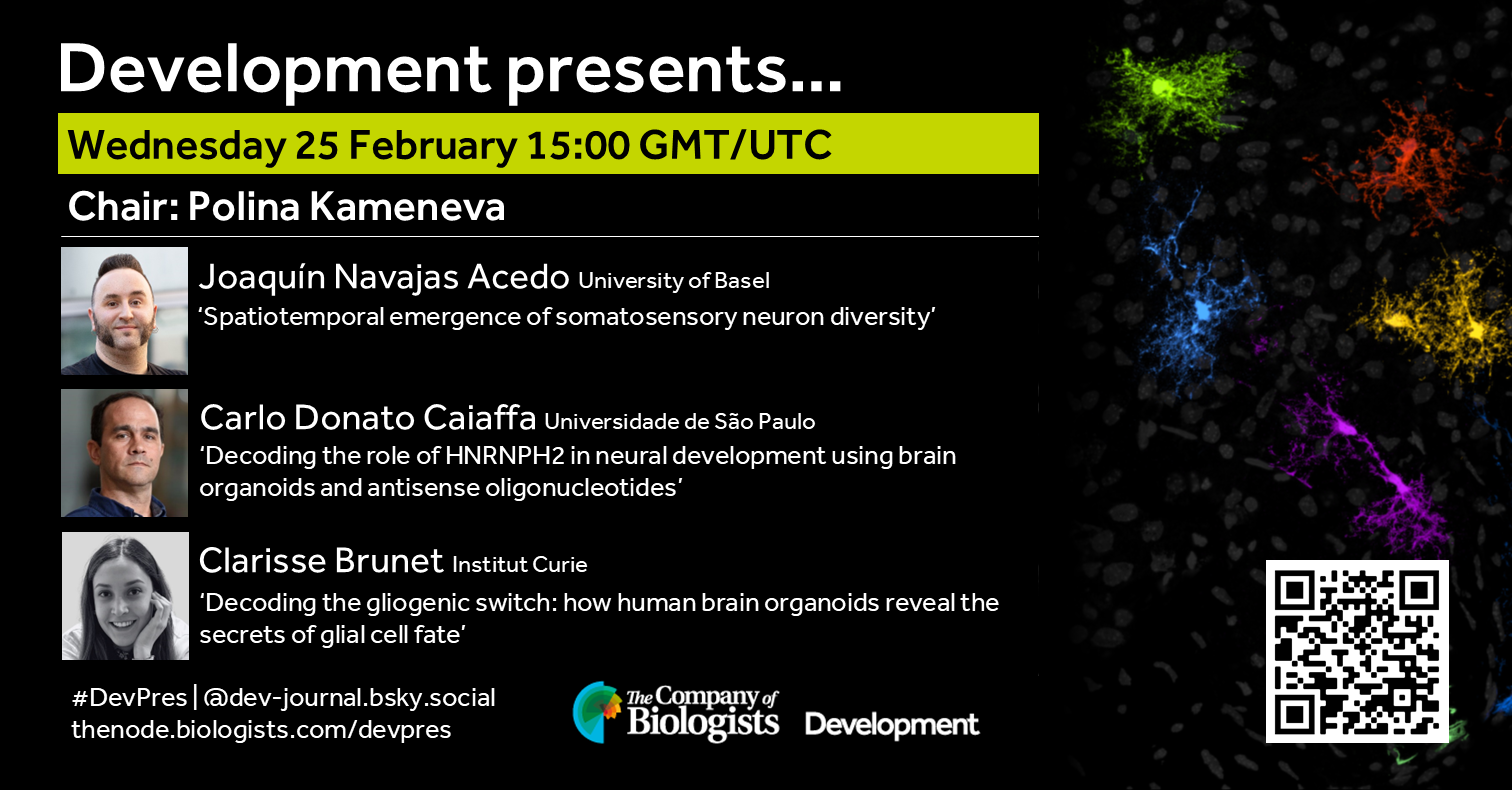
In February, we hear from three early-career researchers studying neural development. Chaired by one of Development’s first Pathway to Independence fellows, Polina Kameneva, Principal Investigator at St. Anna Children’s Cancer Research Institute (CCRI) in Vienna, Austria. Polina’s group uses 2D and 3D human stem cell models to recapitulate adrenal gland cell development to understand the onset of neuroblastoma.
Wednesday 25 February – 15:00 GMT/UTC
Joaquín Navajas Acedo (University of Basel) ‘Spatiotemporal emergence of somatosensory neuron diversity’
Carlo Donato Caiaffa (Universidade de São Paulo) ‘Decoding the role of HNRNPH2 in neural development using brain organoids and antisense oligonucleotides’
Clarisse Brunet (Institut Curie) ‘Decoding the gliogenic switch: how human brain organoids reveal the secrets of glial cell fate’
At the speakers’ discretion, the webinar will be recorded to view on demand. To see the other webinars scheduled in our series, and to catch up on previous talks, please visit: thenode.biologists.com/devpres
 (No Ratings Yet)
 (1 votes)