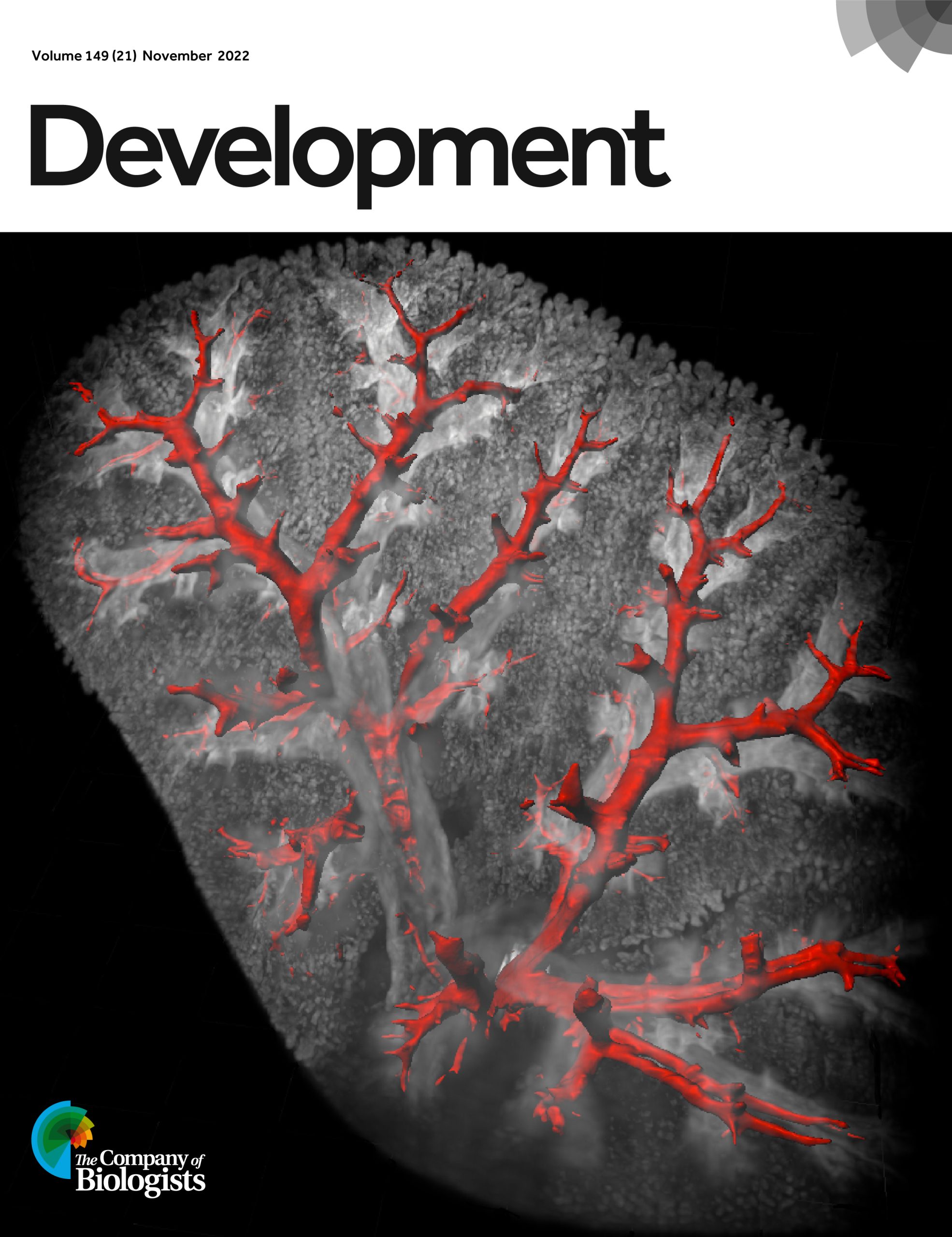
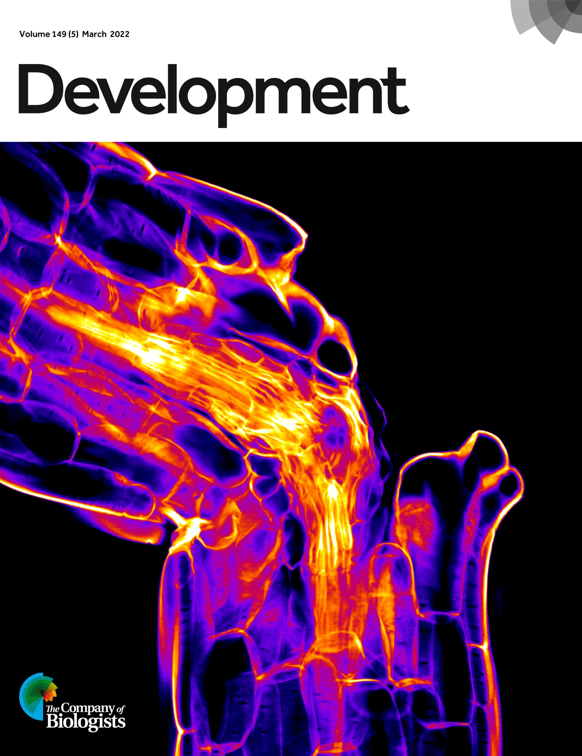
We asked you to select your favourite Development cover from 2022* and after all the votes were counted, the Node community has chosen the mouse lung lobe from Issue 21. The image is linked to the Open Access Research Article from Prashant Chandrasekaran, Nicholas Negretti, Aravind Sivakumar, Jennifer Sucre, David Frank and colleagues on the role of CXCL12 in defining lung endothelial heterogeneity and promoting vascular growth.
Issue 21: Mouse lung lobe
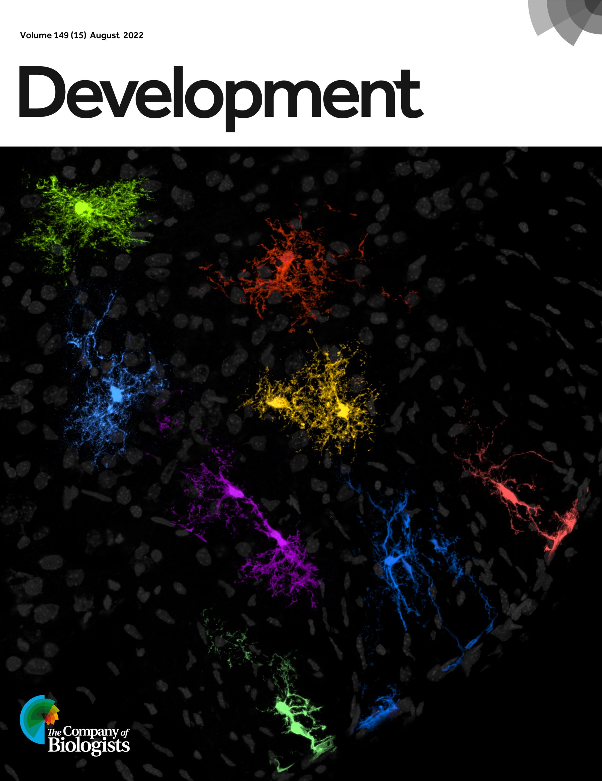
In second place were the astrocytic cells in the mouse spinal cord from Maria Micaela Sartoretti, Carla Campetella and Guillermo Lanuza featured on Issue 15. Third place went to the Arabidopsis hypocotyl graft junction from Phanu Serivichyaswat, Kai Bartusch ,Charles Melnyk and colleagues in Issue 5.
Issue 15Issue 5
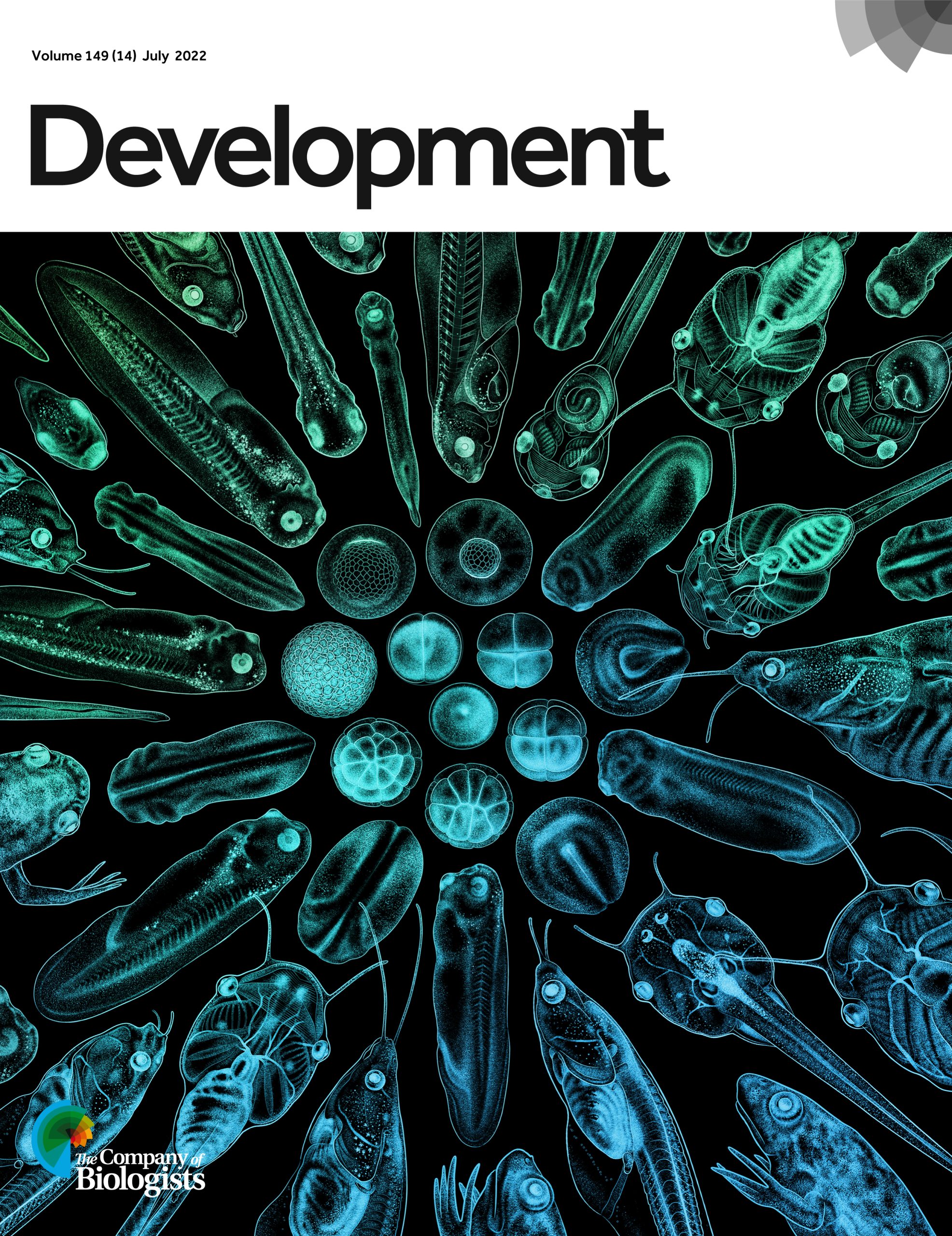
Interestingly, it wasn’t any of these entries that won the in-house team at Development the box of chocolates on offer for the most popular cover from The Company of Biologists in 2022. In this competition, the winning cover image was the stages of Xenopus development from Natalya Zahn, Christina James-Zorn, Aaron Zorn and colleagues in Issue 14.
Congratulations to the winners and thanks to everyone who submitted cover art in 2022. We are delighted to have had another year filled with wonderful cover images and we are now looking forward to seeing what 2023 brings!
During the first year of my course in biological sciences, I became interested in developmental biology. I am mesmerized by the exploration of the beginnings of life through the complex interactions and changes that are taking place from the moment of fertilization. Furthermore, I realized the potential applications that scientific advancements in this area may have for disease prevention and treatment. This is especially interesting to me as a wildlife health student, aiming to conduct research on wildlife conservation in the future.
Research led by Dr. Denis Larkin found a mutation in the amino acid sequence (AA) of the NRAP gene in Yakut Cattle, resulting in a single nucleotide polymorphism (SNP resulting in a Glutamine to Histidine substitution (H100Q) at position 100) (Larkin et al., 2021). Yakut cattle are resistant to the low temperatures of the Siberian arctic and exhibit features such as efficient thermoregulation, a slow metabolic rate, and resistance to diseases. Nebulin-Related-Anchoring Protein (NRAP) is an actin-binding gene responsible for producing proteins to connect actin filaments in skeletal and cardiac muscle (GeneCards, 2022). The NRAP gene AA sequence is evolutionary conserved, however, this specific H100Q mutation was found in other cold-adapted, hibernating species and those entering periods of extreme bradycardia.
The expression of this particular SNP in those specific animal species prompted us to start this study. My summer project aimed to create the H100Q NRAP single-point mutation in the human gene and clone it into a suitable vector for in-vivo expression. This construct will then be used for microinjections into zebrafish embryos to observe potential changes in their heart development, heart rate, and muscle function.
I was keen to start the project and started by gathering information about zebrafish heart development, as well as molecular biology techniques. Together with my team, we created a well-thought-out cloning and mutation strategy to substitute a single nucleotide from the human NRAP gene from CAC (H) to CAA (Q) to recreate the H100Q mutation found in Yakut cattle.
However, I soon learned that theory and practice are two very different sides of the same coin. The world of molecular biology seemed to be unpredictable at first! After the first couple of ligations that didn’t produce any colonies, it was difficult to believe that getting enough material to mutate this gene was even possible. But that didn’t stop us. We troubleshot, experimented with enzymes and buffers, made our own competent cells and bacteria plates, changed reaction conditions, consulted papers, and of course, could always rely on the advice of our helpful supervisors. Having an amazing and experienced team around us at all times was key to our success. Not only could they provide valuable insights through their many years of experience in molecular biology but also offered constant encouragement: “it will be fine” turned out to be right. I was determined to make it work and this experience strengthened my resilience and problem-solving skills.
I acquired many useful laboratory skills which will be very invaluable as a researcher. I learned how to read vector maps, how to clone, use publicly available software, and how to design primers for PCR… just to name a few. Understanding the science behind molecular biology techniques allowed me to modify protocols to troubleshoot efficiently. Finally, the Sanger-sequencing results came back, and excitingly, we validate our single-point mutation H100Q of the human-NRAP gene in pBluescript II SK (+) (Figure 1).
Figure 1: Sanger-sequencing read from nucleotide 253 to 294 by T3 polymerase. CAA mutation highlighted in yellow with C substituted to A at position 285.
Apart from generating the mutation, I learned how to conduct research on live animals during my work in the fish facility. I bred and cared for zebrafish embryos until up to 5 days post-fertilisation period during which they are not regulated under a Home Office licence. I practiced my microinjection skills into one-cell stage embryos. My fascination only grew stronger and I found myself peaking at the embryos throughout the day to witness the one cell splitting into 4-, 8- and 16-cells forming a plate on top of the yolk and was amazed how, in the span of only 48 hours, I could make out the heart structure and observe the heartbeat under a stereomicroscope. Another interesting aspect of this project was to investigate the H100Q-NRAP mutation in Pinniped and hypothesize its origin in Otariids and Odobenidae.
Overall, I was thrilled to discover how different aspects of biology, from molecular biology, and genetics to developmental biology and physiology came together in this project. It reminded me once again of the importance to follow a big-picture approach and that scientific understanding can seldom follow from isolated observations. The point is, however, to put them into the context of constant change interplay with each other and the environment. A continuation of this project would on the one hand be useful to further aid the scientific understanding of cold adaptations and hibernations to conserve ecosystems and species being threatened by climate change and on the other hand understand the physiological implication of heart mutations in humans.
I would like to thank my supervisor Dr. Claire Russell, Dr. Caroline Pellet-Many, Dr. Steve Allen, and Dr. Denis Larkin for their support, insights, well-stocked freezers full of enzymes, and encouragement throughout the whole process. I am also kindly thanking the British Society for Developmental Biology for granting me this fantastic opportunity and opening the doors to the world of scientific research for me. Thank you!
References
Buggiotti, L.; Yurchenko, A.A.; Yudin, N.S.; Vander Jagt, C.J.; Vorobieva, N.V.; Kusliy, M. A.; Vasiliev, S.K.; Rodionov, A.N.; Boronetskaya, O.I.; Zinovieva, N.A.; Graphodatsky, A.S.; Daetwyler, H.D; Larkin, D.M. (2021) ‘Demographic History, Adaptation, and NRAP Convergent Evolution at Amino Acid Residue 100 in the World Northernmost Cattle from Siberia’, Molecular Biology and Evolution, V(38), Issue(8), pp. 3093–3110, https://doi.org/10.1093/molbev/msab078
Now in its sixth year, the Norwich Single-Cell Symposium at Earlham Institute covers single-cell genomics technologies and their application in microbial, plant, animal and human health and disease. Registration is now open!
This event aims to bring together researchers who are curious about applying single cell technologies with those who are experts working at the forefront of the field and across a wide range of species.
The event will feature talks from keynotes, invited speakers and selected abstracts, and we are keen to capture as broad a range of single-cell applications as possible. Our programme is here.
We’re pleased to confirm our two keynote speakers will be:
Seung Yon (Sue) Rhee, Carnegie Institute for Science
Detlev Arendt, European Molecular Biology Laboratory (EMBL), Heidelberg
We will also be opening up doors to Earlham Institute’s Single-Cell labs, so you will be able to see our technology and capability as well as access our expertise through our researchers.
Doing great science depends on teamwork, whether this is within the lab or in collaboration with other labs. However, sometimes the resources that support our work can be overlooked. Our ‘Featured resource’ series aims to shine a light on these unsung heroes of the science world. In our latest article, we hear from Patrick Lemaire and Emmanuel Faure, who describes the work ofMorphoNet.
Patrick Lemaire1 and Emmanuel Faure 2
CRBM, Université de Montpellier, CNRS, 34293 Montpellier, France.
LIRMM, Université de Montpellier, CNRS, 34095 Montpellier, France
What is MorphoNet?
MorphoNet (1) is an interactive morphodynamic web browser designed to help scientists, teachers and students share, analyze and visualize the large 3D morphological datasets that can be generated by modern imaging technology, ranging from live light sheet microscopy of cells and embryos to X Ray tomography of fossils.
Why did you build MorphoNet?
We initially created MorphoNet so that our biologist collaborators can interact, without programming skills, with the 3D + time datasets they obtained by light sheet microscopy of live ascidian embryos followed by segmentation (2). We wanted the system to be web-based so that they do not have to install any sophisticated software on their computers, and to allow data sharing. We therefore chose to use a very popular gaming engine Unity, to show meshed segmented data. This choice allowed us to compress the complex 3D + time datasets sufficiently so that they could be explored, curated and shared through an intuitive graphical user interface, running on a standard web browser. In a similar way as genomic browsers display genetic features and epigenetic or gene expression data as traces onto the primary genome sequence, quantitative and qualitative information can be imported and projected onto individual or grouped segmented objects in MorphoNet. These ‘morphological augmentations’ can be saved and shared with other users (and used in publications), respecting the FAIR philosophy.
Can I use the system with my data?
Of course! It’s open-source, publicly available and we commit not to make any scientific use of your private data! After a free registration to the service, you can upload up to 10Gb of data. This space is sufficient for 20 movies like the one shown in Figure 1 – top left. If you need more storage, please contact us, we will find a solution. There are two ways of uploading your own data to MorphoNet: isolated time points can be uploaded with a dedicated Fiji plugin: https://morphonet.org/help_fiji.
Although users can upload and visualize raw intensity images, MorphoNet’s full potential is unleashed when working with 3D (+ time) segmented data. If you have not yet generated meshes for your segmentations, the system will automatically perform this task for you.
MorphoNet actually offers more flexibility than genome browsers. While the DNA base pair is the universal unit of information in genome browsers, the choice of the relevant morphological unit of information (i.e., the scale of the segmented objects) in MorphoNet is left to the user, from whole organs down to molecular complexes (Figure 1). Like any web-based system MorphoNet has some limitations, which are progressively lifted as the webGL technology is progressing. It is currently difficult to visualize tissues of embryos with more than a couple of thousand segmented cells or objects.
Uploaded datasets are fully interactive. You can rotate, zoom in or out, and click on each individual object. To explore the inside of your segmented structures, you can easily remove entire cell layers, or individually select and hide individual objects. You can also use the scatter view to literally explode all objects radially to clearly visualize each of them (See below).
Can I simultaneously visualise different types of segmented objects (e.g., nuclei within whole cells)?
Yes. You can upload datasets with several segmented channels, obtained from membrane and nuclei recordings, for example. You can also directly add your raw intensity images to your scene. This can be very useful to see gene expression over cell segmentation for example.
Can I annotate my dataset with MorphoNet?
Yes. MorphoNet can be used to add different types of annotations to your dataset: temporal (i.e. to link objects through time), qualitative (for example, to name objects), quantitative (to attribute a value to each individual element, for example cell volume (see below)), a selection value (a given number for each objects) and others (details of the formats used can be found here https://morphonet.org/help_format ). You can either directly label your objects in the scene or upload an annotation file.
Annotating cell volume
Can I assess the quality of my segmentation with the original microscope images?
Yes, you can upload the original microscope images used to segment your dataset and compare them with your segmented data. You can either display the full 3D volume using a threshold value to remove the background or just show some slices in X, Y or Z. You can have a look at this example.
Can I use MorphoNet to visualize Gene Expression Data?
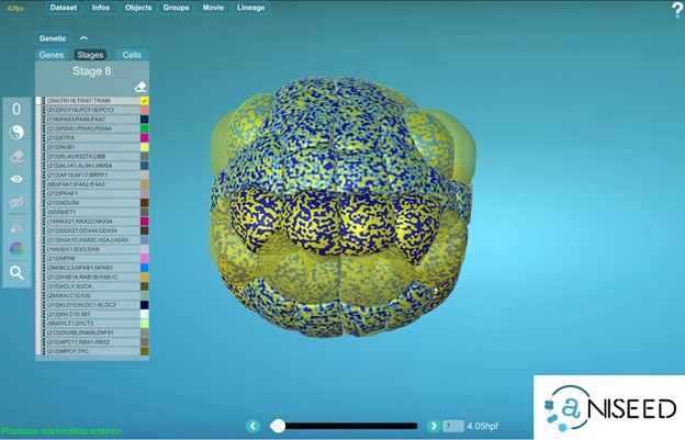
Yes, for ascidian embryos, MorphoNet is interfaced to the gene expression section of the ANISEED database (https://www.aniseed.cnrs.fr) (See Figure 2 and movie below). Other external gene expression databases could be connected to MorphoNet in the future, and we will be happy to work with you on such projects. If no gene expression database is available for your organism, you can directly upload your expression profiles using a simple format (https://morphonet.org/help_format) and then, as we did with the Arabidopsis meristem (3), you can manually curate, if necessary, your gene expression information by labeling individual cells.
To explore gene expression data, each individual gene can be assigned a colour, and the powerful MorphoNet shaders allow you to simultaneously visualize the expression profiles of up to 4 genes. You can also interrogate your data for genes expressed in the intersection or the union of some specific selected cells.
Figure 2: Screen capture of a multiple gene expression data on an ascidian embryo. The gene expression profiles are extracted from the ANISEED database (https://www.aniseed.cnrs.fr).
Can I explore the cell lineage?
MorphoNet is a unique tool which allows you to interact at the same time with your 3D+t dataset and the corresponding lineage. We use a second window to display the cell lineage and each click on a specific cell will also be displayed on the lineage. You can also map supplementary information, such as the cell volume, on the lineage and then order the branches by these values.
How can I make nice figures from MorphoNet?
We recently implemented a new module (inside the tools menu), which allows you to create your own figures in pdf format. You can also create your own movies in MorphoNet. Simply define some key frames and MorphoNet will interpolate the rest.
How can I share data with my colleagues?
You can easily share a specific dataset you own with any colleague who registered to MorphoNet. If you want to share multiple datasets, you can group them into a working group, which can be shared. You can also use MorphoNet to share data publicly, in an article for instance. For this, you can create a session, with a permanent url that can be shared (Just click here as an example https://morphonet.org/hgCbNDIV).
Can I do more if I can code?
You can directly query your data using the python API : https://morphonet.org/help_api. It is very useful when you have many datasets or when you automatically compute some properties. We are also developing a Plot module which allows us to directly visualize your data inside our 3D viewer without any data upload. And you can, of course, contribute! MorphoNet is an open-source software. Please contact us if you are interested. We are having a lot of fun.
What are the next features that you plan to add in MorphoNet?
We have several projects underway. We are currently working on a standalone application which you can directly install on your computer to fully exploit its computational resources when interacting with big datasets.
We are also working on the Virtual Reality representation of your data. This will allow you to interact with your own data with a classical VR headset.
Stay tuned! You can follow us on Twitter @MorphoTweet
REFERENCES:
B. Leggio, J. Laussu, A. Carlier, C. Godin, P. Lemaire, E. Faure, MorphoNet : an interactive online morphological browser to explore complex multi-scale data. Nat Commun. 10, 1–8 (2019).
L. Guignard, U.-M. Fiúza, B. Leggio, J. Laussu, E. Faure, G. Michelin, K. Biasuz, L. Hufnagel, G. Malandain, C. Godin, P. Lemaire, Contact area–dependent cell communication and the morphological invariance of ascidian embryogenesis. Science. 369, aar5663 (2020).
Refahi et al. A multiscale analysis of early flower development in Arabidopsis provides an integrated view of molecular regulation and growth control. Developmental Cell, Elsevier, 2021, 56 (4), pp.540-556.e8. ⟨10.1016/j.devcel.2021.01.019⟩
March 5-10, 2023, Cold Spring Harbor Asia will host a human development meeting in Awaji (Japan). Join us in this beautiful island and meet like-minded, human-centric dev bio and stem biol researchers. Register early for opportunity to present your work orally (twenty slots to be filled), and/or if you need assistance for entering Japan. Check out how to register here: https://www.csh-asia.org/?content/505
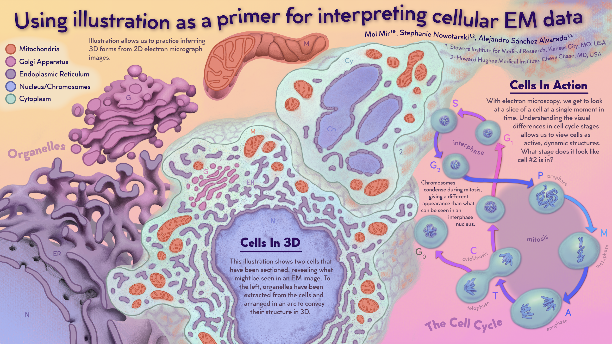
In our first SciArt profile of 2023, we hear from Mol Mir, a Science Visualization specialist in Alejandro Sánchez Alvarado’s lab at the Stowers Institute for Medical Research. Mol shares how a tour of an EM facility turn into a job, and how science and art are deeply intertwined for them.
Where are you originally from and what do you work on now?
I grew up in North Carolina in the United States. I moved to Kansas City, Missouri in 2015, where I attended Kansas City Art Institute and received my bachelor’s degree in Interactive Arts in 2019. I’m currently working at Stowers Institute for Medical Research in Kansas City as a Science Visualization Specialist in Alejandro Sánchez Alvarado’s lab.
As a Science Visualization specialist, my responsibilities are to learn and to share. With a background in art and user-centered design, I get to spend my time solving puzzles about how to present data to both scientists and non-scientists. I mostly work with planarians, using electron microscopy (EM) data to see their cells in 3D, at a high resolution, and within the context of the animal. Planarians are flatworms with an incredible ability to regenerate due to their stem cells, which happen to be the only cells in this animal that divide. I spend a lot of time creating 3D models of dividing cells or differentiated cell types. The amount of time I’ve spent with my eyes on this data makes me an expert resource for other lab members as their research leads them to a particular type of cell or region of the animal.
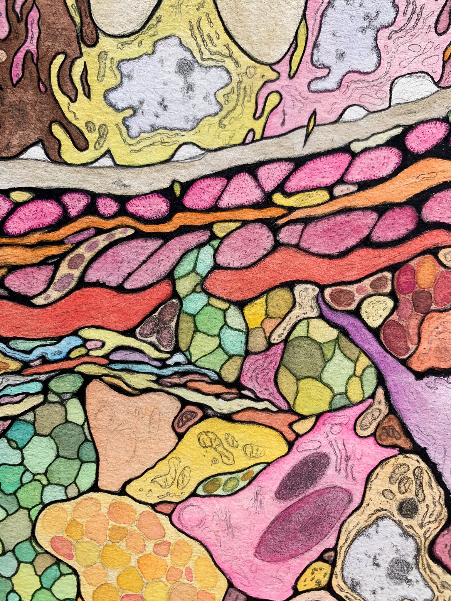
‘Middle Juice’ – Watercolor, graphite, and colored pencil on paper
Were you always going to be an artist?
Yes, I was always going to be a creative of some sort. The first thing I ever said I wanted to be when I grew up is a ‘coloring dentist’ – don’t ask me what I meant by that, because I don’t really know! Although, I imagine it’s actually pretty similar to the work I’m doing now!
‘Rubber Cell Puzzles’ – Urethane rubber cast from a CNC milled HDPE mold
And what about science – have you always enjoyed it, and how did you begin working in the lab?
Yes, I’ve had a love for biology all my life. I thought I missed my chance to be involved in science because I had a difficult time in school (I’m neurodivergent). I headed in the direction of art because I thought that was the only thing I could do. But while I was in school – searching for the reason why I want to make things – I fell in love with the cell! I began making artwork of cells, including a 3D printed cell model, a quilt, and several puzzles. At the end of my third year at KCAI I met my mentor, Steph Nowotarski, during a tour of Stowers in the electron microscopy department. What started as a wonderful internship (where I traded 3D printing knowledge for learning about cell & developmental biology) later turned into my full-time job. I’ve learned so much about science, biology, planarians, and electron microscopy since then.
‘Cell Model’ – 3D printed ABS
What or who are your most important artistic influences?
Growing up, I had this amazing set of books: the Childcraft collection. These books and science museums for children have been huge influences on my work. Recently, I’ve also been inspired by the work of Agnes Pelton, Gemma Anderson, and Ipsa Jain.
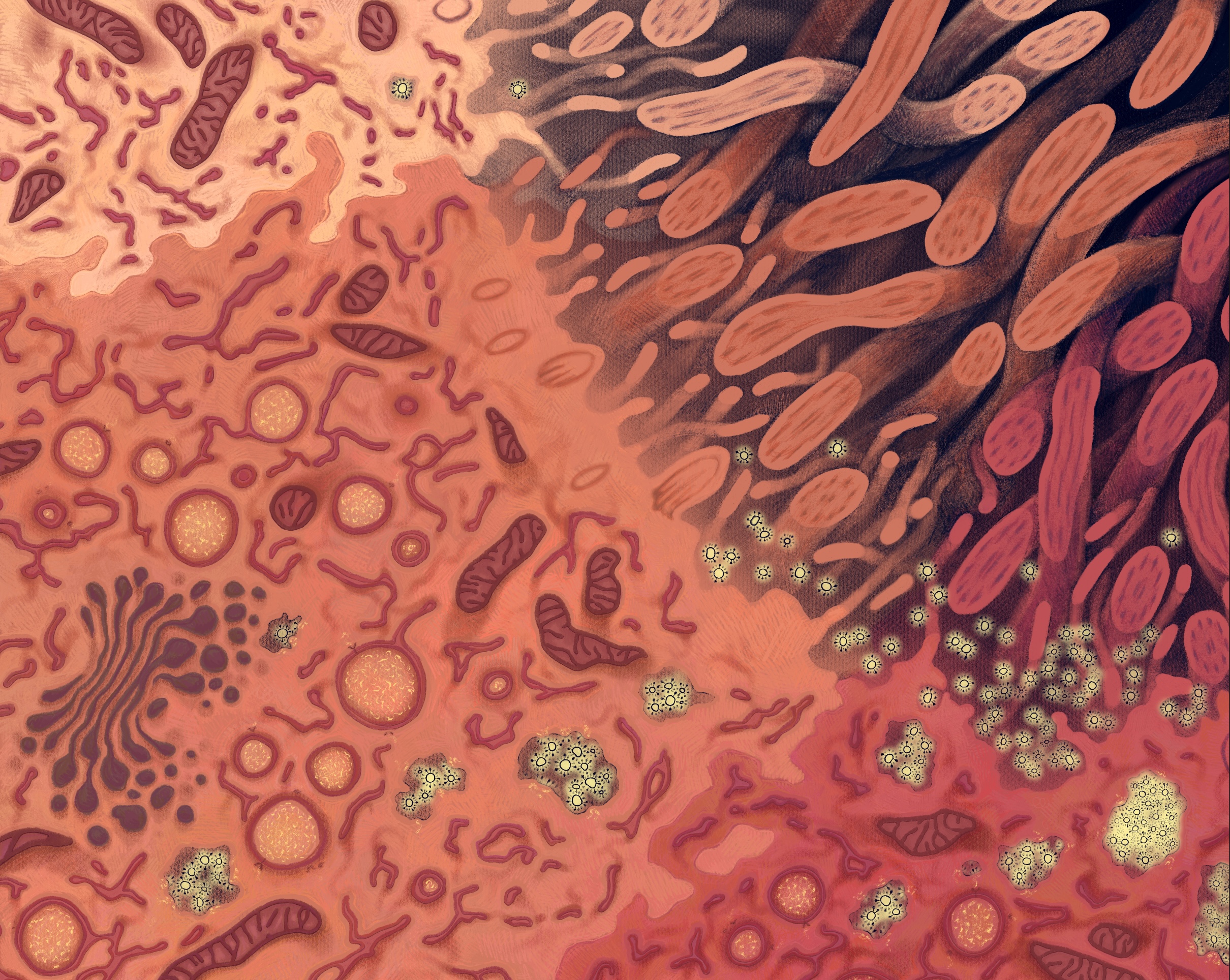
‘Infection Illuminated’ – Digital illustration
How do you make your art?
I really have two categories of art-making: art for communication, and art for expression. My communication artwork centers around a topic I want to explore or share with others. Here, accuracy is very important, and so is ‘testing’. It’s important to test my work with a smaller audience before I set it free into the world, to make sure it’s communicating what it is supposed to communicate! My artwork for expression is something else. It is about making for the sake of making and most of this happens in my sketchbook.
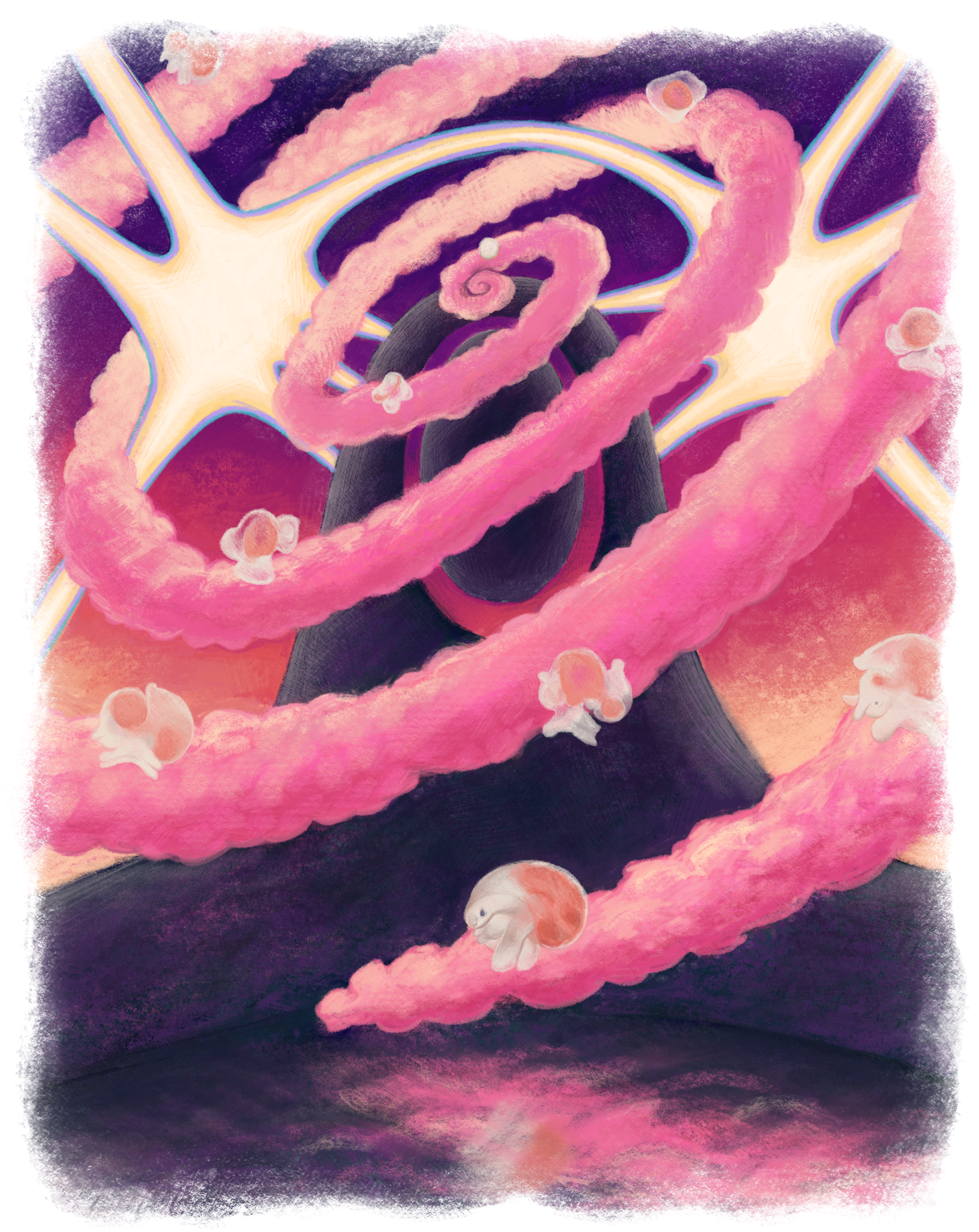
‘Snail Mountain’ – Digital illustration
Does your art influence your science at all, or are they separate worlds?
My art and science are deeply intertwined. Most of my work involves looking at electron microscopy images of cells. This very directly inspires my artwork, as cells are a common subject for me. Sometimes my sketchbook explorations will give me new ideas for data visualization and communication.
So, my art for communication is inspired by science and my science is inspired by my art for expression!
‘Using illustration as a primer for interpreting cellular EM data’ – Scientific poster for VIZBI 2022
What are you thinking of working on next?
My next big project is a book! I’m working on a book that will serve as a collection of planarian cell types through electron microscopy that also helps people to understand EM images and how they are created. This is a project I’ve been thinking about for a long time, so it’s very satisfying to be working towards it now.
Thanks to Mol and all the other SciArtists we have featured so far.We’re looking for new people to feature in this series – whatever kind of art you do, from sculpture to embroidery to music to drawing, if you want to share it with the community just email thenode@biologists.com (nominations are also welcome!)
Exploring how germ cells are metabolically supported in Drosophila testes
During the summer I had the privilege to work in Dr. Amoyel’s lab at UCL, to study the mechanisms providing metabolic support to germ cells in Drosophila testes. As my first lab project, it was thrilling to realize how much I enjoy academic research and feel invested in shining light on the unknown.
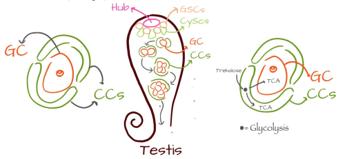
The lab seeks to understand how stem cell interact with their environment and with each other, to influence their fate and maintain tissue homeostasis. In fact, stem cells can either undergo self-renewal or differentiation, to fulfil their functions in tissue maintenance and regeneration. The balance between these two fates is determined by a micro-environment called a niche, which delivers signals promoting proliferation to the adjacent stem cells, while cells that are displaced from the niche differentiate. In particular, the Drosophila testis niche, called the hub, supports two stem cell lineages, namely germline stem cells (GSCs), which differentiate into germ cells, and somatic stem cells (CySCs), differentiating into post-mitotic cyst cells. When differentiating, two somatic cyst cells (CCs) envelop one germ cell (GC), forming a complex called a cyst (See Fig. 1). The enclosed GC then undergoes several mitotic events, increasing its number within the cyst from 1 to 16 cells (See Fig. 2).
Fig. 1 A cyst is composed of two somatic cyst cells (CCs) surrounding one germ cell (GC). Fig. 2 Drosophila testis, containing the hub and two cell lineages. A differentiated germ cell undergoes mitotic events within a cyst. Fig. 3 In a cyst, our theory is that CCs provide glycolysis products to support TCA in both cell lineages.
Since the enclosed GCs are sealed off from the environment by the two somatic CCs, the way they receive the metabolites necessary for the glycolytic and TCA pathways to survive was still unknown. In carbon metabolism, sugars – Trehalose molecules in flies – are broken down through a process called glycolysis, which produces pyruvate and ATP. Pyruvate is then either converted to lactate, or transported into mitochondria to fuel the TCA cycle, which provides most of a cell’s energetic needs. Hence, my project’s objective was to understand whether the two stem cell lineages interact metabolically, exploring the hypothesis that CCs provide glycolysis products to support germ cell metabolism (See Fig. 3). This shuttling of metabolites is already known to happen in the brain, where glia conduct glycolysis and provide lactate to neurons to feed their mitochondria. To test this theory in Drosophila testes, we knocked out glycolytic and TCA enzymes to downregulate the respective metabolic processes. A prediction of our hypothesis is that different metabolic pathways should be required in different cell types. In particular, knocking down glycolysis only in CCs should affect GCs, while knocking it down in GCs should have no effect, while both cell types should rely on TCA cycle enzymes.
During the first week, I learnt about Drosophila handling and husbandry: the basics about fly development and genetics, how to set up crosses, and how to identify markers to select the correct flies for analysis. I was also trained in immunostaining, to identify cell types in the tissue and whether their distribution changed as a result of my genetic manipulations. With plenty of practice and patience, I was soon comfortable enough with the staining procedure to start building my own experiments, and that’s how slowly the puzzle finally started coming together and I found myself in charge of boxes of crosses, and creating my own routine in order to keep crosses and experiments going.
This project was also an opportunity for me to discover and apply the Gal4/UAS-RNAi system, one of the main techniques used to conduct large-scale gene disruption in flies. Gal4 encodes a transcription factor which specifically binds to an enhancer called UAS, and activates expression of target genes downstream of the UAS sequence. Spatial control of expression is achieved by placing Gal4 expression under the control of tissue-specific promoters. When RNA interference (RNAi) sequences are placed downstream of UAS, their expression inhibits the expression of targeted proteins, disrupting gene expression. Therefore, when crossing a female fly carrying a Gal4 transgene with a male carrying a UAS-RNAi for a specific metabolic enzyme, the enzyme will be knocked down in the progeny carrying both transgenes.
To test these hypotheses, I used specific Gal4 drivers, including Traffic jam (Tj-Gal4), which targets CCs, Nanos-Gal4, which is only expressed in GCs, and Tubulin (Tub)-Gal4, which is expressed in all cells. These metabolic genes should all be essential for viability during development, so knocking them down in all cells using the UAS-RNAi system should result in lethality and a failure to obtain offspring carrying both the Gal4 and UAS-RNAi transgene.
My results showed that knocking down all glycolytic and TCA enzymes with Tub-Gal4 led to lethality, except for knockdown of Trehalose transporters (Tret), which did not affect viability. This might indicate either that the many versions of Tret proteins overlap each other in their functions, or that there are other pathways that might lead to the same downstream outcome without the use of Tret enzymes. Together with Holly Jefferson, we went on to show that knocking down glycolysis genes in CCs led to decreased GC survival, while knockdown in GCs had no effect. These results altogether support the hypothesis that cyst cells produce metabolites through glycolysis to support germ cell metabolism.
The sense of responsibility I felt, and the way other lab colleagues, with many more years of experience than me, relied on me to complete the necessary experiments, was in a sense enlightening for me to understand dynamics in a laboratory and in a work environment. A dynamic in which colleagues confide in each other’s’ abilities and strengths, in which every question is a good question, and in which a wrongly dissected testis or a failed experiment is an opportunity to learn and fully comprehend mechanisms without blindly following protocols. This wholesome experience has shown me how working in research, contributing to the scientific community, and entering the world of scientific discovery, is a life full of suspense and serendipity, struggle and competition, failure and accomplishment. It is a selfless life to which my future career aspirations lean fully, allowing me to explore the mechanisms through which life is possible.
Sonia Paoli – UCL, BSc Biomedical Sciences, Cells and Molecules
Supervisors: Marc Amoyel, Diego Sainz de la Maza, Holly Jefferson
The role of canonical Wnt signalling in embryogenesis of the invertebrate chordate Ciona intestinalis.
Ascidians, as the closest invertebrate sister group of vertebrates, are important to study the development and evolution of our own species. Their gene networks are closely related to those of humans, but without the complexities that were introduced via the whole genome duplications that occurred at the origin of vertebrates. A particular conserved mechanism, canonical Wnt (cWnt) signalling is essential to various development processes, especially in patterning along the anterior-posterior axis. Understanding the role of the cWnt pathway in ascidians would be helpful to elucidate the emergence of chordates and their evolution.
The summer project was complementary to ongoing work in the lab of David Ferrier (in the Scottish Ocean Institute, University of St. Andrews), focused on understanding the mechanisms of cWnt controlling their potential target genes in Ciona, such as the ParaHox genes (Gsx, Xlox and Cdx) that are the evolutionary sisters to the Hox genes, with roles in patterning the anterior-posterior axis in the central nervous system and gut. I had the opportunity to try both experiments with my two lab mates (Dr Nuria Torres-Aguila and Anastasia Ellis, see Figure 3) who use different ways to disrupt the cWnt signalling pathway.
Background
The cWnt pathway features the activation of the β-catenin transcription factor and modulation of specific target gene expression. T-cell factor/lymphoid enhancer (TCF/LEF) is the main transcription factor mediating the cWnt pathway. In the absence of Wnt ligands, responsive genes are generally repressed by TCF/LEF due to the transcriptional cofactor β-catenin constantly being degraded by the proteosome. When Wnt ligands interact with the transmembrane receptor proteins, several proteins (Frizzled, LRP5/6 and Disheveled) are brought to form a multimeric complex attached to the membrane, inhibiting the phosphorylation of β-catenin and its degradation. The stabilised β-catenin then converts the former repressor TCF/LEF into a transcriptional activator of the Wnt-target genes (Gilbert and Barresi, 2022).
Heat shock experiment
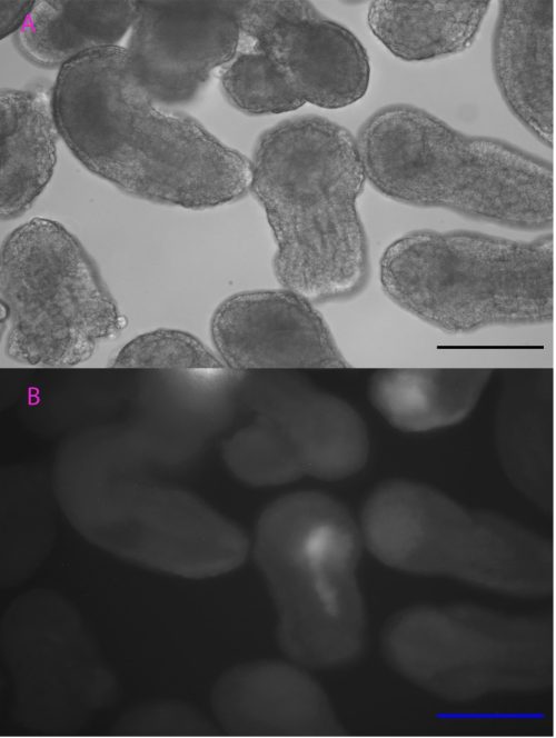
Controlling when and where TCF is expressed is useful to help analyse the function of TCF in cWnt signalling. Based on the heat-inducible cis-regulatory element initially characterized by Kawaguchi’s team (Kawaguchi et al., 2014), we tested the efficiency of a DNA construct with heat-inducible gene Ci-HSPA1/2/6-like and mCherry gene in Ciona embryogenesis to adapt this versatile technique to our species and population (i.e. a temperate Scottish population versus a more tropical Japanese population). Heat shock was initiated at different stages of development with different lengths of time and temperatures before being observed under an epifluorescence microscope.
Although the precise heat-shock conditions need to be further refined, it was encouraging to see the induction of mCherry in embryos with this technique (Figure 1), raising the prospects of using the heat-shock approach to over-express genes like TCF in the near future. In this experiment, the microscope was probably the most exciting but also challenging piece of equipment, the microscope was probably the most exciting but also challenging piece of equipment I used. It took me some time to patiently check every detail like light intensity, exposure time, and magnification to ensure comparability between my images. The experience has been valuable for me to be familiar with this essential equipment for studying developmental biology.
Figure 1. Heat shock-induced mCherry fluorescence under the microscope. 25 degrees 30 minutes heat shock treated embryos. Photos were taken 3 hours after the treatment A: Embryos under white light B: Embryos showed mCherry fluorescence under fluorescent light. Scale bar: 100µm
Chemical treatment
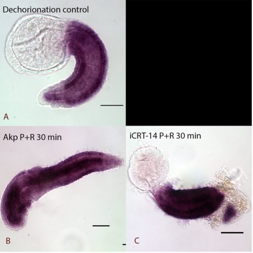
Chemical treatment investigated whether the TCF-dependent ParaHox gene Cdx is potentially directly controlled by the cWnt pathway. Pharmacological agents, iCRT-14 and azakenpaullone (Akp) were used to downregulate and upregulate the level of the cWnt pathway. Embryos were left to develop to the desired stage for recovery as a post treatment to distinguish rapid (potentially direct) responses from slower (possibly secondary) changes. The samples were processed for in-situ hybridization with a probe against the Cdx gene and images were captured by Nomarski microscopy.
The results showed that the expression of Cdx is possibly regulated by a secondary effect instead of directly disrupted by cWnt cascade, as the samples fixed immediately after treatment showed no obvious changes in expression (data not shown) whereas dramatic anterior extension of Cdx expression was seen with a recovery period in cWnt activator treatment (Figure 2). The lack of apparent response to iCRT14-treated samples may be due to TCF not normally being expressed in tail epidermal cells (Garstang et al., 2016). Further work aiming to check the Cdx expression in deeper cells where TCF is expressed will be conducted in the future.
All of this research helped me to appreciate the importance of time management in the lab. I had the opportunity to repeat the in-situ hybridization several times, and each time I got more skilled in the procedure. My first two times of in-situ hybridization turned out with either the embryos being accidentally lost because of I rushed during wash steps, or they were left in the solution for too long, so I stayed in the lab until very late. It is a relief that eventually they were successfully mounted to be observed, but wisely making use of time during short waits and multitasking largely improved the efficiency in my later experiments.
Figure 2. Expression of Cdx in Ciona intestinalis at late tailbud stage I A: Normal expression of Cdx at the late tailbud stage of dechorionated control samples. Expression is observed in the epidermis of the tail except at the very posterior end. B: 30 minutes pulse and recovery samples be treated by cWnt activator (Akp). It had dramatic anterior extension of Cdx expression and lost some of the anterior features. C: cWnt inhibitor (iCRT-14) pulse and recovery treated embryos with extension of Cdx expression to the posterior tip of the tail. Pulse and immediately fixed samples did not show obvious changes in expression. (data not shown) All the embryos are lateral views scale bar: 50 µm
Personal experience
I appreciate that I have experienced the systematic research process as a whole. From collecting sea squirts in the harbour and manipulating the embryos, to finally assaying their responses to various treatments. I have tried many essential techniques in developmental biology and see how the experiments proceed. I learned how the protocols are designed and improved based on the techniques developed from previous research. Even finding the potential mistakes I made by recalling the steps with my supervisors was a valuable and rewarding experience. Thanks to Dave, Nuria, Anastasia and the friendly people around SOI, all of who created a warm and supportive environment. I am more comfortable working in the laboratory and more determined to pursue further studies now. Thanks to the BSDB and Gurdon scholarship for making this opportunity possible. It has been one of my best memories doing scientific exploration with such a great team, besides the beautiful East Sands beach at St. Andrews.
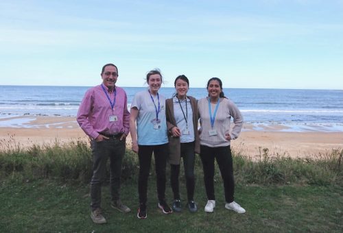
Figure 3. From left to right. Dave, Anastasia, me and Nuria on the seafront in front of Scottish Ocean Institute.
Reference list:
-Garstang M.G., Osborne P.W. and Ferrier D.E.K. (2016) TCF/Lef regulates the Gsx ParaHox gene in central nervous system development in chordates. BMC Evolutionary Biology 16:57.
-Gilbert S.F. and Baresi M.J (2022) Developmental Biology 12th edition Chapter 4 Cell to cell communication. Oxford University Press 109-111
-Kawaguchi A, Utsumi N, Morita M, Ohya A, Wada S. (2014) Application of the cis-regulatory region of a heat-shock protein 70 gene to heat-inducible gene expression in the ascidian Ciona intestinalis. Genesis. ;53(1):170-82.
Role of PAX6 in human cerebral organoid development
The cerebral cortex contains two major classes of neurons: excitatory and inhibitory. The imbalance between them is postulated to underlie some autism spectrum disorders (ASDs) (Rubenstein, 2010; Uzunova et al., 2016). The transcription factor PAX6 is an important regulator of embryonic forebrain development and mutations in PAX6 have been associated with ASDs (Kikkawa et al., 2019; Manuel et al., 2015). It is therefore possible that PAX6 could be involved in the regulation of the inhibitory-excitatory balance, hence playing a role in the development of ASDs. Supporting this hypothesis, recent findings indicate that PAX6 deletion in mice leads to the appearance of ectopic GABAergic (inhibitory) cells (Manuel et al., 2022). While animal data are useful, the mechanisms of this effect, and whether it is present in humans remains unclear, as human neurodevelopment is difficult to study directly. However, human cerebral organoids offer an exciting, new way to directly investigate early human embryonic neurodevelopment, including the effects of PAX6 mutations (Mason & Price, 2016). Hence, as a first step towards uncovering the mechanisms of the possible PAX6-dependent regulation of the inhibitory-excitatory balance in humans, this project aimed to examine PAX6-/- organoids at early developmental stages to investigate the effects of PAX6 mutations.
Methods
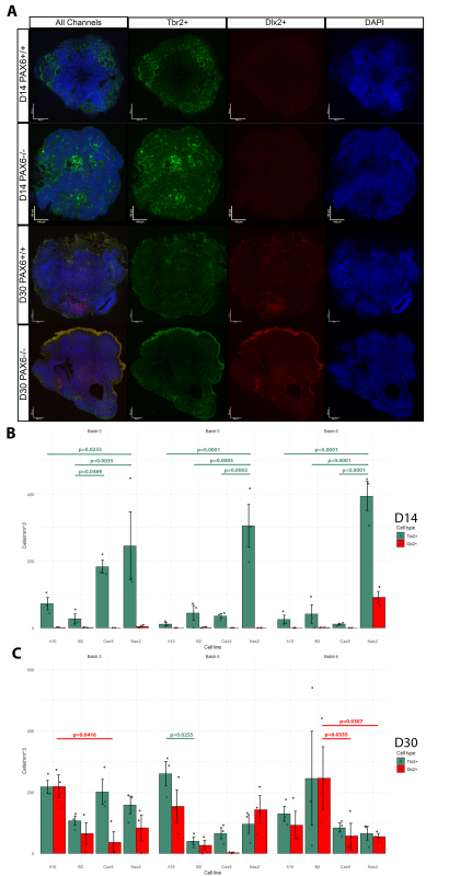
The Mason Lab has previously grown cerebral organoids from PAX6-/- induced pluripotent stem cells and PAX6+/+ controls. At culture day (D)60, the cellular phenotypes of these organoids were tested using scRNA-seq analysis. Indeed, ectopic GABAergic populations were observed in the mutants and not in controls. To examine the earlier stages of PAX6-/- organoid development, I analysed three batches of organoids originating from four cell lines (Controls: Nas2,Cas9; Mutants: A10,B2) at D14 and D30. Organoids were cut into 10μm sections. Inhibitory/excitatory neurons were visualized by tagging Tbr2 (marker of excitatory glutamatergic neurons/progenitors) and Dlx2 (marker of inhibitory GABAergic neurons/progenitors) with fluorescent antibodies. Hence, two distinct cell types could be visualized using fluorescent microscopy (Fig.1A). The number of Tbr2+ and Dlx2+ cells was counted in three randomly selected sections from each organoid using ImageJ software. The cell count was normalized by area of each section and averaged per organoid (Fig.1B-C).
A) Representative immunofluorescent images of control (PAX+/+) and mutant (PAX6-/-) organoids at D14 and D30, counterstained with DAPI. B-C) Counts of Tbr2+ and Dlx2+ cells at B) D14 and C) D30. Each point represents the average cell count from three sections of an organoid, normalised per area (n=3/group, 72 total). 2-Factor (Batch and cell line) ANOVA, evidence for interaction between variables for D14 Tbr2+ counts (F=3.0612, p=0.02278), D30 Dlx2+ counts (F=3.1609, p=0.01984), D30 Tbr2+ counts (F=2.5636, p=0.04615). Significant results of post-hoc pairwise Turkey comparisons presented as lines with p-values above relevant bars.
Results
No D14 organoid (apart from an anomalous batch 6 Nas2 organoids) featured Dlx2+ cells. Hence, ectopic GABAergic cells likely arise after D14, which narrows the window of investigation in potential future research. In many cases, the number of Tbr2+ cells was significantly higher in the Nas2 organoids when compared to other cell lines, including the second control line, Cas9 (Fig.1B). This may suggest the presence of unknown variables which affected organoid development, or possibly that PAX6 has an effect on the speed/timing of Tbr2+ neuron emergence.
Both mutant and control organoids at D30 exhibited comparable counts of Tbr2+ and Dlx2+ cells, with significant differences in cell counts present between only a few groups (Fig.1C). This is surprising, as prior literature suggests that ectopic GABAergic cells (in PAX6-/- mice) appear at some point during development due to environmental signals such as Shh found in cerebrospinal fluid (CSF) and/or immigrating interneurons (Manuel et al., 2022). It seems that in human cerebral organoids (in the absence of CSF or immigrating interneurons) GABAergic cells emerge as a normal part of development whether PAX6 is present or not, but disappear by D60 in controls.
PAX6 could possibly be triggering the death of Dlx2+ cells between D30 and D60. PAX6 could also be causing Dlx2+ cells to switch to an excitatory fate by D60. It would be interesting to expand upon these findings by conducting lineage tracing experiments: this would allow for the tracking of the changes in cellular phenotypes to reveal whether the presence of PAX6 affects cell fate decision making.
My experience at Mason Lab
I found this project very exciting. The problem-solving aspect of research following inevitable experimental failures was genuinely great. It was very interesting to brainstorm the next steps when it becomes apparent that the current experimental procedure is not yielding any results. For instance, originally the experiment involved tagging dividing cells with EdU at D13.5, to see whether these cells switch between Tbr2+ and Dlx2+ identities between D14 and D30. However, the experiment had to be modified as the EdU fluorescent signal became too faint at D30 due to dilution during cell division.
The project also showed me the more labour-intensive side of research as I spent many days cutting organoid sections at the cryostat. After the baptism of fire by cutting my finger, I eventually found it a compelling task. As one of the Mason Lab members said, “it builds character”. I was also introduced to fluorescent microscopy and image analysis using ImageJ. The former forced me to become organised and efficient, as one must tame the unruly microscope and take the hundreds of pictures before the booked time runs out. The latter I found challenging as it was my first time using image analysis software, but it will undoubtedly be useful in my future work.
Every part of this project, no matter how menial or monotonous it might have seemed to an observer, I found thrilling. I have obtained experience in numerous laboratory techniques, and again reaffirmed research as my chosen career path. I am very grateful to Mason Lab and BSDB for this opportunity, and I am looking forward to being able to conduct projects of larger scales in the future.
Figure 2. Right Panel: Me, together with my supervisors: Prof. John Mason and Dr. Calvin Chan! Left Panel: me with my best friend, the cryostat. (2 votes) Loading...
A new article on embryos and the beginning of independent human life:
In her January 1 New York Times article, “When does life start? A post-Roe conundrum,” reporter Elizabeth Dias cites some material from a recent paper that I wrote. The article, “Pseudo-embryology and personhood: How embryological pseudoscience helps structure the American abortion debate. Natural Sciences 2022: e20220041″ is open access and can be found at DOI: 10.1002/ntls.20220041 . The paper reviews scientific opinions as to where independent human life begins, and contends:
There is no consensus among biologists as to when independent human life begins
What passes for science is actually a set of outdated myths that are no longer considered valid
This set of myths denigrates birth and promotes fertilization as the site where personhood begins.
I hope the community of biologists will read and discuss the data and conclusions of this paper.
 (No Ratings Yet)
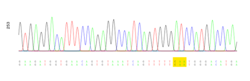
 (3 votes)