Royal Society Publishing has recently published a special issue of Philosophical Transactions B: Extraembryonic tissues: exploring concepts, definitions and functions across the animal kingdom
Compiled and edited by Guojun Sheng, Thorsten E Boroviak, Urs Schmidt-Ott and Shankar Srinivas, the articles can be accessed directly at www.bit.ly/PTB1865
A print version is also available at the special price of £35.00 per issue from sales@royalsociety.org
This is my inaugural Node post. Around 1990, Stephen Jay Gould dedicated my copy of his Ontogeny and Phylogeny (1979), writing, “Thank you for tackling my real book.” As a chemical engineer, I took it seriously when Gould whispered that development is not encoded in the genes and to forget natural selection.
The succeeding thirty years of lab work by scientists and scientific artists attributing morphogenesis to mechanical causes has garnered many papers and hundreds of illustrations covering most complex taxa. Recently, we demonstrated vertebrate origins in self-organized patterns that occur in the predictable geometry of serial, sequential cell division.
The authors hope that the many facets of our work will inspire further investigation. We invite comments and possible collaboration.
The Steinmetz group at the Sars Centre in Bergen (Norway) is looking for a bioinformatician with expertise in analysing high-throughput sequencing datasets (bulk, single-cell, ATAC-Seq). The project aims to characterise the transition from cellular quiescence to cell cycle re-entry, which is induced by re-feeding of starved animals, in the sea anemone Nematostella vectensis. Application deadline is November 25th 2022. Link for more information and to apply for the position: https://tinyurl.com/yc43x7mc
The Symposium was organised by Alison Hawkes, Jonathan Slack, Jim Smith, Claudio Stern, Cheryll Tickle and Neil Vargesson. Kindly supported by the British Society for Developmental Biology, Oxford University Press and UCL.
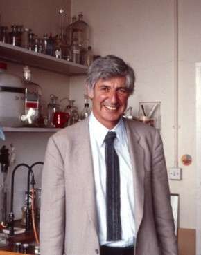
Figure 1- Lewis in one of the labs in the Windeyer Building of The Middlesex Hospital Medical School (circa late 1970s/early 1980s). Picture kindly supplied by Amata Hornbruch.
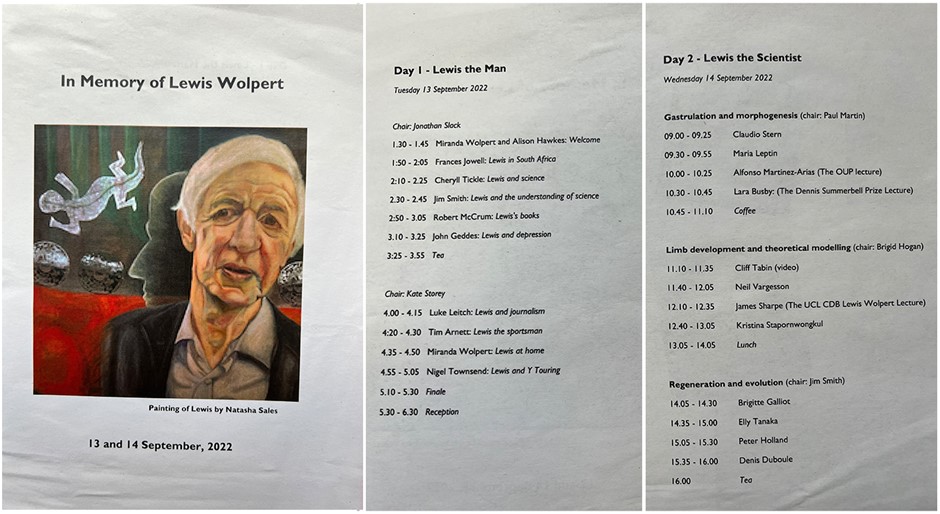
To many readers of the Node, Lewis Wolpert, who died on 28th January 2021, was above all a developmental biologist—one of the most distinguished and influential of his generation (Figure 1). But of course he was much more than this, and the organisers of this meeting in his memory devoted the first day to Lewis the man (Figure 2). We heard from his cousin Frances Jowell about his early life in South Africa, about his aunt Helen Suzman (the politician and anti-apartheid activist), and about Lewis’s own hatred of apartheid. Frances also explained how Lewis arrived in London, having hitch-hiked up Africa and spent some time working as a soil mechanic in Israel. Cheryll Tickle continued the story of Lewis’s early life by recounting how his friend Wilfred Stein introduced him to the mechanics of cell division and how this kindled his interest in developmental biology, leading to the articulation of the French Flag problem and his work on hydra and the chick limb.
Figure 2 – Symposium Programme, including Chairpersons for each Session
Lewis was a charismatic speaker, performer, and communicator, and Jim Smith spoke about Lewis’s contributions to the public understanding of science, illustrating his points with clips from Wolpert’s Medawar lecture of 1999. He was followed by Robert McCrum, former editor-in-chief of Faber and Faber, who discussed Lewis’s writing, and then John Geddes, Professor of Psychiatry in Oxford, who explained how Lewis’s book on depression, Malignant Sadness, had had such an influence on both public understanding and on the field of mental health research. The day finished with Lewis’s stepson Luke Leitch speaking about Lewis’s forays into journalism, with Tim Arnett speaking about Lewis’s sporting life, and especially tennis on Parliament Hill Fields, and then with Lewis’s daughter Miranda speaking about Lewis at home. Finally, Nigel Townsend spoke about one of Lewis’s lesser-known activities, his support for the Y Touring theatre company, which produces plays that encourage schoolchildren to explore the social, moral, scientific, and political questions raised by areas such as stem cell research.
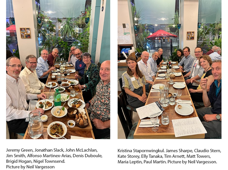
The second day of the meeting focussed on the science in which Wolpert was interested, and speakers included colleagues and collaborators as well as young scientists who had been influenced by Lewis and his work, many of whom said how much they would have liked to have met him. The speakers would have graced any major scientific meeting, and included Claudio Stern, Maria Leptin, Alfonso Martinez-Arias, Lara Busby (recipient of the Summerbell award and introduced by Amata Hornbruch), Cliff Tabin, Neil Vargesson (Lewis’s last PhD student), James Sharpe, Kristina Stapornwongkul, Brigitte Galliot, Elly Tanaka, Peter Holland and Denis Duboule (Figure 3). Areas of research included gastrulation, limb development, modelling, regeneration and evolution, and each speaker described how Lewis had influenced their work and their thinking.
The Symposium made clear Lewis’s influence on the field of developmental biology, his ability to transcend his field and, through his charismatic performances, his power to inspire and inform both scientists and non-scientists. When video clips of him were played at the end of the first day, the audience leant forward as one, listening intently to what he had to say. We also heard that Lewis loved discussing science, especially with the young, and was never slow to explain exactly what your data really meant. As a self-confessed ‘contradictor’ he was always questioning and challenging others, encouraging them to think more deeply about their views and why they differed from his!
We’d like to think Lewis would have enjoyed the meeting. He would have been pleased that his ideas are still informing our experiments and he would have asked lots of questions (something he always asked his PhD students to do). Most importantly, we think he would have been pleased by the great affection in which he was held, and by how pleased we all are that he made the move from soil mechanics to developmental biology.
All too often you find yourself in the audience, watching a scientific presentation with a fully-scrunched grimacing face as you despair slightly at the torrent of PowerPoint slides being projected into your eyeholes.
Whether it’s the classic forty slides for a ten-minute talk, the essay being read aloud to you or the dreaded illegible yellow text, presentation disasters are everywhere.
But it doesn’t have to be like this. You can break the trend. You can be better. You just have to follow a few simple rules.
1. Tell a story
This is super clichéd, but it still seems to be rarer than a paper returned without revisions. You have to tell your audience a story. Have a clear beginning, middle and end. Don’t just rattle through every single experiment you’ve done over the past few months and bombard your audience with every shred of data: tell them a story. Bring them along for the emotional ride we all know lab work is. Research is full of ups and downs, repetitive little failures and surprise successes. Expand on those. Tell people what you were hoping for, what you ended up with and how you felt about that.
When it comes to crafting your science story, here are some helpful prompts:
What’s the question or problem you’re addressing?
Why should anyone care?
What did you do to answer that question?
Were there problems you had to overcome? How did you manage that? (People love a tale of conquered adversity)
Got data? Awesome! What are the main findings?
No data? Awesome? Here’s your chance to open the floor up to troubleshooting (Okay okay, I know it’s not awesome but quite often you’re going to lack data, so here’s a chance to explain what you did and I bet there’s someone who has an idea about why didn’t get the data you expected. Also, unexpected data are the basis of discovery — remember that).
If you’ve thought about what research comes next, leave people with a taster of what you have planned. They’ll be excited by the prospect of a sequel – who doesn’t love a sequel?
And what did you conclude from all this amazing work? Wrap it up in one slide. ONE SLIDE — YEAH YOU HEARD! ONE SINGLE SLIDE.
2. Keep it minimal
This is not your thesis — do not have a ton of text on your slides. You’ll just end up reading it aloud and the audience won’t be listening to you because they’ll either be reading the essay you slapped in front of them or on their phones tweeting about the terrible talk they’re stuck in.
Instead, have a title, a few supporting bullet points and a visual. Talk about what’s on each slide: explain graphs, images and diagrams. You know the data inside out but your audience doesn’t. So walk people through your thinking. Explain what it all means – it keeps them engaged and actively listening to you.
3. Make it pretty
This isn’t superficial: it matters. If your text changes size on every slide, your titles are in a new position every time you hit ‘next’ and your images are blurry, people will be distracted.
An aesthetically pleasing, neat and well-formatted (i.e., consistent!) presentation helps to keep people focused on what you’re saying. It also says you care about the experience of the people looking at your work.
The layout of a pretty presentation may go unnoticed, but the layout of an eye-wateringly ugly presentation is often the only thing people will see.
BONUS PRO POINTS: skip slides altogether. Everyone hates PowerPoint really, so bin it. Rise above it. Try giving your talk with just a blackboard or whiteboard. Stand in front of it. Gesticulate wildly. Draw the data you need to enhance your points. Tell your story. Own that board! This means you really need to know your stuff. But you do. Right? Be the blackboard talk person that everyone remembers.
4. Have a point
This ties in with my first rule about telling a story, but it’s worth reiterating.
What’s the point of your presentation?
Are you talking about one key new finding?
Summing up your last six months of work?
Presenting a problem that you hope to troubleshoot?
Whatever it is, make it the focus of your presentation; don’t try to do everything at once. Again, this isn’t your thesis. You want to leave people with a clear take-home message. You don’t want them coming out, scratching their heads, wondering what just happened and if they can somehow reclaim the last hour of their lives.
5. Be a human
Presentations can be daunting, but if you relax and learn to present with a little bit of personality, maybe even some humour, you’ll find they go a lot smoother.
Take your time. Take longer than you think you need.
Breathe.
Think about the points you’re making. Talk to your audience like you would a friend in the office or the lab. There’s no need to plough through a talk, eyes down, like a robot reading a script.
Get excited by your results. Get exasperated by your failures. A little bit of empathy goes a long way. You can be sure almost everyone in that audience has been through a similar experience, so sharing your feelings here gets them on board. They’ll care. And if they care, they’ll listen and remember your talk.
You might even find you actually end up enjoying it because, all of a sudden, people are enjoying your talk! You’re the expert! And you’re crushing it!
Okay, you got all that? Here’s a summary just in case:
Tell a story with a beginning, middle and end — keep people engaged
Only include essential info on your slides
Make your presentation pretty and consistent
Address one main point
Be a human — be a friendly, potentially entertaining, human if at all possible
There you go: just a few simple rules to make sure your presentations don’t inadvertently bore people to death.
At the Mary Lyon Centre at MRC Harwell we have our 10thGenome Editing Mice for Medicine (GEMM) call open and accepting applications, so we decided it would be a good opportunity to look back at genetically altered mouse lines generated for successful applicants from previous funding calls. We have created some case studies that demonstrate the range of work we have supported and highlight some of the lines that we have available for the scientific community
The GEMM programme offers the expertise and resources required to deliver and validate new and innovative mouse models that contribute to answering vital, pertinent scientific and clinical questions that can only be answered by the use of an in vivo model. Successful applicants demonstrating the scientific and clinical importance of their desired model have their novel mouse line designed and produced free of charge and made available to the scientific community. The first call for applications was made in 2016 and, so far, more than 80 novel genetically altered mouse lines have been generated.
One example line is C57BL/6NTac-Gldcem1H/H, which contains a disease-associated point mutation created by CRISPR/Cas9 in the gene GLDC.
Glycine decarboxylase (GLDC) is one of four enzymes that make up the glycine cleavage system (GCS), a complex that regulates the abundance of the amino acid and neurotransmitter glycine. This is done by catabolising glycine to release carbon dioxide and transfer a one-carbon unit into mitochondrial folate one-carbon metabolism. Mutations in GLDC have been found in patients with a severe inherited metabolic disease, Non-Ketotic Hyperglycinemia (NKH) and neural tube defects (NTDs). GLDC has also been found to be highly expressed in some cancers and it’s thought that sensitivity to GCS inhibition might provide a therapeutic strategy. Triplication of GLDC has been seen in bipolar/schizoaffective disorder, likely due to low levels of glycine impacting its role as a neurotransmitter.
NKH is a rare, life-limiting inherited metabolic disease characterised by accumulation of excess glycine in the body fluids and tissues. It becomes apparent soon after birth with lethargy, breathing difficulties, and neurological symptoms, including seizures. Affected individuals suffer epilepsy and profound delay in development. More than 80% of patients with NKH carry mutations in GLDC. NTDs are severe birth defects of the developing nervous system that affect 1 in 1,000 pregnancies worldwide. Due to the importance of folate for brain development, disruption of folate metabolism is implicated in causing NTDs and folic acid supplements can prevent some, but not all NTDs. The GLDC missense mutation S951Y has been identified both in patients with NKH and with NTDs.
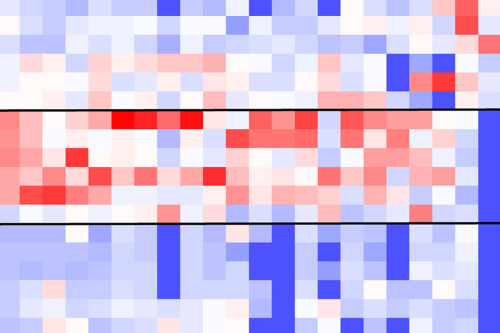
To provide a pre-clinical model for these diseases, Nick Greene, a group leader at the UCL Great Ormond Street Institute of Child Health, applied to the GEMM programme to generate a mouse model for the S951Y variant (S956Y in mice). He and his team were then able to generate mice carrying the S956Y variant in combination with a deleted copy of GLDC, as a means to genetically copy the combination of variants present in an NKH patient. Using this model, they were recently able to show mild but significant elevation of plasma glycine, suggesting that the mutant protein retains some function, when compared with the glycine elevation seen in a knockout model. Despite only mild elevation of plasma glycine, there was still significant glycine accumulation in the brain, as well as a trend towards accumulation of related compounds that are also thought to cause epilepsy. Importantly, they were also able to demonstrate that glycine levels in the brain could be normalised by activation of glycine conjugation via administration of cinnamate, which could suggest a future therapy.
Heatmap and fold-changes for glycine and glycine derivatives in liver of adult wild-type and GldcGT1/GT1 mice with/without administration of benzoate
Prof Greene’s team plan to use the model to test disease treatments, including via gene therapy, and the model could also be useful for testing of putative genetic interactions with “second hit” variants that may increase the likelihood of NTDs.
We are currently accepting applications for the 10th GEMM call and invite you to nominate ideas and designs for your own genetically altered mouse lines.
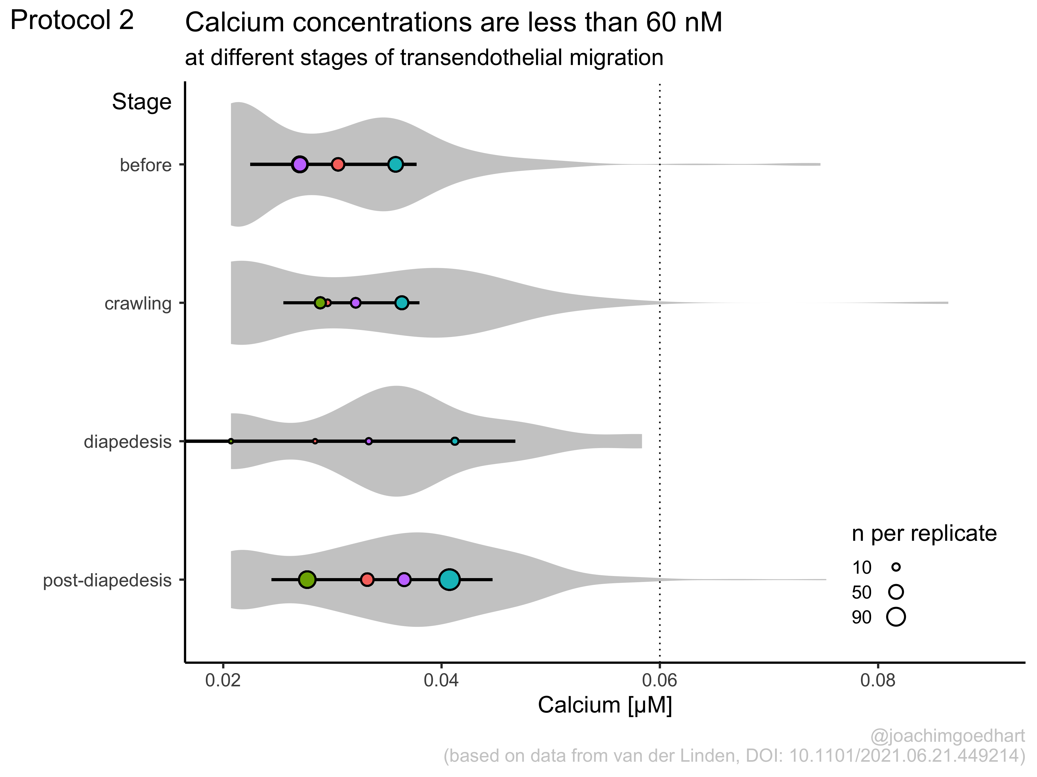
Scientists are familiar with protocols that describe in a step-by-step fashion how an experiment is performed. But they are usually less familiar with code or scripts for handling data. Yet, experimental protocols and computer instructions have a similar structure and purpose. Therefore, it should be within reach for experimental scientists to add coding skills to their toolkit. This is a very valuable skill to have as it enables automated, reproducible data processing and visualization. To lower the barrier for using R and the ggplot2 package for data visualization, I have written a book that is available online: https://joachimgoedhart.github.io/DataViz-protocols/
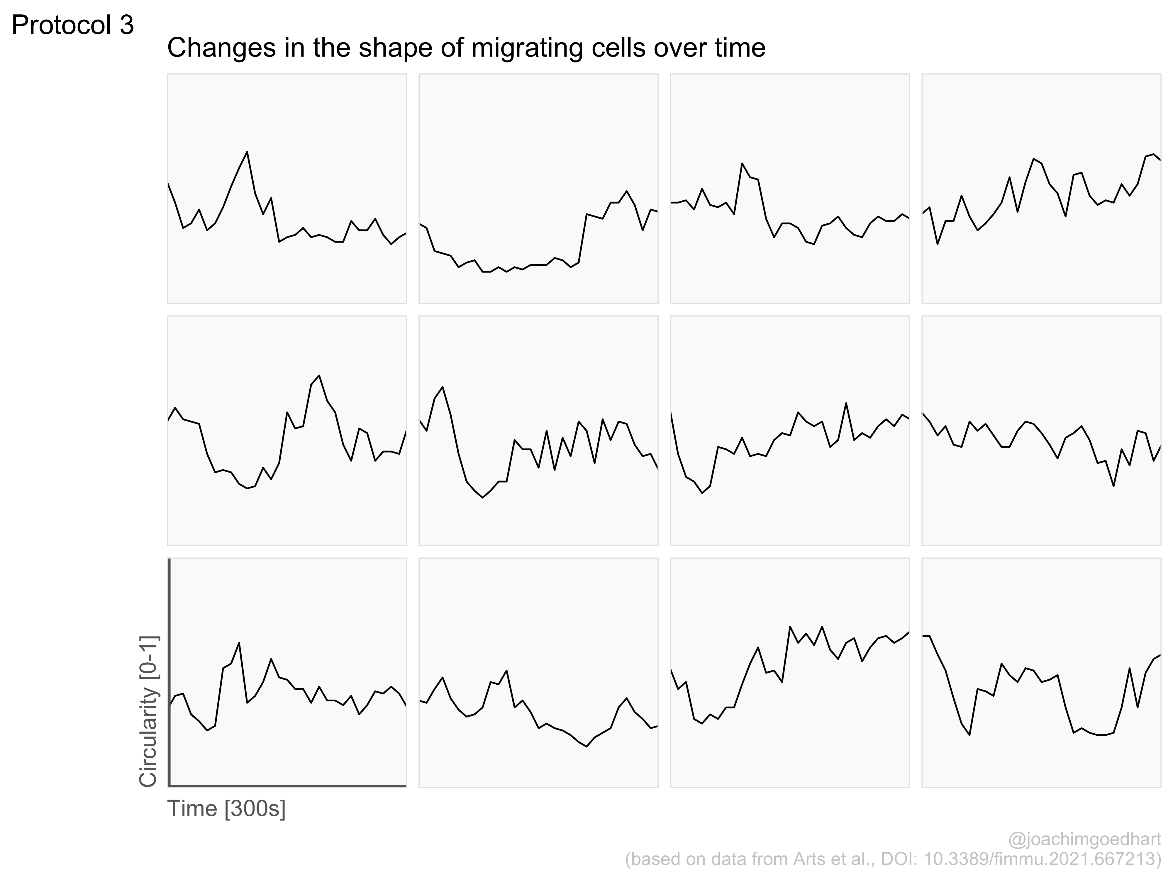
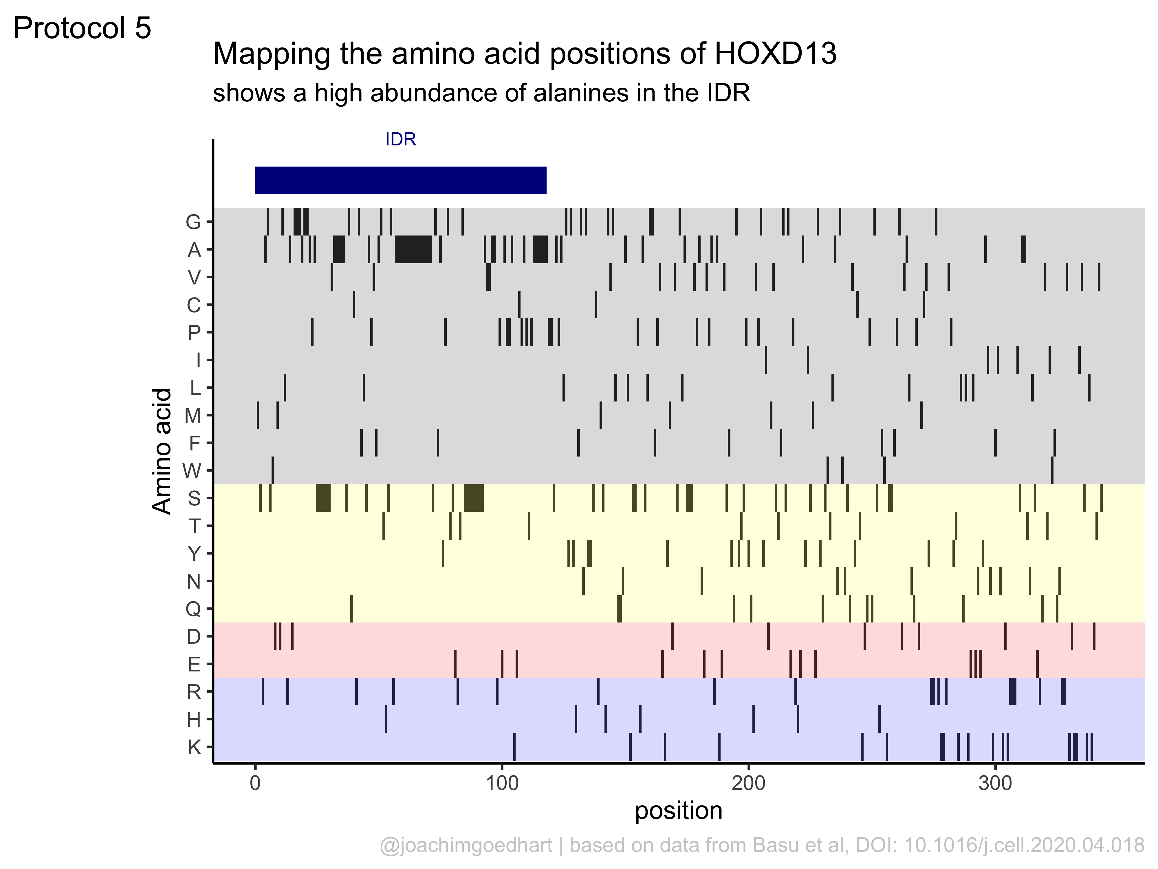
In my opinion, the best part of the book is the section with full, dedicated dataViz protocols. Examples of the output of three protocols is shown below. The protocols use realistic experimental data and provide step-by-step instructions that readers can reproduce or repurpose for their own use.
Example output of three different dataViz protocols (click to enlarge)
The book is work in progress, so this is not a final version as it will be updated. Especially the chapter with dataViz protocols will be extended. I will announce the addition of new protocols on twitter. I hope that the book is useful and that it provides a solid foundation for anyone that wants to use R for the analysis and visualization of scientific data that comes from a wetlab. I look forward to seeing the results on twitter (and please tag me: @joachimgoedhart), in meetings, in preprints or in peer reviewed publications.
Looking back
Previously, I have authored a number of blogs on the Node that provide step-by-step instructions on how to do data wrangling or plotting in R. The first blog was about the conversion of ‘spreadsheet’ type data into tidy data. I wrote this blog because I had a hard time understanding the tidy format. Blogging about it helped me to understand the concept and I thought it would also serve others who want to learn R. I continued to post step-by-step guides whenever I figured out something new (to me) and the enthusiastic responses from colleagues were very rewarding. At that time, however, I lacked an important skill which is called ‘literate programming’. This approach to programming combines styled text with chunks of code. It is a great way to explain and show what code is doing in a step-by-step way. After I learned literate programming in R with Rmarkdown, I decided to convert the blogs into this format. From there on, it was a logical step to compile the different topics into a book.
Looking forward
The advantage of Rmarkdown as the framework is that it is easy to maintain, edit and update. Using Rmarkdown, new protocols can be written independently and added to the book as individual chapters. At this moment, there are 12 complete protocols and I’m preparing another 8. I take inspiration from nice data visualizations that I see and I also do remakes of figures that we have published. If you have any ideas for a remake of a plot or if you have seen a nice dataViz which could use a protocol, please let me know! Also, I welcome any feedback on any aspect of the book.
Below you’ll find each of the talks and Q&As hosted by our Associate Editor, Matthias Lutolf (EPFL).
Alexandra Wehmeyer (M.D. thesis student) and Sebastian Arnold (Acting Director, Institute of Pharmacology, University of Freiburg) ‘Chimeric 3D-gastruloids – a versatile tool for studies of mammalian peri-gastrulation development’
Ansley Conchola (MSTP MD/PhD candidate in Jason Spence‘s lab at the University of Michigan Medical School) ‘Stable iPSC-derived NKX2-1+ lung bud tip progenitor organoids give rise to airway and alveolar cell types’
Sham Tlili (CNRS research investigator at the Marseille Developmental Biology Institute (IBDM) in Aix-Marseille University) ‘A microfluidic platform to investigate the role of mechanical constraints on tissue reorganization’
This week, #SciTwitter has been focussed on discussions around publishing, starting with prestige-signalling, followed by a big announcement from eLife, which received mixed reactions on Twitter. Going forward, eLife will no longer be accept or reject papers after review, but will publish a version of record (VOR) alongside public peer reviews. Check out their announcement for more details. We’ve picked out some tweets on both topics, and as always, click on the tweet for the full discussion and the opportunity to fall into a Twitter rabbit hole!
Prestige-signalling
We're doing a thing! Speakers @HHMINEWS Science Meetings will no longer put journal names on their talk slides. Instead display first author, year, PMID (or DOI) like @ardemp Why? -end prestige-signaling -people can actually find reference#nojournalnamesontalkslidespic.twitter.com/POe1M45pf6
I’m sympathetic to this idea, but it really makes most sense if the slides are available for download. Otherwise not so productive for me. Taking note of a DOI or PMID would detract from talk (Authors, year and journal name I can just about cope with). https://t.co/Hqt5i9zSgF
Today, we’re introducing a new model that eliminates accept/reject decisions.
By publishing every paper with eLife reviews as a Reviewed Preprint, we plan to restore autonomy to authors, ensuring that they will be judged by what, not where, they publish. https://t.co/OAsiOVFStIpic.twitter.com/lhnNElQp7y
Now that I've had a chance to see peoples' questions about @eLife's new publishing model, time for a thread for a thread with some details and answers. https://t.co/tD1BovOzLd
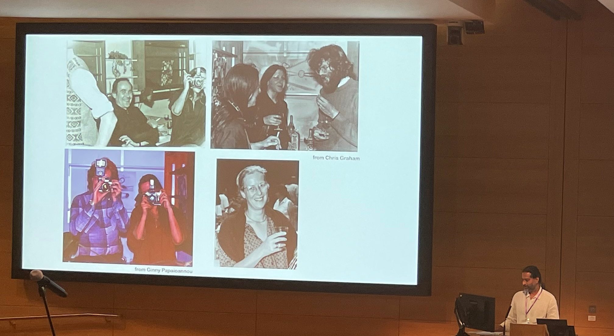
The 2nd Crick-Beddington Developmental Biology Symposium took place at The Francis Crick Institute between 9-10 October 2022. The hybrid symposium was generously funded by the MRC Rosa Beddington fund, which was established to uphold her memory and support curiosity-led science. Although a little later than originally planned, the conference maintained a similar ethos to the 1st symposium in 2019 (see an overview of that meeting from Alex Gould and Vicki Metzis and my previous Meeting Summary); many of the plenary speakers had direct links to Rosa and the talk topics reflected Rosa Beddington’s love of embryos and microscopy, as well as her meticulous description of morphogenesis. Shankar Srivivas, a former postdoc with Rosa and co-organiser of the meeting, opened the symposium with memories and photos of Rosa, mentioning her work on transplanting the mouse node (Beddington, 1994), how she believed in unity between all model organisms and how she often felt more comfortable behind the camera than in front of it.
Shankar Srinivas (organiser) begins the symposium by sharing some memories and photos of Rosa Beddington.
Patrick Tam (University of Sydney) began the meeting by discussing how he and Rosa had a common interest in mouse gastrulation and were each other’s first collaborators in the late 1970s. Patrick presented a spatial transcriptomic atlas of the gastrula mouse embryo that provides insights into the molecular activity underpinning lineage trajectory during gastrulation. Matthew Stower (Srinivas lab, University of Oxford) also discussed mouse gastrulation, using light-sheet imaging to reveal coordinated, collective migratory behaviours in the dorsal visceral endoderm. In their first talk as an independent group leader, Diana Pinheiro (Research Institute of Molecular Pathology) employs computational modelling to understand the collective internalisation of the mesendoderm in response to morphogen signalling during zebrafish gastrulation. Yanlan Mao (University College London) also uses computational modelling to understand how mechanics generates the three major folds in the Drosophila wing disc and how structures are maintained under stress, such as when larvae are constricted. On this theme of mechanics and morphogenesis, Caren Norden (Gulbenkian Science) discussed optic cup development, revealing the importance of the extracellular matrix for the migration of a population of ‘rim cells’ into the retinal neuroepithelium and Toby Andrews (Priya lab, Crick) zebrafish heart morphogenesis and the role of mechanical stretching for trabecular development. Similarly, Golnar Kolahgar (University of Cambridge) explained how vinculin-dependent mechanosensory activity in Drosophila enterocyte progenitors maintains homeostasis of the intestine. Irene Miguel-Aliaga (MRC London Institute of Medical Sciences) presented a different perspective on the Drosophila gut, discussing how inter-organ communication creates sex differences in intestine geometry and function, revealed using microCT. Simon Bullock (MRC Laboratory of Molecular Cell Biology), a former graduate student with Rosa Beddington, uses Drosophila as a model to understand how dynactin-dynein complexes asymmetrically localise RNA transcripts, including Gap genes. Drosophila segmentation was the focus of Jacques Bothma‘s talk (Hubrecht Institute) as well. Jacques has developed ‘LlamaTags’ for rapid imaging of transcriptional dynamics to reveal how the genome encodes and interprets time. Alain Chédotal (Vision Institute) similarly works on the leading edge of technology, employing virtual reality for the characterisation and 3D analysis of cleared human embryos. Jan Huisken (University of Göttingen) concluded the imaging theme by presenting Flamingo, a low-cost, customisable, modular light-sheet imaging microscopy set-up that can be shipped around the world and operated remotely.
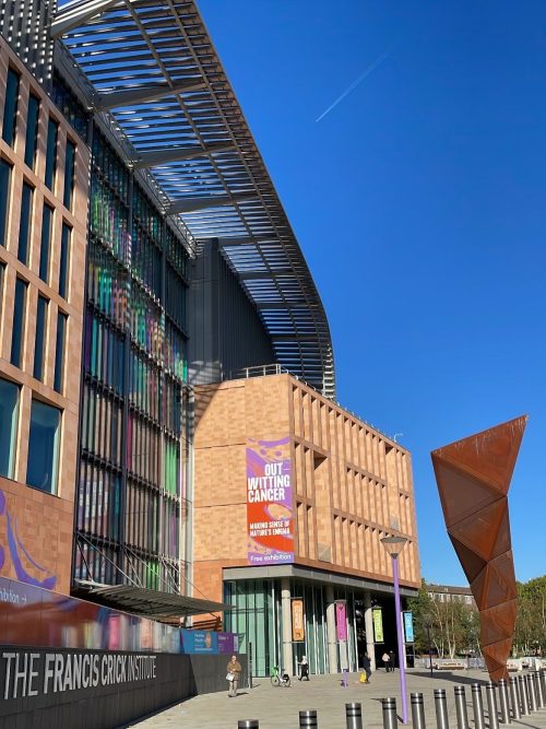
The outside of The Francis Crick Institute.
Several of the talks reflected on metabolism. Josh Brickman (reNEW), a former postdoc with Rosa, looked back at their time together with humour, reflecting how Rosa had insisted he work on transcription factors, to which he now credits his career. Josh explained how transcription factor residence on enhancers supports plasticity in foregut stem cells and lipid metabolism alters gene expression via sirtuin-dependent acetylation. Aydan Bulut-Karslioglu (Max Planck Institute for Molecular Genetics) uses mTOR-inhibited embryonic stem cells as a model to show that fatty acid degradation sustains mammalian diapause. Moving from development to cancer, Salvador Aznar-Benitah (Institute for Research in Biomedicine) discussed how metastasis-initiating cells have high fatty acid metabolism and are sensitive to dietary fats, such as palmitic acid. Meritxell Huch also showed how lipid metabolism can reprogram differentiated liver tumour cells and discussed the importance of TET1-mediated demethylation for de-differentiation during liver regeneration. Selin Jessa (Kleinman lab, McGill University) presented the developmental origins of paediatric gliomas, focusing on the links between histone 3 lysine 27 methylation mutations and 3′ Hox genes.
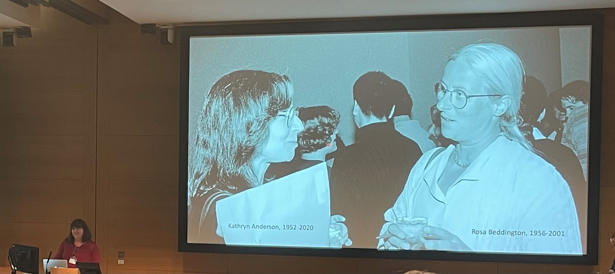
Another emergent theme from the meeting was developmental principles across scales. At the population level, Hugh Ford (Chubb lab, Laboratory for Molecular Cell Biology) uses Dictyostelium to study cAMP signal propagation and cell migration. In tissues, Danelle Davenport (Princeton University) explained how the periodic pattern of skin appendages is established via polarising morphogenesis and Jakub Sumbal (Koledová lab, Masaryk University) presented insights from single-cell RNA-sequencing data of terminal end buds during mammary gland epithelial branching. Val Wilson (University of Edinburgh), a former postdoc in Rosa’s lab, presented research on two populations of axial progenitors in mice (neuromesodermal progenitors and lateral-paraxial mesodermal progenitors) and how signalling controls fate decisions within these populations. At the cellular level, Tamara Caspary (Emory School of Medicine) began her talk by explaining how Kathryn Anderson and Rosa Beddington catalysed her research in mouse ‘hat’ gene mutations, which were later mapped to ciliogenesis and Hedgehog signalling. Tamara showed how these two processes could be uncoupled through the activity of ARL13B in the cilia versus the cytoplasm. Rita Sousa-Nunes (King’s College London), a graduate student with Rosa, similarly aims to uncouple the proteome and transcriptome. Rita began her talk by sharing how Rosa once said to her “if she did it all again she’d [work on] flies and she’d [work with] with Daniel St Johnston”. Using Drosophila neural stem cells as a model, Rita explained how quiescent cells retain untranslated transcripts in their nuclei. Finally, Alex Schier (University of Basal) reminisced about how he once sat next to Rosa before she gave a talk, remarking to him, “I hope I don’t faint”. Alex presented on how the cell develops a specialised form and function by expressing gene ‘modules’, using the differentiation of zebrafish notochord and hatching gland as a model system.
Tamara Caspary told the story about her work was catalysed by the motivation of Rosa Beddington and Kathryn Anderson (1952-2020).
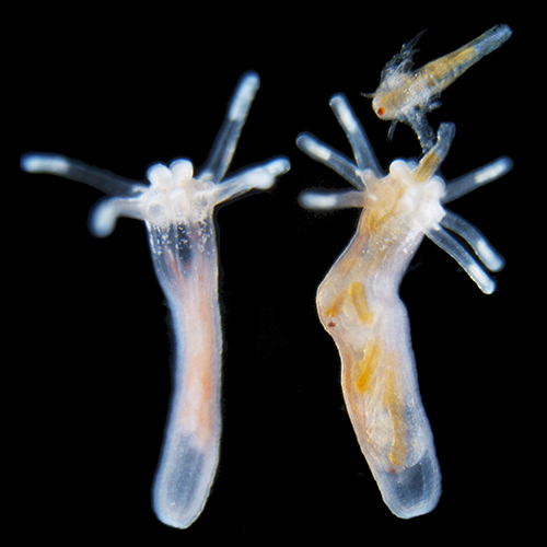
A new addition to this year’s programme was two sessions of flash talks by a selection of the poster presenters. Equipped with just three minutes each, the flash presenters covered a wide range of different topics within developmental biology. They did a fantastic job of enticing the audience to visit their poster during the breaks and poster sessions. Several speakers use live cell imaging to answer their development questions: Sunandan Dhar (Saunders lab, National University of Singapore) studies cell fusion in skeletal muscle; Jean-Francois Derrigand (Spagnoli lab, King’s College London) is exploring exocrine pancreas morphogenesis; Michael Smutny (University of Warwick) is investigating global tissue reshaping zebrafish gastrulation; Markus Schliffka (Maitre lab, Curie Institute) studies the fluid dynamics of microlumens that form in the mammalian blastocyst; and Cerys Manning (University of Manchester) follows dynamic decisions in the development of the eye. Also studying eye development, Jana Sipkova (Franze lab, University of Cambridge) investigates the role of Eph/Ephrin signalling and mechanics in neuronal migration between the eye and the optic tectum. Making the link between mechanics and development, Eirin Maniou (Galea lab, University College London) uses 3D printing to measure mechanical forces during chick neural tube closure in vivo and Sera Weevers (Tsiairis lab, Friedrich Miescher Institute for Biomedical Research) studies how osmosis-driven mechanical stretching underlies the establishment of the Wnt organiser in Hydra. Further understanding the connections between signalling, transcriptional regulation and cell fate, Sergio Menchero (Tuner lab, Crick) aims to uncouple transcriptional and morphological changes by comparing opossum and mouse organogenesis. Similarly, Joaquina Delas (Briscoe lab, Crick) studies how cell fate decisions in the neural tube are regulated at the chromatin level by differential binding and differential element availability and Vicki Metzis (Imperial Collge London) investigates how the posterior neural fate is acquired via CDX2. Furthermore, Lisa Thomann (Lemaire lab, CRBM) uncovers the role of PI3K signalling in ascidian notochord development. Finally, at the subcellular level, Azelle Hawdon (Zenker lab, Australian Regenerative Medicine Institute) studies sub-cellular mRNA asymmetry in mammalian embryogenesis and Chantal Roubinet (Baum lab, University College London) spoke on the asymmetric inheritance of nuclei during Drosophila neuroblast divisions.
As well as the plenaries, selected talks and flash talks, there were also presentations from ThermoFisher and 10X Genomics. For ThermoFisher, Sarawuth Wantha presented the Amira software, suitable for large lattice light-sheet microscopy data, transmission electron microscopy, AI-based segmentation and high-content screening. On behalf of 10X Genomics, Nicola Cahill introduced the Chromium, Visium and Xenium platforms for single-cell sequencing, spatial transcriptome and in situ applications, respectively.
A final variation from the first meeting was the hybrid aspect. I was fortunate to attend most of the meeting in person; however, I didn’t miss out by having to duck out a little early to catch my train. I was able to quickly log in to the live stream on Zoom to watch the concluding remarks. I hope those of attended virtually also found the hybrid format helped accessibility. The switching, too, between in-person and video presenters was seamless and it was great that those who could not attend in person were still able to share their work with us.
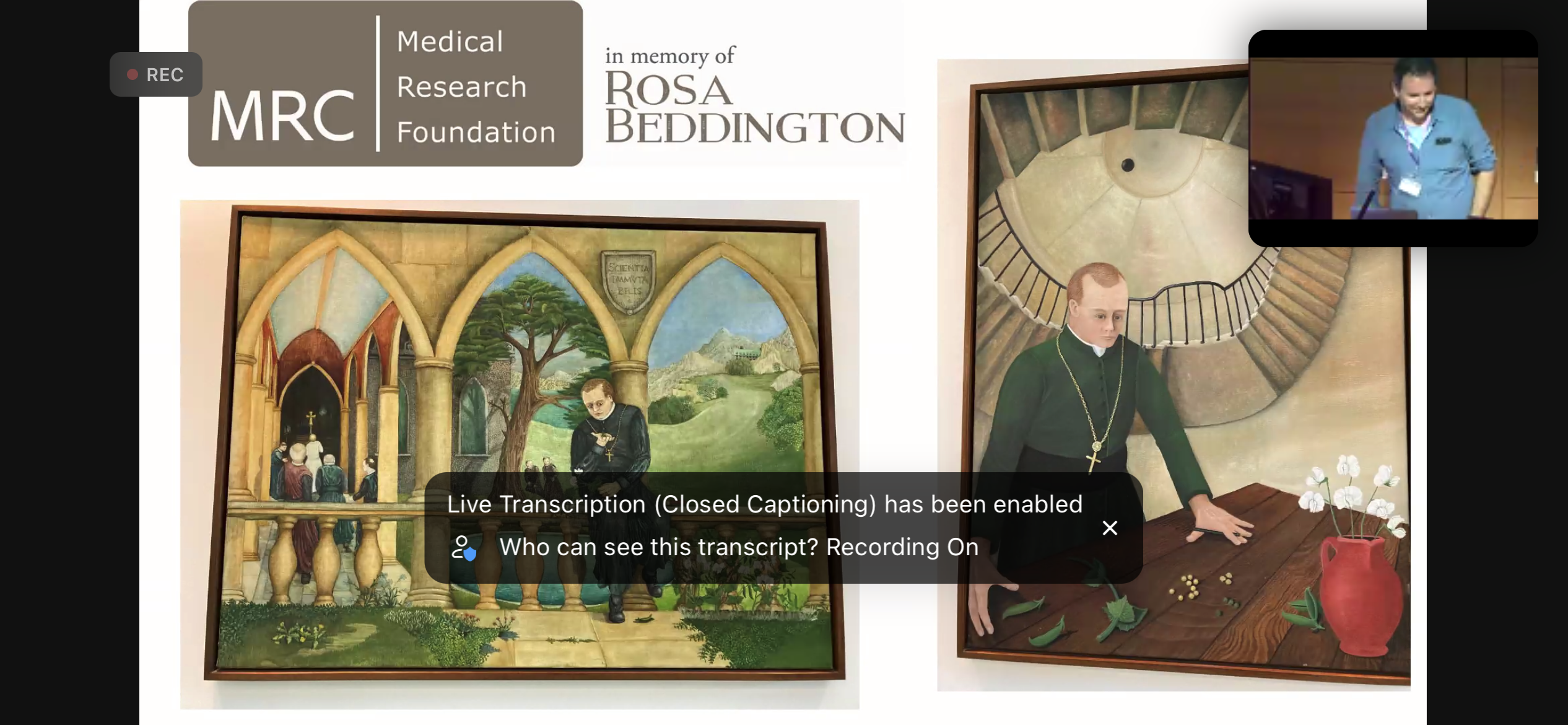
Concluding remarks from Nic Tapon (organiser) sharing some of Rosa’s paintings of Mendel, appropriate because this year marked the 200th anniversary of his birth.
Nic Tapon concluded the symposium by thanking the speakers, the Crick Events team, sponsors and co-organisers; Shankar Srinivas, Alex Gould and Caroline Hill. Nic explained how two of Rosa’s paintings of Mendel that hang in the Crick were especially appropriate this year because it’s the 200th anniversary of Mendel’s birth.
Thank you to the organisers for their effort in assembling the symposium together, chairing sessions and for the smooth execution of the programme. I also thank all the speakers for sharing their stories, both of their science and their memories of Rosa Beddington.
 (No Ratings Yet)
 (9 votes)