We’ve had a great response so far to our tenth birthday user survey, which we hope will guide us going forwards and give us ideas for how we can better serve the developmental biology and stem cell community. If you haven’t taken it yet, it closes Thursday midday BST. Even if you’re a less-than-occasional reader, we’d love to have your input:
Hi there! Today’s highlight is focusing on a major question for the haematology community: what is causing blood stem cell heterogeneity? Please do not hesitate to let me know your thoughts in the comments here or on Twitter (@BioRugby)!
Highlight #2: Chromatin accessibility is the main source of heterogeneity in foetal haematopoiesis
We have a limited understanding of the haematopoiesis in the human foetus. Particularly, it is unclear whether stem cell differentiation depends on transcriptional priming (lineage-specific transcription factors) or a broader epigenetic one. This preprint delves into this question, presenting a single-cell resolution study of foetal human blood stem and progenitor cells. The authors analysed the transcriptome and the chromatin accessibility of over 8000 cells from different organs, confirming the high heterogeneity of the stem cell compartment. They inferred some differentiation trajectories, although the most-well know transcription factors were not consistently identified. In addition, the authors combined the datasets, and with the exception of the progenitors linked to red blood cells and platelets, they have been unable to unequivocally associate chromatin accessibility to defined transcriptional signatures. Therefore, they concluded that stem cells in the developing foetus are minimally primed towards differentiation, and this priming is not due to specific transcriptional programs associated to mature cell types. Finally, they compared different organs, confirming the higher cycling tendency of stem cells from foetal liver, compared to bone marrow. In the future, it will be interesting to garner a better understanding of the signals that trigger differences between haematopoietic sites.
Ranzoni AM, Tangherloni A et al. “Integrative Single-cell RNA-Seq and ATAC-Seq Analysis of Human Foetal Liver and Bone Marrow Haematopoiesis”
doi: https://doi.org/10.1101/2020.05.06.080259
Perhaps the most exciting aspect of attending any scientific meeting is the privilege of becoming aware of novel research findings in our fields of interest, prior to their appearance in published literature – and this begins as soon as we have the abstract book in hand! Sitting in my hotel room in Suzhou, and browsing through the abstracts of the first Cold Spring Harbor meeting on cilia and centrosomes which was held in the spring of 2017, I chanced upon work from Chengtian Zhao’s group (Ocean University, Qindao, China) that described the analysis of gene expression changes in zebrafish embryos deficient in zymnd10, a gene encoding a ciliary dynein assembly factor (1). Since my lab had also performed similar analyses with two other cilia mutants, ccdc103 and lrrc50 (also encoding dynein assembly factors (2,3)), I decided that I must meet up with Chengtian and discuss whether we should collaborate or at the least, co-ordinate our studies. That evening, as I settled down for a sumptuous dinner in the esteemed company of two prominent cilia researchers, Hiroshi Hamada and Cecilia Lo, I was pleasantly surprised when a Chinese gentleman came forward and introduced himself to me as Chengtian Zhao, and asked if he could join us for the meal. Conversation flowed effortlessly and the rest, as they say, is history.
Over the years, a number of studies had observed that zebrafish embryos with mutations in many different cilia genes exhibit ventrally curved body axis. More recently, work from the Burdine and Ciruna labs had implicated defects in cilia motility, causing abnormal cerebrospinal fluid (CSF) flow, in curvatures of the vertebral column in adult zebrafish (4), making this system a good model for uncovering the etiological basis of the common human spine disorder idiopathic scoliosis (IS) that affects up to 3% of children and adolescents world-wide (5). Abnormal 3D curvatures of the spine in IS can cause a considerable degree of morbidity in patients, manifest in symptoms like difficulties in breathing, postural issues and gait problems. Despite these important findings, the molecular mechanism operating downstream of cilia-driven CSF flow that ensures proper spine development had remained unclear. With the collaboration that sparked off at the dinner table of the Suzhou meeting, Chengtian’s and my lab published a collaborative paper just before Christmas of 2018, describing a possible molecular pathway by which ciliary motility, within the brain ventricles and spinal canal of the zebrafish, ensures that they develop with a straight body axis (6). We showed that cilia beating transports catecholamines (like epinephrine) in CSF, and they stimulate the expression of Urotensin-related peptides (Urp) in CSF-contacting neurons (CSF-cNs) that differentiate along the spinal canal. The Urp family consists of small cyclic neuropeptides, homologous to Urotensin II, which is known to function as a potent vasoconstrictor (7). Indeed, it was the significant decline in expression levels of the urp genes in our respective gene expression analyses of cilia mutants that prompted us to follow up on Urp signalling to uncover the mechanism acting downstream of cilia-driven CSF flow. Our current model posits that Urp peptides, secreted from CSF-cNS, brings about a certain degree of tension or contraction of the somitic slow-twitch muscles of the dorsal somites, and this is the biomechanical force for axial straightening. In keeping with this view, we also found that a Urp receptor, Uts2r3, a G-protein coupled receptor, is expressed in the dorsal slow muscles, and when it was rendered non-functional using a targeted lesion at the locus, the adult uts2r3 mutants developed with severely curved spines, resembling humans with IS (6).
More or less concurrent with our publication, Claire Wyart, Pascal Bardet and colleagues reported a very intriguing story that ascribed, for the first time, a definitive biological role for the enigmatic Reissner fiber (RF) (8). Discovered in the 19th century by the German anatomist Ernst Reissner, RF is a filamentous structure that remains suspended in CSF along the brain ventricles and the spinal canal in all vertebrates examined, including humans. RF is polymerized from a large glycoprotein, SCO-spondin (Sspo), secreted from the subcommissural organ (hence SCO) and the floor plate. RF is thought to participate in many functions of the nervous system, but the lack of genetic mutations in Sspo has precluded a firm association of RF with any of these proposed roles. Wyart et al. showed that in zebrafish embryos, RF is required for proper axial development since sspo mutants developed ventrally curved body axes (and perished at the early larval stage), closely mimicking the curved bodies of cilia mutants (8). They also demonstrated that cilia motility is required for RF biogenesis, by somehow facilitating the polymerization of the protein into the fiber. Since biochemical studies with mammalian RF had already shown that it can efficiently bind and facilitate the transport of catecholamines present in CSF (9), it dawned on me that cilia, RF, and CSF catecholamines could all be functioning via Urp signaling from CSF-cNs.
The idea that I wanted to explore is whether sspo mutants develop curved bodies because in the absence of RF, they are unable to bind CSF catecholamines and present to CSF-cNs to activate urp gene expression. It is with this view in mind that we approached Claire and Pascal, who most generously shared with us the sspo mutants. In a paper that we have just published in Biology Open (10), we now show that consistent with our expectations, sspo mutants exhibit a loss of urp gene expression from CSF-cNs: urp1 is significantly reduced, while urp2 is almost completely absent. As we had demonstrated previously for cilia mutants, culturing sspo mutants in the presence of exogenously added epinephrine in embryo medium, restored urp expression in CSF-cNs and also rescued their ventrally curved axial defects. However, one issue that continued to confound us was whether abnormalities of the embryonic axis in sspo mutants have any connection with scoliosis of the spine in adult zebrafish. If there is, our work will have relevance for furthering our understanding of the etiology of IS. The Burdine and Ciruna et al. paper, that had initially linked cilia motility and CSF flow with spine curvature, used a clever strategy to bypass the severe embryonic body curvature and associated lethality of the cilia mutants that they studied (4). They injected the corresponding (in vitro synthesized) wild-type sense mRNA into mutant eggs: this rescued the axial defects as well as lethality, and the mutants developed into adults but they exhibited severely curved spines. Unfortunately, we could not utilize this strategy to rescue sspo mutants as the Sspo protein consists of more than 5000 amino acids, and in vitro synthesis of an mRNA to encode such a large protein is not feasible. To circumvent this problem, we prematurely dechorionated the sspo mutants with the hope that their severe ventrally curved axis could be partially rescued when freed off the confines of the spherical, non-elastic chorion. Indeed, this simple trick ameliorated the strong axial curvature of sspo mutants to varying degrees, and many of them matured into adults with severe spine malformations reminiscent of cilia mutants rescued of their embryonic lethality and mutations in the Urp receptor. Thus, in the zebrafish, ventral curvature of the embryonic axis and scoliotic malformations of the adult spine represent linked morphogenetic anomalies. Most satisfyingly, we found that restoring expression of Urp2 exclusively in CSF-cNs, rescued not only the embryonic and larval body curvature of sspo mutants, but also allowed one such mutant to develop into an adult with an apparently normal spine! Additional findings that we report in our Biology Open paper include data showing that it is a definite threshold of Urp signaling that is critical for the morphogenesis of a straight body axis. Too little signaling causes ventral curvature as in sspo and cilia mutants, while exaggerated signaling (for instance, by over-expression of the urp gene or the protein in the muscle cells themselves) causes the converse effect of profound dorsal curvature of the axis. Finally, using mutations in Smoothened (Smo), an essential component of the Hedgehog pathway that directs slow-twitch muscle cell differentiation in the zebrafish somites, we could show that lack of the slow muscles make these mutants refractory to Urp signaling (10). Smo mutants have strong ventrally curved bodies that could not be rescued by over-expression of the Urp proteins.
Image of an sspo mutant adult zebrafish showing curvatures of the trunk and tail (top panel). Micro-CT scan of an sspo mutant revealing 3D curvatures of the vertebral column, resembling spines of IS patients.
How do we take these findings forward, especially for furthering our understanding of the mechanistic basis of pathogenesis in IS? First, with reference to the zebrafish, we need to better understand how Urp signaling-induces activity of the slow-twitch fibers of the somites, and how this activity feeds back to the growing spine to ensure that it develops along a straight axis. Secondly, while RF and CSF-cNs exist in mammals and Urp signaling has also been shown to operate there, we will need to examine whether the circuitry that we have been able to dissect in the zebrafish is also conserved. In this regard, one caveat that we need to bear in mind is that traditional experimental mammals like mice and rats are quadrupeds, and they have not turned out to be effective for modeling human spine disorders (hence the promise of the zebrafish)(11). Finally, based on what we glean from all of these investigations, we can begin to parse the underlying mechanisms driving spine malformations in IS. There is already accumulating evidence that ciliary dysfunction could be causative of the disease (12,13). Moreover, presence of RF has been reported in human embryos and a teenager (14), implying that defects in this structure could be responsible for IS in some of the individuals afflicted with IS. And of course, the real benefit of all this research effort will be if we can invent effective therapeutic strategies for IS by pharmacologically manipulating the Urp pathway, since current treatment options are largely limited to managing the disorder with physiotherapy and braces, and in severe cases, the rectification of severe spine deformities with invasive surgery.
Morphogenesis is a complicated network of processes that involve cell shape changes, cell movements and division patterns. While these are part of the intricate game that gives rise to tissue shapes, there is another element that is now emerging as a major player. A supra-cellular physical scaffold that directs sculpting at the tissue level; the basement membrane (BM).
To put this statement to the test, in our recent paper [1] we have investigated how the developing mouse embryo takes shape through a ‘primitive’ and newly forming basement membrane that surrounds the embryo, at a time when it’s exponentially growing.
Once the mouse embryo implants, it transforms into what we call an ‘egg cylinder’ which refers to its elongated cylindrical shape (Figure 1).
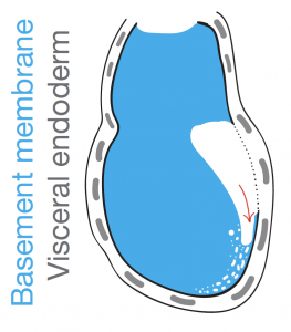
Figure 1:A schematic of the pre-implantation blastocyst and post-implantation egg cylinder. epiblast, primitive/visceral endoderm, extraembryonic ectoderm, basement membrane
The elongated shape of the egg cylinder is maintained while it grows in size. The very fact that shape remains virtually unaltered during growth baffled us however. We suspected that there must be a mechanism in place that actively maintains the shape somehow. Our suspicions started to lean towards the BM when its complete dissolution with Collagenase IV affected the embryo shape.
We know quite a bit about the signalling niche the BM creates. It’s a laminin- and collagen-rich extracellular matrix that surrounds the embryo and induces polarisation of the cells it’s in contact with through integrin-mediated signalling. In turn this drives processes like cavity formation and extension that gives egg cylinder its characteristic hollowed appearance (Figure 1)[2]. But how about its physical properties and its potential role in physically guiding (and maintaining) shape?
The ‘net-like’ basement membrane
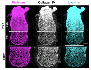
Inspired by the work of Kenneth Yamada and Donald Ingber on the architecture of BM we went on to investigate its appearance [3,4]. What we discovered was a striking difference in how BM is laid down in the two halves of the embryo along the proximo-distal axis (long axis). Specifically, the embryonic half (EPI) is heavily perforated in contrast to the extraembryonic half (ExE) (Figure 2). This architecture has been observed before in growing limb buds and was suggested that this allows directional bud growth and subsequent branching morphogenesis. Similarly, in this study we argued that this perforated morphology of the BM in the embryonic portion of the embryo allows embryo growth in a directional manner i.e. growth towards the side of least resistance (in this case the distal portion of the embryo) while maintaining at the same time the overall BM integrity and the embryo’s elongated shape.
Is it possible that this ‘net-like’ morphology of the BM is a way of decreasing physical constraints at the side of ‘desired’ growth direction? To answer this, we would need to ‘homogenise’ the BM by ‘filling up’ these perforations and re-examining embryo growth.
But first we would need to abrogate whatever mechanism creates these perforations in the first place.
Figure 2: Early post-implantation mouse embryo (emrbyonic day (E) 5.5), stained for basement membrane components perlecan, collagen iv and laminin and projected at maximum intensity. Embryonic portion, epiblast (EPI) is visibly perforated compared to the extraembryonic portion; extraembryonic ectoderm (ExE)
Finding an AVEnue to elucidate the mechanism
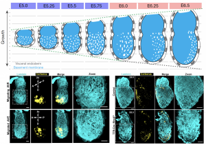
The natural process itself gave us our most valuable hint towards understanding the underlying mechanism of perforation generation. The natural process I am referring to is the establishment of the anterior-posterior axis, which takes place during these early stages of development. A specialised population of cells in the outer layer of the embryo (the visceral endoderm; VE) emerge at the distal tip and start migrating to one side to establish the anterior portion. These cells are called (you guessed it) anterior visceral endoderm (AVE). To our surprise, the BM underlying these cells loses its perforated morphology and by the time AVE has populated the future anterior side of the embryo, the perforation distribution is heavily skewed towards the posterior. Abrogation of AVE movement in a RhoA VE-specific knockout also affects the asymmetric redistribution of BM perforations, in line with the suspected impact of AVE on the underlying BM (Figure 3). This was our breakthrough in not only understanding the mechanism of forming the perforations but also their role in the pre-gastrula stages. But first let us consider the mechanism and how this observation led us to solving it.
Figure 3: Top panel shows a schematic of appearance of perforation distribution from early post-implantation stages to the pre-gastrula stages. Bottom left panels show the AVE (yellow) at two different stages of its migration and the appearance of basement membrane (cyan) perforation distribution. Bottom right panels show that failure of AVE migration in the RhoA-VE konckout (TTR-Cre;RhoA fl/-) perturbs perforation redistribution.
The AVE brings about its anteriorising and posteriorising effect by modulating Nodal activity. It secretes Nodal inhibitors (namely Cerberus and Lefty) in its vicinity confining Nodal activity to the future posterior. In turn, Nodal drives mesendodermal specification in the posterior and kicks off later development with the formation of the three germ layers. In the context of our question, however, it suggested that Nodal inhibition may be behind the loss of BM perforations under AVE. Both pharmacological inhibition and genetic knockout of Nodal presented the same phenotype; a solid unperforated BM.
Nodal is not-all there is to it: MMPs are the MVPs
Next question was to identify the downstream effectors of this process. Naturally, we suspected matrix metalloproteinases (MMPs) might be playing a role in this, due to their collagenolytic properties and dissolving extracellular matrix to facilitate processes in cancer pathogenesis like metastasis. Knowing that BM perforation was a Nodal-dependent mechanism we established MMP candidate eligibility criteria based on: 1) temporal expression (during stages of development we were investigating i.e. early post-implantation to pre-gastrula stages), 2) spatial expression (embryonic portion of the embryo and posterior expression at pre-gastrula stages) and 3) response to Nodal.
Thanks to the incredibly useful single cell sequencing and ChIP-seq databases available from previous studies we were able to mine them for the MMP candidates meeting these criteria [5,6]. This gave us two candidates, MMP2 and MMP14.
To upgrade their “candidate” status to the “player” status, we confirmed that they fit all three criteria by RNAscope (FISH), directly looking at their distribution and response to Nodal inhibition. To functionally confirm them, we inhibited these MMPs using a cocktail of MMP inhibitors and phenocopied Nodal inhibition and perturbation. BM lost its perforated morphology and the embryo growth was significantly reduced in response to it.
To condense all this in one sentence: “The growth of the embryo is regulated by modulating the physical constraints, imposed by the surrounding BM, through perforations, in a Nodal-dependent MMP-mediated fashion”.
But what’s the connection of this process with gastrulation?
Revisiting the ‘Run in the stocking’ model
Gastrulation is a super-process, if you will, that involves a number of processes taking place simultaneously and co-ordinately, to achieve the formation of the three germ layers and the extraembryonic mesoderm for the extraembryonic membranes. While most of us would recognise the onset of gastrulation at the time when we observe the first mesendoderm cells forming at the posterior of the embryo, it’s merely the preparation for gastrulation.
What kicks things off, is the breaking of the BM under the mesendoderm cells and initiation of epithelial-to-mesenchymal transition with subsequent ingression in the layers below. So, we can argue that the cells in the posterior epiblast are specified into mesendoderm in preparation for their journey to the lower layers to establish the germ layers. Similarly, we suggest that the BM may go through a similar preparation phase becoming prone to breakage and ultimately be breached at the right time when mesendoderm cells have been specified and ready to ingress.
Figure 4: Basement membrane progressive breaching through a “run in the stocking” model
The suggestion of a BM pre-patterning for gastrulation came with the observation of perforation redistribution to the future posterior upon establishment of the anterior-posterior axis. As described earlier, the anterior signalling centre, AVE, through local inhibition of Nodal activity, creates a posterior “perforation domain” in the BM. This domain is established even before mesendoderm specification and constitutes now a first and novel posteriorising characteristic. We hypothesised that an asymmetrically perforated BM will preferentially be breached on the side of least resistance and thus the posterior BM. By investigating the successive steps of gastrulation and primitive streak extension with progressive breaching of BM we saw that the perforations persist even during the process of gastrulation, “paving the way”, in a sense, for a controlled and ordered ingression of mesendoderm cells.
What we saw was consistent with a “run in the stocking” model, as first proposed by Donald Ingber [4] in which a “weakened” BM domain will expand in response to forces imposed by cell proliferation and tissue growth. In our case this weakened BM is the pre-patterned posterior BM which, much like a tear in a stocking, will progressively break as cells force their way through the breach (Figure 4). To confirm the importance of the perforation domain along the prospective primitive streak, we inhibited MMPs prior to gastrulation to homogenise the BM. We noticed mesendoderm cells being specified but failing to ingress to the extent of our controls, trapping them in the epiblast and in some cases seeing them being extruded and dying in the pro-amniotic cavity.
Final thoughts
The discovery of BM perforations in the early post-implantation mouse embryo has given a new, more physical, perspective of early morphogenesis. The groups of David Bilder, Barry Thompson, Magali Suzanne and Kenneth Yamada are some of the leading teams looking at BM-directed tissue morphogenesis and have published exciting research that stretches from fruit flies to mice [3,7,8,9,10]. While the mode of basement membrane remodelling in different species and tissues can be different (perforations, collagen fibril arrangement or complete dissolution) it seems that the principle of this morphogenetic mechanism is evolutionary conserved. Further study promises more insight in how complicated structures and shapes have evolved. This is an area where computational modelling of BM dynamics in shape-forming can supplement the cellular dynamics field of study in morphogenesis.
The current work in mouse has translational value as well. A similar mechanism could be in operation in human embryos during implantation, a stage when embryos rapidly expand and transform but also a stage when many human pregnancies fail.
I am looking forward to see how this area develops and discuss with anyone interested.
Illustration of the perforated basement membrane (cyan) and the layer of visceral endoderm on the embryonic portion of the mouse embryo. Artwork by Andreas Karpasitis
[1] Kyprianou, C., Christodoulou, N., Hamilton, R. S., Nahaboo, W., Boomgaard, D. S., Amadei, G., & Migeotte, I. (2020). Basement membrane remodelling regulates mouse embryogenesis. Nature, (March 2019). https://doi.org/10.1038/s41586-020-2264-2
[2] Christodoulou, N., Kyprianou, C., Weberling, A., Wang, R., Cui, G., Peng, G., … and Zernicka-Goetz, M. (2018). Sequential formation and resolution of multiple rosettes drives embryo remodeling after implantation. Nature Cell Biology. https://doi.org/10.1038/s41556-018-0211-3
[3] Harunaga, J. S., Doyle, A. D., & Yamada, K. M. (2014). Local and global dynamics of the basement membrane during branching morphogenesis require protease activity and actomyosin contractility. Developmental Biology, 394(2), 197–205. https://doi.org/10.1016/j.ydbio.2014.08.014
[4] Moore, K. A., Polte, T., Huang, S., Shi, B., Alsberg, E., Sunday, M. E., & Ingber, D. E. (2005). Control of basement membrane remodeling and epithelial branching morphogenesis in embryonic lung by Rho and cytoskeletal tension. Developmental Dynamics, 232(2), 268–281. https://doi.org/10.1002/dvdy.20237
[5] Cheng, S., Pei, Y., He, L., Peng, G., Reinius, B., Tam, P. P. L., … Deng, Q. (2019). Single-Cell RNA-Seq Reveals Cellular Heterogeneity of Pluripotency Transition and X Chromosome Dynamics during Early Mouse Development. Cell Reports, 26(10), 2593-2607.e3. https://doi.org/10.1016/j.celrep.2019.02.031
[6] Wang, Q., Zou, Y., Nowotschin, S., Kim, S. Y., Li, Q. V., Soh, C. L., … Massagué, J. (2017). The p53 Family Coordinates Wnt and Nodal Inputs in Mesendodermal Differentiation of Embryonic Stem Cells. Cell Stem Cell, 20(1), 70–86. https://doi.org/10.1016/j.stem.2016.10.002
[7] Haigo, S. L., & Bilder, D. (2011). Global Tissue Revolutions in a Morphogenetic Movement Controlling Elongation. Science, 331(6020), 1071–1074. https://doi.org/10.1126/science.1199424
[8] Crest, J., Diz-Muñoz, A., Chen, D. Y., Fletcher, D. A., & Bilder, D. (2017). Organ sculpting by patterned extracellular matrix stiffness. ELife, 6, 1–16. https://doi.org/10.7554/eLife.24958
[9] Del-Carmen Diaz-De-La-Loza, M., Loker, R., Mann, R. S., & Thompson, B. J. (2020). Control of tissue morphogenesis by the HOX gene Ultrabithorax. Development (Cambridge), 147(5), 1–13. https://doi.org/10.1242/dev.184564
[10] Proag, A., Monier, B., & Suzanne, M. (2019). Physical and functional cell-matrix uncoupling in a developing tissue under tension. Development (Cambridge), 146(11). https://doi.org/10.1242/dev.172577
This piece recently featured on The Company of Biologists’ WeChat Channel. For more information about our efforts to engage Chinese researchers, read Annabel Nicholson’s post from last month.
The Zeng Lab
Dr Yi Zeng is a member of the Development Editorial Advisory Board and her lab at the Shanghai Institute of Biochemistry and Cell Biology studies adult stem cells in various organ systems. Dr Cissy Yu is a Research Associate in the Zeng lab. Her research on vascular endothelial stem cells focuses on understanding their cellular behaviour and molecular dynamics during physiological development, as well as pathological progression.
Cissy Yu
Here, Cissy gives a personal account of how the lab navigated lockdown in China:
“Back in mid-January 2020 we were all excited about the upcoming Spring Festival break. Our lab traditionally celebrates a year’s hard work with an annual party, before travelling home for 2 weeks to reunite with our families across China. Everyone was glowing and cheerful during the party dinner as life carried on without concern.
A few days into the holiday, the escalating COVID-19 crisis led to the nationwide lockdown. On Chinese New Year’s Eve my close medical doctor friend told me she had volunteered to go to Wuhan. I rushed to give her my supply of N95 masks, and hugged her goodbye with feelings of worry and admiration.
Throughout lockdown our lab WeChat group was flooded with updates, news, safety, and jokes on what staying home meant or words to lift spirits. We shared confusion, frustration, sorrow, joy and enlightenment. We felt even more connected even though we were apart.
When we were told that we could not return to work for almost two months, we realized we had to figure out how to continue with lab maintenance. Our institute announced strict campus access only for healthy personnel without recent travel history. A small group of us managed to support the ongoing animal experiments originally conducted by other lab members, so that their year-long experiments wouldn’t be wasted.
We had uncertainty and confusion on whether liquid nitrogen could be filled, whether the CO2 tank would be delivered in time, whether cell culture reagents would be out of stock, whether there would be alternative approaches? We had to make hard decisions.
Students who had their thesis defence scheduled for after the holiday or those in the middle of a paper rebuttal were most affected. Fortunately, the science community around the world has been very kind and remarkably supportive in all ways. We received warm encouragement and gracious extension, which also greatly relieved our stress.
My friend safely returned home last week and our lab is gradually reviving. Nevertheless, wearing masks, extensive hand washing and regular temperature checks have become the new norms.
The rest of the world is now where we were two months ago. My blessing goes to those who suffered.”
Chan, Z., Santella, L., Tuszynski, J. and Gordon, R. (eds.) (2020) Waves in Fertilization, Cell Division and Embryogenesis [special issue of BioSystems, In preparation] [WAVS].
Hello everyone! My name is Alessandro, I am a postdoc in love with science, stem cells and blood! I am also a big fan of the Node, The Company of Biologists generously granted me a travel grant back in 2016, that allowed me to discover the beautiful Cambridge (the old one). As the lockdown starts to ease a bit around the world, I have decided to start a little writing workout: I will try to regularly post a highlight of science that grabbed my attention, as a paper or preprint. Feedback is more than welcome, as are suggestions. I hope you will enjoy my writings, you can find me also on Twitter (@BioRugby)!
Highlight #1: Inhibition of cell cycle regulators alters the EHT efficiency of endothelial-to-haematopoietic transition
Endothelial-to-Haematopoietic Transition (EHT) is the key mechanism behind the formation of the first haematopoietic stem cells. Albeit first described in the mid-80s, we still do not understand some of the cellular mechanism that regulates it. In their preprint, Canu and colleagues used human pluripotent stem cells to characterize the EHT. Extensive use of single-cell transcriptomics revealed the existence of multiple endothelial and haematopoietic populations; one of the latter was able to generate myeloid and erythroid colonies, revealing a certain multipotency. Interestingly, the transcriptomic analysis revealed different cell cycle activity between populations, prompting the authors to investigate the cell cycle impact on EHT. Slowing down cell cycle progression disrupted the occurrence of EHT, increasing the proportion of endothelial cells without modifying their viability. Moreover, a similar phenotype was observed by inhibiting cyclin-dependent kinase 1 (CDK1) and CDK4/6, which suggests that the EHT disruption is not caused by the decreased proliferation of endothelial cells. Taken together, this manuscript proves a major impact on differentiation for CDKs, beyond their canonical role as cell cycle regulators. The proposed link between cell cycle and differentiation has major implications on our understanding of haematopoietic development. The authors successfully proved it in a robust and human-relevant experimental system, combining single-cell transcriptomics, flow cytometry and functional assays. It will be very intriguing to delve further into the molecular intricacies of EHT, to reveal their impact on this fascinating developmental transition.
Canu G et al. “Analysis of Endothelial-to-Haematopoietic Transition at the Single-Cell Level identifies Cell Cycle Regulation as a Driver of Differentiation”.
I wanted to take a couple of minutes to update you on how things are going at Development. I imagine many of you will be reading this on a laptop in your home office/kitchen/bedroom in between Zoom calls, homeschooling and/or sourdough baking. In much of Europe and North America, as well as other parts of the world, it’s been like this for the best part of 2 months and will continue for at least the next few weeks. Even when quarantine measures begin to be eased and our labs and institutes start to open up, it won’t be straight back to how it was before. Physical distancing requirements, travel restrictions, disrupted supply chains, and potentially new financial constraints will affect us for months to come and might permanently change how we do science.
New colleagues settling into a Development home office
At Development, we’re more fortunate than most. As Academic Editors, although our labs are affected, we’re already used to doing our editorial work remotely and communicating with each other electronically. The professional staff based in the Company of Biologists offices in Cambridge have been able to adapt quickly to working from home and you can see some of their new locations on our Instagram. Regular electronic meetings between Development staff and regular seminars involving all Company staff have kept communication going and morale high. It’s been great to see the team pull together despite the difficulties and I’m pleased that Development has continued to run smoothly.
As the lockdowns and shelter-in-place orders spread around the globe the Development editorial team discussed the effects it might have on our community and what we could do to support researchers. We put together a statement. This included asking reviewers or authors unable to meet journal deadlines to contact us for extensions. We’ve been able to do this for everyone that has been in touch. Of course, our scoop protection policy means that authors do not have to worry about being scooped once a manuscript is submitted. Moreover, we do not necessarily reject a new submission if a similar paper has recently been published: if this happens, please contact us to discuss the case.
We also emphasised that reviewers should limit requests for revisions to those that are essential to support the conclusions of the paper. This has always been the case at Development, but it’s even more important in these times when the ability to conduct experiments is severely restricted. We want to ensure that papers meet the high standards our readers expect, but we do not want to see publication delayed unnecessarily. As editors we try to provide guidance on the revisions that are necessary when we send the reviewers reports and we encourage authors to discuss revision plans with us, if that would be helpful, before embarking on extensive new experiments. More generally, we’re conscious that – with opportunities to meet the community severely restricted this year – there won’t be the occasions to talk about your science or the journal that we normally have. So if you’ve got anything you’d like to discuss, please feel free to drop us a line.
Katherine Brown, our Executive Editor, has been keeping a close eye on any changes in the rate or type of submissions we’ve received. We’re pleasantly surprised that we haven’t noticed any obvious change in the number of research papers submitted, at least not yet. The numbers in both March and April were in line with previous years. As there have been concerns that working from home disproportionately disadvantages women, we’ve also tried to gauge whether there has been a decrease in number of papers submitted with female corresponding authors. This doesn’t seem to be the case for Development, at least from our very preliminary assessment. Approximately 40% of submissions in the last couple of months have a female corresponding author, similar to previous months. Nevertheless, it is something we will continue to monitor and something that will need a more thorough analysis once the immediate crisis is over.
Seema Grewal, our Senior Editor, and Alex Eve, Reviews Editor, have been receiving more proposals for review articles than normal and they have been busy assessing these and advising potential authors. So it looks as if some researchers are using time away from lab to take a deep dive into the literature and write the review they’ve been meaning to. We’re not able to consider all the proposals we receive, but are always happy to discuss ideas. If this is something you’re thinking about, don’t forget to check out Seema’s Introduction to Writing Review Articles. Alternatively, if you want to communicate your thoughts more immediately or practice a different style of writing, take a look at Node Editor Aidan Maartens’ suggestions for Writing for the Node.
I’m also pleased to say that reviewers are continuing to support Development and all the editors are very grateful for this. Understandably, some reviewers are finding it’s taking longer than normal to return their reports, if this happens to you please let us know so that we can keep authors informed. In their reports, many reviewers are acknowledging the difficulty that proposed revisions pose to authors and the discussion amongst reviewers, using cross-referee commenting, can help establish what is really essential for the authors to address. It’s good to see reviewers adopting this constructive approach and I hope this becomes the new normal from now on.
We’ve also begun to think about how scientific life will change beyond the next month or two. Travel is likely to be greatly restricted for a long time and may never return in quite the same way. This will of course have a major effect on conferences and workshops. It is likely to accelerate changes that were already underway because of the contribution of international travel to Climate Change. We’ve seen conferences switch to online (including TAGC and SDB) as an immediate response to the crisis and this trend is set to continue. As an organiser of conferences and workshops, Development and the Company of Biologists have started to consider how we can adapt formats and use new technology to continue to get most of the benefits of conferences without the travel – we’re currently considering options for our upcoming ‘From stem cells to human development’ meeting and expect to have more news on this in the next few weeks. This is not straightforward as conferences are more than a series of talks. They also provide the opportunity for spontaneous encounters that spark unexpected conversations, ideas, collaborations; for early career researchers to present posters and talk informally with each other as well as leaders in a field; for social interactions that result in new friendships as well as new science. It is these less obvious ingredients of a good conference that builds a field, supports the careers of junior colleagues and enhances scientific dialogue. How we replicate or replace these features is a challenge we need to take on. Nevertheless, the developmental biology community has always been innovative and we’d love to hear your thoughts on what can be done.
Anyway, I have a Zoom meeting scheduled and I can smell my banana bread cooking so let me finish by thanking you all – authors, reviewers and readers – for your continuing support of Development. Your suggestions and opinions are always welcome and I look forward to hearing from you, and one day seeing you again in person.
Hello! Recently, I’ve been tweeting writer’s advice from @jbwallingford using the hashtag: #DevBiolWriteClub. I’m psyched that The Node is now letting me add a little depth to this venture. In this first post, I’ll start by managing some expectations.
If you’ve followed #DevBiolWriteClub on Twitter, you might recall that one of my earliest tweets said: “For now, I will focus on how to be a better writer. This is different from how to write better.” Personally, I like that sentence. I think it’s clever, and it conveys some crucial information. That said, one could also make the argument that it sucks. After all, it’s kind of confusing, and by forcing the reader to think hard about just a few words, it risks failing to convey any information at all.
Regardless, the details of well-written and poorly written passages is NOT the point of #DevBiolWriteClub. Rather, I want to use this forum to address one of the most common and intractable misconceptions in writing, especially among busy scientists. The issue is this: If you are serious about better writing, DO NOT start by thinking harder about sentence structure and grammar. Instead, start right now by focusing on your practice of being a writer.
Writing is like a sport. You only get good at it if you practice, with intent, every day. When I’m not doing science, I’m a rock climber, and one of my favorite coaches is Steve Bechtel (Climbstrong.com.), and he once lamented that he always wanted to write an article titled “500 weeks to stronger fingers,” but that no one would read it. I fear the same for these blog posts, because he’s exactly right. No one likes to hear that there are no shortcuts.
The difference, of course, is that most scientists relate to sports (or art, or baking) as a hobby, as this other thing we do sometimes, and we hope to get better at. Ultimately, though, if we don’t get better, it’s not that big a deal. The problem is that many scientists take the same view of writing. But not improving as a writer is a big deal in science. It’s a very big deal, actually. If you are a scientist and you want to succeed, you mustbecome a writer. And the only way to do that is to practice, day in and day out. For years.
So, that’s the bad news. The good news is that the process for becoming a writer is pretty simple, and you can start today. There are only five rules:
Do the work.
Do the work.
Revise and edit. Again, and again, and again.
Read with intent.
You can’t do it alone.
Now, it’s essential that you understand these rules, especially #1 and #2:
Of course, writers need to write, but what I mean by “do the work” is broader. I want you to create a new habit of mind. Take time out of every workday to practice the craft of being a writer by following any one of the five rules. What you do in each session is less important than doing something each workday.
Maybe you will actually write new words in a session, but if you don’t, that’s OK! Some days are stacked with experiments, so maybe you can only find a few minutes to revise something you’re working on (#3). Or maybe all you can manage is to spell-check something you wrote yesterday (also #3). Or maybe you just read (#4). Or maybe today’s the day you have the courage to show what you’ve written to a friend and get their feedback (#5). All of these examples can fall under Rules #1 and #2. But here’s the thing: You need to approach every session intent. Set aside time to do it; try hard; and when you’re done, reflect on your performance. This is the work. Let’s get to it.
In the coming posts, I’ll write more about each of these rules and I’ll provide advice on how to follow them.
In the latest episode of Genetics Unzipped, Kat Arney sits down with leading evolutionary geneticist Professor Sarah Tishkoff from the University of Pennsylvania to talk about her work mapping the genetic diversity of African populations. Sarah talks about the challenges of carrying out ethical fieldwork in Africa and explains why its so important to us more genetically diverse data to inform the development of precision medicine.
We also hear from Garrett Hellenthal and Lucy van Dorp from the UCL Genetics Institute, who are unearthing hidden histories and cultural complexities hidden within DNA. From the mighty Kuba Kingdom to the curiously exclusionary Ethiopian Ari people, the genomes of these populations tell rich, detailed stories about people and places.
If you enjoy the show, please do rate and review on Apple podcasts and help to spread the word on social media. And you can always send feedback and suggestions for future episodes and guests to podcast@geneticsunzipped.com Follow us on Twitter – @geneticsunzip

 (No Ratings Yet)
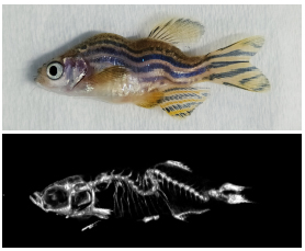

 (5 votes)
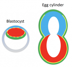


 (12 votes)
 In the latest episode of Genetics Unzipped,