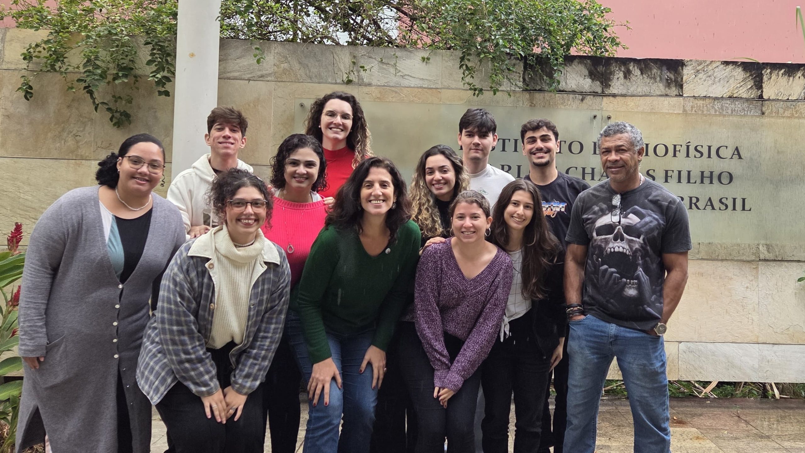
Lab meeting with the Silveira Lab
Posted by the Node, on 24 June 2025
This is part of the ‘Lab meeting’ series featuring developmental and stem cell biology labs around the world.
Lab introduction
Mariana S. Silveira: I am the head of LINDes, my current laboratory established in 2023. Prior to that, I shared another research space, the Neurogenesis Lab, with colleagues and my former advisor, Dr. Rafael Linden. I held the position of Associate Professor at the Institute of Biophysics Carlos Chagas Filho, part of the Center for Health Sciences at the Federal University of Rio de Janeiro, Brazil. Notably, this institution is the first university founded in our country, previously known as the University of Brazil, and our Institute is now approaching its 80th anniversary. In Brazil, this represents an esteemed and traditional institution, as our country is relatively young. Our Institute’s Program of Biological Sciences–Biophysics, one of the first doctoral programs in Brazil, has a history spanning 62 years.
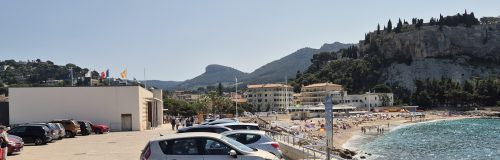
The lab is in Rio de Janeiro and our city boasts unparalleled natural beauty and a vibrant cultural scene.
Research summary
Our current research focuses on leveraging advancements in retinal development and cell reprogramming to explore potential therapeutic strategies for vision loss. Specifically, we focus on retinal ganglion cells, the projection neurons of the retina, working in collaboration with both Brazilian and international research groups. Additionally, we are investigating the retinal microglia in a collaborative project aiming to design innovative tools employing machine learning for morphological categorization.
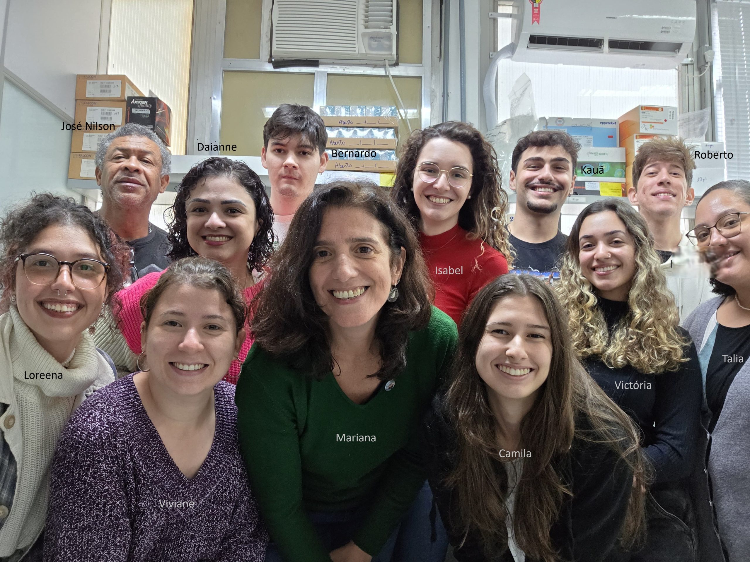
Lab roll call
José Nilson dos Santos – He’s been the go-to technician ready to help anyone in the lab ever since the lab head was a PhD student in the Neurogenesis Lab. Always there to tackle any kind of problem, it’s awesome to have him around!
Mariana S. Silveira – Associate professor and lab head. Honestly, leading this fresh and young team is both a challenge and a joy. My goal is to put all my energy into making sure they have a productive and happy experience in the lab.
Viviane Oliveira Valença – Postdoctoral researcher, Viviane serves as my right hand. Having recently completed her PhD; she is currently assisting in training the group and moving forward some new collaborations while finalizing manuscripts. Her presence is truly invaluable to the lab.
Daianne Torres – A combination of lab manager and technician, Daianne is consistently available to address both administrative and technical challenges.
Isabel Guedes Ferreira – A highly skilled Master’s student currently focusing on the study of retinal microglial morphology and the development of computational tools to enhance and optimize these analyses.
Bernardo Benincá – He has recently started his Master’s program, where his research project focuses on stimulating the reprogramming of Müller glial cells in vitro through the overexpression of transcription factors.
Kauã Mourão – An undergraduate student in Biological Sciences who joined the lab as a trainee in 2024. He is currently exploring various techniques to evaluate the potential for reopening the window of retinal ganglion cell generation and the synaptic integration of newly formed neurons.
Victoria Mattos – An undergraduate student in Biomedical Sciences who joined the lab in 2024. She is currently acquiring the essential foundational techniques needed to initiate her own project. Presently, she assists other students with their work and participates in routine laboratory activities.
Camila Barbosa – An undergraduate student in Microbiology and Immunology who joined the lab in 2024 and is currently enhancing her technical skills.
Loreena Klein – A PhD candidate who has recently joined the laboratory after completing her Master’s in the field of pain regulation, is currently investigating the regenerative potential of Müller glial cells for the generation of retinal ganglion cells in vivo.
Roberto Matias – An undergraduate student specializing in Biophysics, Roberto contributes to Isabel’s research by assisting in the morphological analysis of microglial cells across various functional states and investigating their correlation with neurodegenerative processes.
Talia Pontes – An undergraduate student in Biomedical Sciences, has recently joined the laboratory with the intention of pursuing a graduate program in the future.
Favourite technique, and why?
Mariana: In my opinion, microscopy remains an invaluable tool, particularly with advancements in resolution and the ability to detect multiple antigens simultaneously. This approach provides precise in situ information when combined with cell morphology, defining in a remarkably accurate way, cell identity within tissue. Nevertheless, employing a combination of various methodologies is always the optimal strategy. Recently, I have also become fascinated by scRNA-seq as a very relevant tool.
Mariana, apart from your own research, what are you most excited about in developmental and stem cell biology?
Mariana: The emerging field of organoids and assembloids, particularly for investigating early stages of brain development and the underlying mechanisms of diseases, is truly captivating.
Mariana, how do you approach managing your group and all the different tasks required in your job?
Mariana: I must admit it’s not an easy task. My approach involves holding regular meetings with the team, where we not only discuss relevant literature related to our projects and related fields but also hold individual and group follow-ups to review the goals set for each member. Despite time being limited, I always keep my office door open for them.
What is the best thing about where you work?
Mariana S. Silveira – Although science funding in Brazil remains quite limited, which makes competing for international grants an essential challenge, I truly appreciate working at such a prestigious institution. Here, we are often supported and encouraged to strive for quality and excellence, especially in training the next generation of researchers. While the number of young individuals pursuing this demanding career is gradually declining, it’s rewarding to discover talented individuals and witness their scientific growth and development. Celebrating small achievements serves as a motivation to keep moving forward.
Viviane Oliveira Valença– We are at one of Brazil’s top universities, which is definitely the highlight for me, as numerous scientific contributions are made here. Even though we lack good infrastructure, safety, and other resources, being inside the university and surrounded by students from different fields fosters interaction and knowledge exchange.
Daianne Torres – The best thing about our workplace is the people who make up our lab. Even when we’re stressed about failed experiments, having supportive and caring teammates always makes the effort worthwhile.
Isabel Guedes – The best thing about our lab is the camaraderie and sense of community. We genuinely look out for each other, and that support makes even the hardest days a lot easier to get through.
Bernardo Benincá – It is the people. The scientific environment is challenging and can often be frustrating, but we take great pride in the quality of the work we produce despite numerous hardships and limited funding. Another important factor is how we always support each other, creating a welcoming and inclusive space. It’s an honor to be part of our lab.
Kauã Mourão – The people. They are not only my lab group with whom I talk about research and papers, but also my friends whom I know I can count on in this crazy work routine. It is really great to share my workspace with them because I learn more every day. They are truly skilled at what they do.
Victoria Mattos – It’s all about the connections we build with people and what we learn. Joining the lab helped me grow, both as a student and as a person. Being part of this environment and contributing to our research is very rewarding.
Camila Barbosa – The great thing about working in this lab is the constant exchange with the whole team, which always helps me learn something new, whether during experiments or in our meetings. Outside the lab, we have the privilege of being close to renowned professionals, and we get to attend various lectures and conferences, which also helps broaden our scientific perspectives.
Loreena Klein – The best thing about where I work is the strong sense of community. Everyone is very supportive and willing to help, which has made a big difference for me as a new PhD student adjusting to the environment.
Roberto Matias – I can say for sure that the best thing about where I work is the patient, dedicated and fun people who guide me in everything I need to learn, whether they are colleagues or teachers. The Institute is a peaceful and friendly place capable of comforting anyone in difficult times.
Talia Pontes – Honestly, the best thing about working here is getting to learn hands-on lab techniques from experienced researchers right at my own university. Plus, it feels great to know I’m helping push the boundaries of retinal studies.
What’s there to do outside of the lab?
Mariana S. Silveira – Besides enjoying the quiet pleasure of reading a captivating romance, I’m a big fan of Brazilian music. I enjoy attending live shows and participating in groups that use samba school instruments to explore the diverse rhythms of our rich musical culture.
Viviane Oliveira Valença – Here in Rio de Janeiro, there are plenty of things to do outside the lab, such as hiking in various places where you can enjoy breathtaking views of the city. Besides that, you can go to the beach, soak up the sun, and spend quality time with friends.
Daianne Torres – During my free time, I enjoy curling up on the couch with a good TV series to unwind or diving into the pages of a new book to escape into a world of fantasy.
Isabel Guedes Ferreira – Outside of the lab, I like to unwind by reading, playing games, and spending time with my friends. It helps me recharge and keep a healthy balance.
Bernardo Benincá – Sometimes it is important to take a break from the routine and do things to relax. Hobbies like watching movies, reading, playing video games, going out with friends, or simply going to the garden and looking at the trees.
Kauã Mourão – We are talking about Rio, so I must mention the beaches! They are amazing! There is nothing better than heading to the sea and unwinding from everything.
Victoria Mattos – Outside of the lab, I enjoy spending my time with my family and friends, it’s always a lot of fun to be surrounded by good people in a nice place. I like to go to the beach, enjoy samba and visit new restaurants.
Camila Barbosa – In my free time, I enjoy spending time with friends, going hiking, connecting with nature, watching series, and reading.
Loreena Klein – Outside of the lab, I love spending time with my friends, whether we’re going out or just hanging out together. I also enjoy staying home and watching some TV shows, which help me relax and recharge.
Roberto Matias – Rio de Janeiro offers many interesting activities, such as modern museums, restaurants with unique and exotic cuisine, and adrenaline-pumping amusement parks. With such a beautiful city full of activities and people, it is impossible to decide what to do.
Talia Pontes – Outside of the lab, I enjoy watching movies, reading, and spending quality time with my family. I also dedicate time to studying extracurricular subjects and learning new languages—currently, I am studying Spanish.
 (3 votes)
 (No Ratings Yet)