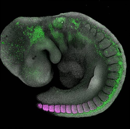
The Development and Transcriptional Control group is interested in defining how cellular diversity arises during development, and the roles that regulatory elements play in this process. By developing quantitative methods to probe cell identity, we are defining how diverse cellular outcomes are generated in the head versus the trunk of the embryo. For further details, please visit our lab website.
Your role and opportunities
You will be eager to work at the intersection of developmental biology and the latest technological advancements in genomics and genome editing. You will have the opportunity to train in a range of techniques including embryonic stem cells, mouse genetics, and computational approaches, with access to state-of-the-art core facilities and infrastructure. In addition, you will enjoy the freedom to develop and carry out your own research within the group’s area of interest.
How to apply
For further details about this role, and to apply, please examine the job description and complete the online application. Application deadline: Sunday, March 1, 2020.
For informal inquiries, please contact Vicki Metzis.
Our understanding of lineage decisions in early human development has been greatly aided by embryonic stem cell lines, which avoid many of the practical and ethical difficulties of in vivo material. A new paper in Development exploits naïve human embryonic stem cells to generate in vitro models for the extra-embryonic endoderm. We caught up with first authors Madeleine Linneberg-Agerholm and Yan Fung Wong, and their supervisor Josh Brickman, Professor of Stem Cell and Developmental Biology at the Novo Nordisk Foundation Center for Stem Cell Biology (DanStem) in Copenhagen, to hear more about the work.
Yan Fung, Madeleine and Josh (L-R)
Josh, can you give us your scientific biography and the questions your lab is trying to answer?
JB Since the beginning of my PhD, I have been focused on the transcriptional basis for cell identity. Following a brief foray into the music industry as both a DJ and journalist, I began a PhD under the guidance of Mark Ptashne at Harvard University, where I worked on general mechanisms of transcriptional synergy and cooperativity. By the end of my PhD, I felt the need to take this work into a more biological context. To this end, I trained as a post-doctoral fellow with Rosa Beddington at the National Institute for Medical Research in London, where I began to work with a combination of early embryos (mouse and frog), and embryonic stem cells (ESCs), to explore the means by which transcription controls anterior specification. In 2001, I started my own group at the Institute for Stem Cell Research (now the MRC Centre for Regenerative Medicine), University of Edinburgh, where I used a combination of ESC models and early embryos to deconstruct the transcriptional basis for lineage specification and potency, focusing on endoderm induction and patterning. In those early years, we used Xenopus embryos with parallel experiments in ESCs as a rapid means to understand conserved mechanisms regulating both pluripotency and differentiation. However, with time, my lab has unfortunately lost touch with its amphibian roots as the lure of stem cells became too much for my students to resist.
One of the most important observations we made in those early years was that ESCs could be used as means to model early development in the primitive endoderm and to trap spontaneously arising transcriptional states in which cells were reversibly and functionally primed for differentiation. This led us to the notion that self-renewing cell culture models could be used to trap intermediate, or uncommitted, transcriptional states in differentiation. We see these states as analogous to transition states for lineage specification, and we have used these models to identify mechanisms governing these reversible transcriptional changes. At the same time, we also began to view karyotypically normal, embryo-derived cell culture as a means to trap decision points in differentiation with a capacity for proliferation. We exploited this idea as a way to isolate and expand lineage-restricted progenitors from differentiating ESCs in both the definitive and later primitive endoderm lineages.
In 2011, my lab relocated to the DanStem at the University of Copenhagen, where we continue to focus on the transcriptional basis for cell fate choice. In particular, we’re interested in the basic mechanisms regulating transcriptional heterogeneities in early embryos and differentiation, how gene regulatory networks can be used to explain the differentiation competence and self-renewal of stem and progenitor cells, and how transient transcriptional states become committed in differentiation. Of course, a number of these questions concern the interface of gene regulatory networks with signalling and this has been a major focus of our recent work, including this new paper.
Before concluding, I think I should tell a short story about the origin of this work. About ten years ago, we had some translational funding to apply our work on mouse endoderm differentiation to human ESCs. I used this money to support a student (Maurice Canham) who was finishing his work in the lab on mouse primitive endoderm priming and wanted to take on this translational project. At the time, all the available human cells were primed pluripotent cells. While he was adapting our culture conditions to human ESCs, he decided to dump human endoderm differentiation media on mouse ESCs and see what happened. He observed this remarkably homogenous differentiation to a cell type he thought resembled a slice of pizza and, therefore, referred to them as pizza cells. At the time we were convinced that ‘pizza cells’ were probably primitive endoderm, but it took another PhD student (Kathryn Anderson) years to prove this was the case, to test the activity of these cytokines side by side on primed and naïve cells, and to work out the conditions for the passaging of naïve extra-embryonic endoderm (nEnd). Years later, naïve human pluripotent cells became available, and we were finally able take this work back to the human cells that it started with.
Madeleine and Yan Fung, how did you come to work with Josh and what drives your research today?
ML-A Although originally from Denmark, I did my undergraduate degree in the UK. I became really interested in early mammalian development as a result of my bachelor’s thesis project in Ryohei Sekido’s group at the University of Aberdeen, working on Y-linked sex-specific epigenetic modifications in mouse ESCs. After four years abroad, I got homesick, but luckily found Josh’s group in Copenhagen, and was able to return to begin a master’s degree under his supervision. In Josh’s group, I was trained by Fung who became my day-to-day supervisor and taught me hESC culture. I was quite fortunate to join when I did, as it was an exciting time both for the group, as they were about to publish the story of the context dependence in mouse endoderm differentiation (Anderson et al., 2017), and also in the field, as a number of human naïve ESC papers had recently come out. I think what drives my research today is trying to fill in the most fundamental steps in human development and reconcile what we know in other species with ourselves.
YFW I finished my PhD in Hong Kong where I studied gene regulatory networks and organ patterning using C. elegans as a model organism. I then applied to work with Shinichi Nishikawa at the RIKEN Center for Developmental Biology, where I used human cell lines and primary cells as disease models to study epigenetic regulation. During that time, I had the chance to meet stem cell biologists from all over the world, including a former PhD student of Josh’s, Kathryn Anderson (one of the authors in this paper), and she convinced me to think about going to his lab. I then applied to Josh’s lab and met with him in Washington DC, after which he encouraged me to visit the lab in Denmark. In 2013, shortly after the group had relocated from Edinburgh to Copenhagen, I came to visit the new centre, DanStem. Attracted by the passionate people in the group, the newly established research institution, and Josh’s impressive work using stem cell culture systems as models to understand the transcriptional basis for lineage choice, I joined the lab.
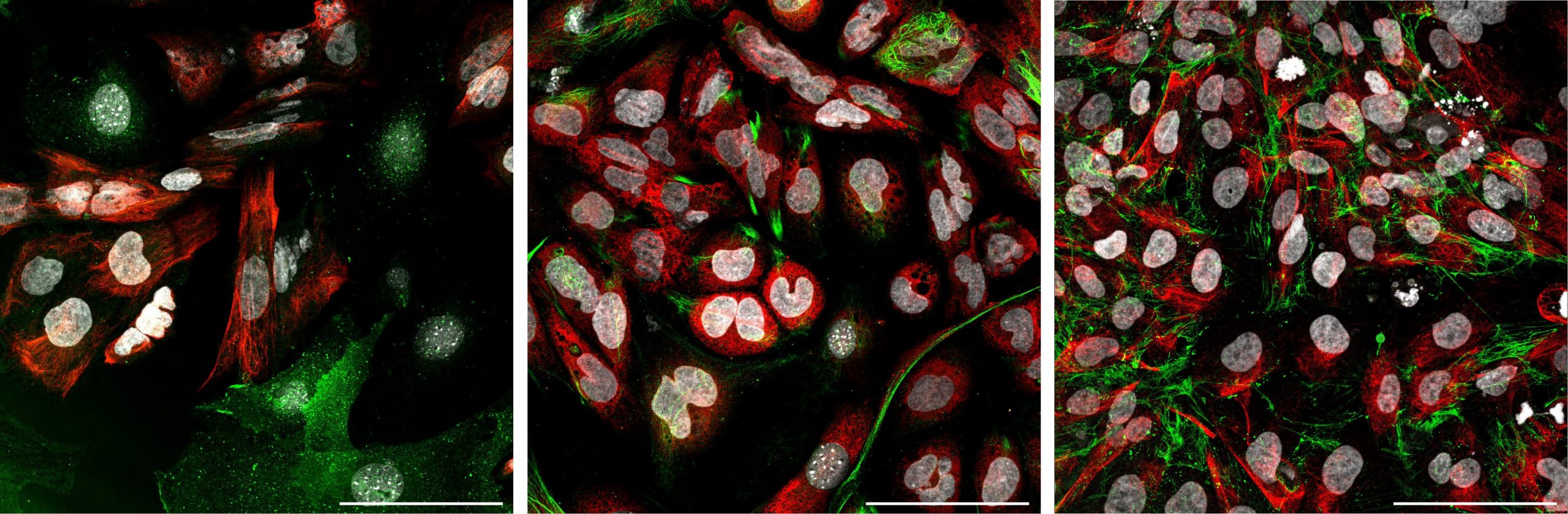
Immunofluorescence of human nEnd stained for endoderm and basement membrane markers. Left: E-cadherin (green), vimentin (red), GATA6 (white). Middle: AFP (green), collagen IV (red), GATA6 (white). Right: fibronectin (green), vimentin (red), GATA6 (white).
What makes endoderm induction in the mouse context dependent, and before your study what was known about its conservation in humans?
JB, ML-A & YFW We believe that the context dependence we originally saw in mouse was determined by changes in the enhancer landscape between naïve and primed pluripotency. The interaction of Wnt and Nodal-related TGFβ signalling with the set of enhancers primed in these cell types would determine the trajectory of differentiation. In our mouse nEnd paper (Anderson et al., 2017), we found that there was a correlation between enhancer accessibility and definitive endoderm versus primitive endoderm lineage differentiation. This is a remarkably similar idea to our recent thoughts on specificity of FGF/ERK signalling (Hamilton et al., 2019). Here, we found that ERK directly regulates enhancers, but that the activity of ERK on enhancers is likely to depend on pre-bound transcription factors, that don’t in themselves activate transcription, but prepare the available differentiation trajectories a cell can take when exposed to a signal. We believe a similar mechanism must be at work here with respect to endoderm enhancers that respond to Nodal/Wnt signalling in either naïve or primed pluripotency, with these pathways acting on different pre-wired transcriptional circuits that are stimulated by the same signalling pathways, but in different pluripotent states.
At the time we started, it was known that it was possible to culture human naïve cells and that their culture was usually dependent on FGF/ERK inhibition. However, the role of FGF/ERK described extensively in mouse primitive endoderm and epiblast segregation appeared not to be conserved in human embryos. As we believe that inhibiting primitive endoderm differentiation was a primary function of ERK in naïve ESC culture, we wondered how one could reconcile these observations.
Can you give us the key results of the paper in a paragraph?
JB, ML-A & YFW We found that the context dependence we observed in mouse, in which activation of Wnt/Nodal and LIF signalling could promote lineage-specific endoderm differentiation (i.e. primitive versus definitive) based on the developmentally proximal state of the starting culture, was conserved in human. Thus, human naïve pluripotent cells, which resemble the pre-implantation embryo, differentiated to primitive endoderm in response to these pathways, whereas primed pluripotent cells, which resemble the pre-gastrulation-stage epiblast, gave rise to definitive endoderm. We were then able to use this primitive endoderm differentiation model to show that the role of FGF/ERK in specifying this early lineage, at least in vitro, was conserved. Importantly, we were able to establish conditions for the expansion of these in vitro-derived cells to establish a culture/stem cell system for human hypoblast (as the human primitive endoderm is known). As trophoblast stem cells have recently been produced in human and naïve ESCs are thought to represent epiblast, this new culture system means that there are now human cell lines/in vitro models for all three lineages of the blastocyst.
What changes between the naïve and primed states to direct what kind of endoderm ESCs can give rise to?
JB, ML-A & YFW This was discussed above with respect to mouse. We believe it is the gene regulatory network in these different states that provides that platform on which the signalling pathway acts. The transcription factors expressed in these different stages of pluripotency could be sitting on distinct enhancers preparing cells to adopt different trajectories of differentiation in response to the same signal. It’s as if the transcription factors are laying down a road along which the cells can progress in differentiation in response to these signals. When cells transition from naïve to primed pluripotency the road is diverted and signalling pushes cells in this new direction.
What pressing questions do you think your nEnd cells will be particularly suitable for addressing?
JB, ML-A & YFW We think these cells will be particularly useful for the study of human primitive endoderm patterning and differentiation. They will be an excellent tool for studying how regulatory networks become stabilised in self-renewal in the endoderm and how these can then initiate patterning. As nEnd represents the third cell type from the blastocyst, they will also be very useful in experiments designed to determine the self-organising properties of early embryonic cells in order to generate embryoids. Finally, they provide a system in which to study the differentiation of the primitive endoderm and understand how it compares to the definitive endoderm.
When doing the research, did you have any particular result or eureka moment that has stuck with you?
ML-A For me, there were three moments that really stood out for different reasons. The first one was when I saw the first naïve colony after chemical resetting from primed hESCs. It was the first ‘big’ experiment that I did, both on this project and also in my time in the group, so that was a big moment of success for me. The second was when I saw the first patch of primitive endoderm after my first ever differentiation from naïve hESCs. That was when we knew the project was going somewhere, and it was likely that endoderm specification between mouse and human was conserved. The third was when we figured out the expansion conditions for primitive endoderm to make nEnd, which I was stuck on for easily half a year. When the expansion worked, I started to see the future potential of what I was doing beyond this paper and all the exciting experiments that it could lead to.
YFW Expansion, and the excitement of getting expansion working!
When the expansion worked, I started to see the future potential of what I was doing beyond this paper
And what about the flipside: any moments of frustration or despair?
ML-A A lot! It was really challenging having to learn the most basic aspects of doing research at same time as having such an ambitious project, from cloning and doing my first RT-qPCR to learning R (thank you, Stack Overflow). But I think that just made it all the more rewarding, or at least that’s how I feel now.
YFW When I found out that I could not detect HHEX expression in differentiating primitive endoderm from human naïve ESCs, I thought we had a problem with the cells. However, based on the single cell transcriptome data on the human blastocyst it turned out to be true.
So what next for you two after this paper?
ML-A I graduated with my master’s degree this summer and now I’m taking a year ‘off’ working as a research assistant in the lab. My plan is to start my PhD with Josh next year. I am continuing with human nEnd projects, but I’ve started working with mouse endoderm as well, as it offers a whole new world of experimental possibilities.
YFW Besides this work, I am finishing other projects related to foregut endoderm expansion and differentiation to visceral organs, including pancreas and liver. The main focus is to understand how extrinsic signals influence transcriptional networks or chromatin accessibility. I am interested in how these networks impact the choice these progenitor cells make between self-renewal and lineage specification. I hope this work will bring us one step closer to understanding human embryonic development and perhaps translating this knowledge into strategies for regenerative medicine.
Where will this work take the Brickman lab?
JB As a lab we are very excited about these cell lines. We are excited by the potential of exploiting nEnd to explore the self-organising properties of human primitive endoderm, both on its own and when recombined with other cell types. We are also excited about using nEnd as a model to understand human visceral endoderm patterning.
Since I first started my lab, I have worked on gastrulation-stage endoderm patterning and using ESC differentiation as a model for this. While we have just begun this sort of work in human models, nEnd will complement them nicely. We are looking forward to using these cells to explore ‘extra-embryonic’ endoderm differentiation in human.
Finally, the in vitro model we describe here for human primitive endoderm differentiation will provide us with an excellent platform to collect evidence for our ideas about signalling context. How does the enhancer state or gene regulatory network in naïve and primed pluripotency determine signalling response?
Finally, let’s move outside the lab – what do you like to do in your spare time in Copenhagen?
ML-A Just like in the UK, it definitely depends on the weather. If it’s nice, I like going for walks with my dog in a forest north of Copenhagen called Dyrehaven, which is actually a UNESCO World Heritage Site. On rainy days, I like to try and find the best ramen place in Copenhagen (currently Ramen To Bíiru) or stay at home watching ‘90s rom-coms and playing video games.
YFW Hygge with family and friends, discussing the big and small things in life.
We are pleased to announce a call for applications for a post-doctoral researcher position in the lab of Professor Jonathan Chubb in the MRC Laboratory for Molecular Cell Biology (http://www.ucl.ac.uk/lmcb/research-group/jonathan-chubb-research-group). To understand how cells decide their fates, during development and reprogramming, the lab develops and implements powerful new technologies to directly monitor gene activity in single cells (eLife e13051, Curr Biol 27:1811-17, PNAS 115:8364-8369). This position is an ideal platform for developing an independent research career in a rapidly expanding sector of the life sciences.
Candidates are expected to be exceptional, highly motivated scientists with a strong track record of research in a relevant area of the life sciences. We will also consider applicants with a PhD in physics, engineering, mathematics or computer science with a strong interest in biology.
Work will be carried out at the MRC Laboratory for Molecular Cell Biology http://www.ucl.ac.uk/LMCB/. The LMCB is a focal point for molecular, cell and tissue biology in the UK and is situated in the main UCL campus, in the heart of central London.
Please contact j.chubb@ucl.ac.uk for informal enquiries. Deadline 28th February. Apply using this link:
How do trees find their sense of direction as they grow? Researchers are getting to the root — and the branches — of how the grandest of plants develop.
By Rachel Ehrenberg
There’s a place in West Virginia where trees grow upside-down. Branches sprout from their trunks in the ordinary fashion, but then they do an about-face, curving toward the soil. On a chilly December day, the confused trees’ bare branches bob and weave in the breeze like slender snakes straining to touch the ground.
I’m visiting an orchard at the Appalachian Fruit Research Station, an outpost of the US Department of Agriculture nestled in the sleepy Shenandoah Valley. Here, at Dardick’s workplace, the disoriented plums are but one in an orchard of oddities, their outlines, seasonally stripped of leaves, standing out in stark relief.
There are trees with branches that shoot straight up, standing to attention in disciplined rows, with nary a sideways branch. There are trees with branches that elegantly arch, like woody umbrellas; others with appendages that lazily wander this way and that.
Dwarf trees crouch, sporting ball-like crowns akin to Truffula trees. Compact “trees” poke from the ground in clumps of scraggly, knee-high sticks. Apple trees with some hidden predicaments grow in a greenhouse nearby: Their roots reach sideways rather than down. The topsy-turvy growth of all of these trees comes from genetic variations that cause the dialing up, dialing down or elimination altogether of the activity of key genes controlling plant architecture.
Understanding these misfits has real-world applications: It could help grow the next generation of orchards that, densely packed with trees, produce more fruit while using less land and labor than today. But Dardick is also trying to answer a fundamental question: How do different trees get their distinctive shapes? From the towering spires of spruce and fir, the massive spreading limbs of an oak to the stately arching canopies of an elm, the skeletal shapes of trees offer signature silhouettes.
Dardick’s work and that of other researchers also could help to explain how the shapes of individual trees are far from fixed. Trees, much more than we can, will morph in response to their literal neck of the woods. Limbs in the shade reach toward spots of sunlight. Trees on windswept hills bend trunk and branches into gnarled architectures.
Work by breeders, biologists and botanists have revealed sizable pockets of knowledge about the hormones, genes and processes that yield the diverse shapes of trees and other plants, between species and within species. It has not been easy: Two of trees’ most appealing attributes — their long lives and large sizes — make them intractable research subjects.
But as scientists pursue these questions, commonalities are emerging between vastly different species. The puzzle of shape diversity and adaptability turns out to be tied to the fundamentals of being a plant: grappling with gravity, fighting for sunlight, all while anchored in one place for a lifetime.
“Plants are stuck. The best they can do is grow toward something,” says Courtney Hollender, a former postdoc of Dardick’s who now runs her own lab in the Department of Horticulture at Michigan State University in East Lansing. “That’s all they’ve got; they can’t run, they have to adapt to their environment. And they’ve developed brilliant ways to do it.”
Available at all branches
Scientists have a word for the ability to adapt so readily: plasticity. In plants, this feature is both obvious and astounding. Most animals are born in specific shapes then just grow larger, but plants are modular — they grow in various iterations of two building blocks: shoots and roots.
It is the first of these — where and when a shoot grows or doesn’t grow — that governs the basic form a tree takes.
Some aspects are hardwired. Leaves emerge in a pattern that is usually fixed throughout the tree’s life, with structural arrangements that tend to be shared by members of a given plant family. And shoots emerge where leaves meet the stem. So, for example, plants in the maple family, which have leaves set opposite each other, have branches in the same format. Members of the beech family have leaves, and thus branches, that alternate up the stem.
But the interplay between physiology and external forces also plays a large part. Take your standard-issue plant with a main central stem that grows upward and has few side branches. Most plants, from basil to birch, start out this way, a growth habit that probably evolved because it enables them to quickly reach the light — more rapidly than the competition. Called apical dominance (the tip of the plant is the “apex”), this is largely under the purview of the plant hormone indole acetic acid, also known as auxin. Made in the tip, auxin diffuses downward and blocks the growth of side branches.
This is why pinching the tips off of basil or geranium makes them bushy — you are removing the source of that bossy auxin, freeing buds on the stem’s sides from the prohibition and allowing them to grow. (Though auxin is mighty, it’s not the only player here. Other plant hormones, along with light intensity and access to nutrients, also wield power.)
Another related and less-understood phenomenon occurs in some tree species. Called apical control, it also is imposed by the tip of a tree and probably also by auxin. But rather than operating at the scale of a branch, it commandeers the whole dang tree.
Think of a pine. At the top, there’s a pointy tip, then upper branches that tend to reach skyward. Moving down, the branches become more horizontal, growing out more than up. But unlike a basil plant, a pine tree does not become bushy when you lop off the top. Instead, a new bud near the top grows upward, becoming the new leader. Or an existing branch reorients to grow up and become the new dominant tip.
These two principles are always in the back of arborists’ minds as they work. “They have to consider, ‘If we cut a branch here, that bud below is going to break and we’ll just get a branch in basically the same spot,’” Dardick says. “All of their rules of what to prune and where are based on these physiological factors that contribute to tree shape.”
A natural reaction
Physiology also underpins the plastic responses trees have to more extreme situations they may face. A tree on a high mountain peak or windswept coast must contend with exposure to mechanical forces that could topple and kill it. To survive, such trees become short and stocky, their bent, asymmetric crowns reducing drag and presumably protecting a tree from violent gusts. The driver is the wind’s very touch — a response now called thigmomorphogenesis that has been observed for hundreds of years.
How it works is still unclear, but over the past decade researchers have made some headway. They’re actively studying force-sensing proteins and processes that may be involved. And recent work suggests an important role for hormones such as jasmonate, which accumulates in all kinds of plants in response to damage and mechanical stress. In experiments with a weedy mustard called Arabidopsis, plants became stunted when researchers bent their leaves back and forth twice a day. Mutants that couldn’t make jasmonate, though, grew normally.
Sometimes, wind does more than gust against a tree: It blows the whole tree over, and that tree, if still rooted, must reorient the growth of its branches and buds toward the sky. Avalanches, erosion and landslides deal similar fates. And trees in all sorts of circumstances must grow around obstacles, away from competitors and toward the light. To get these jobs done, trees make a special kind of wood called reaction wood.
Trees may become contorted in challenging physical environments, such as this ridge in the Rocky Mountains. The touch of wind and other forces prompt physiological responses by the plant that yield a shorter, stockier stature, gnarled asymmetric shape and the development of specialized wood. This characteristic tree form is called a krummholz (German for “crooked wood”). CREDIT: BRYCE BRADFORD / FLICKR
Hardwoods such as maple, beech, oak and poplar form this tough stuff (in this case called tension wood) on the upper side of their stems. Incredibly, it creates a tensile force that pulls the stem upward. “If you walk around the woods, you can see that most species, if not all species, have this kind of reaction wood response,” says Andrew Groover, a research geneticist with the USDA Forest Service’s Pacific Southwest Research Station in Davis, California.
The hardwood tree first discerns that it is off-kilter using specialized gravity-sensing cells. Where these cells reside in trees — the woody stem? the tip of new shoots? — was unknown until Groover and colleagues detected them in woody and soft tissues of poplar, a few years back. The cells contain organelles called statoliths that sink down in the cell and indicate to the plant that it’s leaning one way or the other. This, in turn, causes that influential auxin to mobilize, triggering the growth of tension wood on the top. Cellulose with a peculiar gelatinous layer is thought to act as the “muscle” that generates the pulling-up force.
In this experiment, young, potted poplar trees were placed sideways to investigate the plants’ gravity-sensing machinery. The poplar in this time-lapse movie, taken over two weeks, responded to being tipped on its side by reorienting its growth upward. The plant hormone auxin is key to this response. Mutants that cannot respond appropriately to auxin’s signaling instructions do not right themselves this way. (This particular poplar also received a dose of a chemical called gibberellic acid that interacts with auxin, so that scientists could learn more about its role.) CREDIT: ANDREW GROOVER AND SUZANNE GERTTULA, US FOREST SERVICE, PACIFIC SOUTHWEST RESEARCH STATION DAVIS CA
When genes defy gravity
Much of the knowledge about the architecture of plants is rooted in millennia of human efforts to alter crop shapes to make them more suitable for cultivation, and modern science is now revealing the genetic changes that lie behind these creations. The lessons, it turns out, apply broadly across the plant kingdom, to herbaceous and woody species alike.
It is hard to overstate the importance to human history of some of these plant-shape changes, says plant molecular geneticist Jiayang Li, who details some of their genetic underpinnings in the Annual Review of Plant Biology. A classic example is the transformation of the ancestor of corn (maize) into a key staple crop for much of the world. It arose from a species of the Central American grasses called teosintes — bushy plants with many branches. Domestication, among other things, abolished that branching, yielding the single-stalked upright corn we plant today.
Similarly, explains Li, who works at the Chinese Academy of Sciences’ Institute of Genetics and Developmental Biology, the green revolution of the 20th century ushered in compact, dwarf varieties of wheat and rice . By modifying the height and thickness of the stems of these grasses, breeders developed varieties that could carry more grain without toppling over in wind and rain.
Much of Li’s own research has focused on architectural variation in rice, although the work turns out to have implications for the architecture of plants in general, from lowly mosses to towering trees. Like other grasses, rice grows shoots called tillers — specialized, grain-bearing branches that emerge from the base. In cultivated rice, the angle at which these tillers grow varies widely: Some varieties are squat and wide-spreading, others have shoots that are more upright. Breeders are interested in altering tiller angle because upright plants can be grown more densely, giving farmers more bang for their acreage.
In a key advance, in 2007, a team including Li reported they’d discovered the genetic cause of the spread-out architecture trait. The scientists named the responsible gene TAC1, short for “tiller angle control.” A functional TAC1 gene increases rice’s tiller angle, leading to open, widely branching plants. Mutations in TAC1 lead to the opposite: plants with erect shoots that reach up, instead of out.
That same year, Li’s team and a group in Japan both reported another major achievement: finding a long-sought gene behind a curious trait in some rice varieties that gives plant branches a scruffy, lounging look. The trait, known as “lazy,” had intrigued plant breeders and geneticists since the 1930s, when researchers described its extreme manifestation in corn: “The lazy plants grow along the ground, following the unevenness of the surface.”
The cause, it turns out, was errors in a gene that normally makes branches shoot straight up. Li and his colleagues surveyed some 30,000 mutant rice plants to pin down that gene, now called LAZY (names of genes, confusingly, often refer to what happens when a gene is mutated and doesn’t work, rather than when it is functioning properly). And they provided convincing evidence for an idea batted around for decades — that lazy plants have muddled perceptions of gravity and that auxin is centrally involved.
A common test for whether a plant’s gravity-perception machinery is working is to lay the plant on its side. If it knows up from down, it won’t continue to grow sideways, but will start to grow up again, akin to the reaction-wood response of a toppled tree’s branches. An important step in this reorienting involves auxin pooling on the bottom side of the shoot. But in lazy mutants, proteins that help ferry auxin around the plant are malfunctioning, so instead of shoots growing in the correct direction, they’re prone to casually sprawl about.
Scientists now know that LAZY genes come in multiple versions. Some appear to operate in plant roots, telling them which way is down, probably using similar, auxin-related signals. If those genes are absent or inactive, confused roots grow upward. And though the genes were first found in monocots, a branch of the plant kingdom including rice and corn, researchers now know that LAZY genes exist in numerous plants, including the plums growing in the fruit research station in West Virginia.
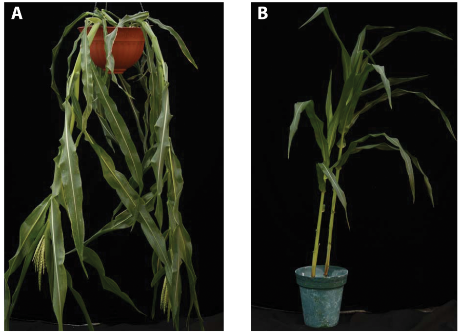
A lazy mutant of corn (left) compared with normal corn (right). Such corn mutants were described nearly 100 years ago, but it took 21st century molecular biology to nail down the growth habit’s cause: genetic malfunctions that meddle with responses to gravity. CREDIT: T.P. HOWARD III ET AL / PLOS ONE 2014
Reaching upward and outwards
As our boots crunch along the uneven ground, Dardick points at an errant orchard cat watching our tree tour from a distance. One row of trees stands so upright that a fencepost at the end of it is enough to block the row from view. These regimented trees are “pillar” peaches, and they are favorites of landscapers (one reason: it’s easy to get around them with a lawnmower). They also were key to uncovering genes like LAZY and TAC1 at the Shenandoah Valley station.
By comparing ordinary peaches to pillar peaches, and drawing on decades of work by former lead scientist Ralph Scorza, a team of station scientists and others in the US and Germany discovered the cause of the pillar trait: mutations in the peach version of TAC1.
The team also found that LAZY was at work in many of their misfits. Just as with the corn plants described nearly 100 years ago, mutations in LAZY made plums grow topsy-turvy, their branches seeking the soil. Apple trees with LAZY mutations have similarly disoriented roots. And when multiple copies of LAZY genes malfunction in the weed Arabidopsis, its roots grow up, its shoots down.
In the last decade, researchers have found that TAC1 influences branch angle in plums, poplar trees, the grass Miscanthus and Arabidopsis, and it appears to affect leaf angle in corn. But LAZY genes have even deeper roots. They’re found in all manner of plants, including the evolutionarily older Loblolly pine and even more ancient mosses.
This finding suggests a very old role for LAZY: It may have allowed plants to grow up, literally, when they first colonized land. Plants got their start in water. There, rootless and leafless, they were buoyed, unconcerned with gravity. The transition to land spurred the development of proper roots and stems, and plants then had to figure out up from down. LAZY seems to have allowed plants to orient their above-ground growth away from gravity and up toward the sun.
Scientists think that TAC1 evolved somewhat later, providing a counterpoint to LAZY — ensuring that branches don’t only grow straight up, but also reach out. Together, these genes laid critical groundwork for the diversity of plant forms we see today, all seeking sustenance in their own ways.
“Once you start to grow up as a vascular plant, you need to maximize your resources, you need to capture as much sun as possible,” says Hollender, who has been working on yet another gene, called WEEP, that — when nonfunctional — lends plants a weeping, waterfall-like structure seen here and there in trees of ornamental gardens. (But it’s probably not responsible for the shape of weeping willow trees.) “Modifying your shoot angles is an important adaptive trait for plants that allows them to capture light. It’s essential for them to survive.”
This kind of research has broad economic implications. Fruit and nut trees bring $25 billion annually in the US alone and there are hefty costs associated with pruning, bending and tying branches; spraying hormones; and the manual labor of picking fruit from an unruly cacophony of limbs. Understanding the genetic controls behind tree architecture could help scientists breed trees that make the whole fruit-farming enterprise more efficient and environmentally friendly.
“Orchard systems are not the most sustainable in the world,” Dardick says. “The idea is, if we can modify tree architecture, if we could reduce their size and limit the amount of area they take up, then we could plant them at higher density and potentially increase their sustainability.”
And there may be odder outcomes than friendlier outdoor orchards: In collaboration with NASA, the USDA team is investigating genetic tweaks that might even help bring fruit to space. On that December day, Dardick takes me to a greenhouse tucked in a corner of the lab. In it are plum and apple trees whose shape is so transformed that they look more like the love children of shrubs and vines. This strange growth habit is a side-effect of efforts to breed plants that flower and make fruit sooner and then do so continuously, rather than flowering after growing for several years, and then only in the spring.
The genetic tweaks that sent the trees’ developmental program into overdrive have also transformed their architecture. In the greenhouse, these precocious “trees” sprawl, draping lazily along wire trellises, happily flowering and heavy with fruit. “They’re growing almost like tomatoes,” Dardick says. “So we’re broaching the concept of, can we bring an orchard indoors?”
Those ambitions aside, Dardick has his hands full trying to answer numerous basic-science questions about how trees do what they do. Researchers still don’t know how different tree species set the angles of their branches — going wide like an oak, or arching like an elm. They don’t know how trees alter those angles during the course of mature growth, as branches sprout from branches sprouted from branches, until some of them finally point down. Trees are both kindred and foreign to us, their various forms so familiar, but their architectural rules still in so many ways opaque.
“I find myself looking at trees all the time now in a new way; they fill space so beautifully and efficiently,” Dardick says. “They are the biggest organism we have that’s visible, that’s in our face all the time. But there’s so much we don’t know.”
10.1146/knowable-013120-1
Rachel Ehrenberg is the associate editor of Knowable Magazine.
This article originally appeared in Knowable Magazine, an independent journalistic endeavor from Annual Reviews. Sign up for the newsletter.
The Institut de Génomique Fonctionnelle de Lyon (IGFL) is a unique scientific environment. Teams investigate basic research questions at the interfaces of development, physiology and evolution. The main focus is integrative, organism-level research on animals.
We have an opening for an independent group leader (junior or established) and encourage talented scientists leading research falling within our scientific scope to apply. As we’re seeking to increase the number of women team leaders we will particularly welcome applications from women scientists.
The deadline to submit an application is 11 April, 2020.
DanStem is seeking a highly motivated and self-starting Cell Culture Specialist.
Responsibilities: The Cell Culture Specialist will manage the Stem Cell Culture Platform (https://danstem.ku.dk/platforms/stem-cell-culture/) at DanStem, a self-contained shared-resource facility dedicated for the maintenance of experimentation with mouse and human stem cells. Specific responsibilities include:
Work closely with DanStem scientists, platform specialists, laboratory manager and management to maintain and develop the cell culture laboratory facility and services
Introduce, maintain and enforce clear operating guidelines and safety in GMO1 and GMO2 cell culture labs
Inform on and support stem cell culture activities for facility users, e.g., introduction to basic cell culture techniques, troubleshooting, safety considerations, aliquoting and preparing reagents, testing for mycoplasma, karyotyping, maintaining equipment, monitoring and refilling stocks, defrosting freezers, and resolving conflicts between users
Order materials, equipment and furniture, negotiate and follow-up with vendors and service providers, and handle invoices
Establish new service agreements, annual service of equipment, and equipment upgrades
Organize and maintain cell cryostorage and backup storage system
Evaluate operations and coordinate upgrades and repairs and follow-up, including communication with the building operations department and assessment of proposed repairs
Respond to alarms, providing assistance by phone or in person
Substitute for the DanStem Laboratory Manager in case of holidays or illness
Qualifications, competences and experience:Candidates are expected to have at least a Master’s degree in natural or health sciences. A PhD degree is advantageous. In addition, we are seeking a candidate who has experience in embryonic stem cell and/or iPS cell culture, genetic manipulations and differentiations, and
Has a scientific background in developmental or stem cell biology, experience with embryonic stem cells and/or organoid culture as well as working in GMO1/2 laboratories
Has demonstrated success working in research service facilities; managerial experience in this or another type of organization is a strong advantage
Has experience in project management and a strong ability to prioritize and handle multiple tasks and frequent deadlines
Develops good relations in a multi-cultural research environment, and excellent oral and written communication skills in English
Works successfully with persons from a variety of organizations and professional levels
Is proactive, innovative, analytical, goal- and solution-oriented
Enjoys new challenges
Employment conditions: We proudly offer a stimulating, multifaceted and international environment of high scientific and societal impact; the possibility for continued education and training; collaborative and creative colleagues; and the opportunity to work with departments and centers at the University and greater community.
The employment can begin in Maj 2020 or upon agreement with the chosen candidate. The place of work is at DanStem, University of Copenhagen, Blegdamsvej 3B, Copenhagen. The position, at the University of Copenhagen, will be in accordance with the provisions of the collective agreement between the Danish Government and AC (the Danish Confederation of Professional Associations). To the salary is added a monthly contribution to a pension fund according to the collective agreement, and a supplement can be negotiated, depending on the candidate’s experiences and qualifications.
Inquiries are welcome to Executive Director Henrik Semb (henrik.semb@sund.ku.dk).
To apply
To apply, please submit your application, in English, via http://employment.ku.dk/administrative/ by clicking “Apply Now”. Applications must include a statement of motivation, curriculum vitae, and copies of relevant diplomas. Only complete applications in English, submitted online by the deadline will be accepted.
DanStem highly values diversity and encourages applications from people of all backgrounds.
The closing date for applications is March 15 2020
We are seeking a Postdoctoral Research Scientist to join research projects investigating the basis of neurodevelopmental disorders in the laboratory of Kristen Kroll at Washington University School of Medicine. We work in collaboration with Washington University’s Intellectual and Developmental Disabilities Research Center, (https://sites.wustl.edu/krolllab/cellular_models/), using directed differentiation of human pluripotent stem cells (embryonic stem cells and patient-derived induced pluripotent stem cells), mouse models, and a wide range of cellular, molecular, biochemical, and genomic approaches, to elucidate gene regulatory networks that control the specification and differentiation of specific human neuronal cell types, such as cortical interneurons. We are defining roles for transcriptional and epigenetic regulation in controlling these networks and identifying mechanisms by which their dysregulation contributes to neurodevelopmental disorders, including autism spectrum disorder and intellectual disability syndromes, and pediatric epilepsies. For additional information about our ongoing work and research interests, please see: https://sites.wustl.edu/krolllab/
Setting/Salary/Benefits: Our laboratory is in an academic setting in the Department of Developmental Biology at Washington University School of Medicine (St. Louis), an internationally recognized research institution with a dynamic research environment and extensive infrastructural and core facility support. Postdoctoral appointees at Washington University receive a starting salary based on the NIH NRSA guidelines and a generous benefit package.
Complete information on the benefit package is located on the WUSM Human Resources Benefits Website (http://medschoolhr.wustl.edu). The St. Louis area combines the attractions of a major city with family-friendly and affordable lifestyle opportunities (https://explorestlouis.com/).
Qualifications: Candidates should hold a PhD with preference given to applicants with a strong interest in and research training relevant to the areas of neural development, stem cell biology, and transcriptional or epigenetic regulation. Interested candidates should send a CV/names of references by email to kkroll@wustl.edu or by regular mail to Kristen L. Kroll, Washington University School of Medicine, Campus Box 8103, 660 S. Euclid Ave, St. Louis, MO 63110.
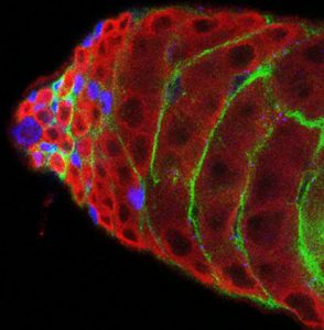
A PhD position is available in the lab of Fani Papagiannouli (@Medway School of Pharmacy, Universities of Kent and Greenwich) to study soma-germline communication and the role of cortical polarity in signaling regulation during Drosophila spermatogenesis. The position is funded by a Medway School of Pharmacy Post Graduate Research Scholarship.
Stem cells are critical for multi-cellular development, since they supply the cells that build up our bodies and replenish, as we age, worn out, damaged, and diseased tissues. Stem cell therapies use the power of stem cells to generate treatment for medical diseases by replacing lost or damaged cells. Due to the high degree of conservation to humans, Drosophila has led ground-breaking discoveries in genetics, cell biology, human disease and stem cell-related processes.
Research in Papagiannouli’s lab focuses on understanding how the communication between the germline and the squamous somatic cyst cells encapsulating them is established and maintained, in particular how the somatic cyst cells support the developmental decision of the germline in the Drosophila testis (1).
You will be integrated in the lab of an enthusiastic investigator who will support you to develop the skills required for your career development. We will employ a highly innovative proximity biotinylation assay coupled to mass spectrometry, along with cutting-edge array-tomography with scanning electron microscopy (AT-SEM) to elucidate the basic principles of squamous epithelial cell function and cross-communication with the germline. Excellent training in state-of-art Drosophila genetics, histological and molecular approaches, innovative genomic and proteomic techniques and high-resolution microscopy (1, 2), will help you investigate key stem cell and germline organizing principles. Studying the relatively simple somatic cyst cells will provide insights on the underlying causes that drive human squamous epithelia to develop squamous cell carcinomas. The regulatory strategies uncovered here will unravel fundamental mechanisms of stem cell function beyond Drosophila and will aid the identification of new approaches in regenerative medicine and infertility.
You will be part of a dynamic working environment at Medway School of Pharmacy and the Universities of Kent and Greenwich at Medway Campus, and will have access to shared facilities such as advanced microscopy and proteomic tools. You will also work alongside our world-renowned collaborators from Stanford University and the University of Lausanne with complementing expertise.
We look for an enthusiastic, talented and motivated student with the ability to work both independently and as part of a team that can quickly integrate into an interdisciplinary environment. The successful candidate should have knowledge on basic molecular and protein work, however experience in confocal microscopy, immunohistochemistry, genome wide and proteomic techniques or Drosophila genetics would be an advantage. Excellent writing and communications skills in English are necessary.
Papagiannouli, C. W. Berry, M. T. Fuller (2019), The Dlg-module and clathrin-mediated endocytosis regulate EGFR signaling and cyst cell-germline coordination in the Drosophila testis, Stem Cell Reports, May 14, 12: 1-17 (doi.org/10.1016/j.stemcr.2019.03.008)
Papagiannouli, L. Schardt, J. Grajcarek, N. Ha, I. Lohmann (2014), The Hox gene Abd-B controls stem cell niche function in the Drosophila testis, Developmental Cell 28(2):189-202
Apical tip of an adult Drosophila testis. The squamous somatic cyst cells (green; membrane-GFP) encapsulate the germ cells marked with Vasa (red). The nuclei of the stem cell niche (also called hub) and the nuclei of early somatic cyst cells are marked with Traffic Jam (TJ; blue).
Deadline for application is 31st of March 2020. The Scholarship is available to both UK and EU nationals. Self-funded applicants and those who have access to international scholarship applications, please contact directly Dr. Fani Papagiannouli (f.papagiannouli-227[at]kent.ac.uk).
As an equal opportunities institution we welcome applicants from all sections of the community regardless of gender, ethnicity, disability, sexual orientation and transgender status. All appointments are made on merit.
New research by the Serup group shows how the Notch signalling pathway works when the pancreas forms as the fetus develops. This discovery may lead to new opportunities to cure people with diabetes and understand how pancreatic cancer develops.
Imagine doctors in the near future being able to cultivate stem cells that turn into the insulin-producing beta cells in the pancreas – and then implanting these in people with diabetes to replace their damaged beta cells and thus cure them.
This dream has just come a step closer, after researchers from DanStem have revealed how a signalling pathway that guides the development of the pancreas works.
The discovery means that researchers now understand much better what they need to do to cultivate insulin-producing beta cells in a petri dish with the goal of curing people with diabetes.
“The interesting perspective is to take fetal stem cells and direct them to become insulin-producing cells. This requires knowing how nature does this normally, and we have come a step closer to understanding this,” says Palle Serup, Professor, Novo Nordisk Foundation Center for Stem Cell Biology, DanStem, University of Copenhagen.
Curing people with diabetes using home-grown beta cells
Phase 1 clinical trials around the world are already trying to cure people with diabetes by inserting laboratory-grown insulin-producing beta cells into people’s pancreases.
So far, the trials have been oriented towards ensuring that this procedure is safe, but the idea is to be able to cure the first people with type 1 diabetes within a few years.
Researchers from the Serup group are at the forefront of this, and leading researchers can also determine how to optimally improve the various procedures. This applies to the procedures the researchers use to develop the insulin-producing beta cells they implant in people.
The current laboratory-grown beta cells do not respond as well to glucose as they should, and the yield of the cultivation process is also relatively low.
“One reason is that we have not yet been able to fully replicate the natural process in the laboratory,” explains Palle Serup.
Current protocols do not exploit signalling pathways fully
Palle Serup and colleagues studied how the fetal pancreas develops. Many signalling pathways play a role in the process of inducing the stem cells to become the various cells of a pancreas.
These signalling pathways ensure that insulin-producing beta cells, blood vessels and the ducts that secrete digestive enzymes are produced where they are needed. The signalling pathways are communication tools between neighbouring cells, and the Notch signalling pathway that Palle Serup has now mapped is very important for the natural development of the pancreas.
“We did not know very much previously about this signalling pathway, and the protocols we use in cultivating pancreatic cells in the laboratory are therefore not very good at using the regulation of this pathway,” says Palle Serup.
Signal molecules oscillate
Notch has previously been linked to pancreatic development, and the new study explains this. The research shows that the concentration of the signal molecule DLL1 oscillates from high to low and back again, with a 45-minute interval per direction. Similarly, the oscillation activates the HES1 gene in the neighbouring cell, so the expression of this gene also begins to oscillate.
This is complicated, but Palle Serup’s research also shows that manipulating the oscillations causes the pancreas to grow more slowly. “This gives us insight into how the cells act when the pancreas is formed, and we have to recreate that activity in the petri dishes,” explains Palle Serup.
Several signal molecules guide pancreatic development
The research also shows that DLL1 is not alone in controlling pancreatic growth during fetal development. The signal molecule JAG1 also plays a role.
Both molecules target the same receptors on neighbouring cells, but DLL1 stimulates pancreatic growth by promoting cell division in neighbouring cells, whereas JAG1 inhibits growth.
JAG1 also plays a role in the paths the cells take in their development. All pancreatic cells originate from two small groups of stem cells that can develop into all the different types of pancreatic cells. During fetal development, cells develop in one direction or another. JAG1 influences the direction in which the cells develop.
When the researchers remove JAG1, too many cells develop towards cells that secrete digestive enzymes, and too few of the other types are formed. When JAG1 is present, a more appropriate number of the cells develop into insulin-producing beta cells.
To their surprise, the researchers could change the cell types by manipulating the oscillations. Attenuating the fluctuation in HES1 concentrations was equivalent to losing JAG1, whereas the opposite happened if the interval was increased from 45 to 60 minutes. “Our experiments showed that removing JAG1 or artificially inhibiting oscillations makes the pancreas develop almost no insulin-producing beta cells. This is important to know for growing pancreatic cells in the laboratory,” says Palle Serup.
Improving protocols for developing pancreatic cells
Palle Serup says that the researchers are already looking towards the next step in investigating the role of the signalling pathways in developing the pancreas. They want to confirm that these oscillations also occur in human pancreatic cells and not just in mice. Then they will investigate the extent to which they can manipulate the oscillations to control cell development.
Specifically, the researchers would like to accelerate the first cell divisions that lead to a fully developed pancreas. This will make the process in the laboratory more efficient when the cells divide more than they do today. Then the researchers will learn how to manipulate the individual steps in the process so that the finished product will resemble a natural pancreas as much as possible.
“Once the stem cells have become pancreatic cells, we need to determine whether we can make them divide more frequently and rapidly and become more normal types of cell compared with what is currently possible,” explains Palle Serup.
Discovery may also be relevant in cancer research
The research on pancreatic cells from mouse embryos also indicates new understanding of how pancreatic cancer develops. Pancreatic cancer is very rare, but the mortality rate is very high.
Researchers know from studies of people with cancer that the JAG1, DLL1 and HES1 signalling pathway is important in developing pancreatic cancer. This signalling pathway is shut down as the pancreas matures into adulthood, but among people with cancer, it is reactivated and causes uninhibited growth of cancer cells in the pancreas – and a hallmark of cancer cells is uncontrolled growth.
“Cancer may be caused by various mutations in components of this signalling pathway, and we have now begun collaborating with another researcher from the University of Copenhagen to try to understand how the signalling pathway specifically influences the development of pancreatic cancer. We do not know whether the oscillations play a role, but we will investigate these,” explains Palle Serup.
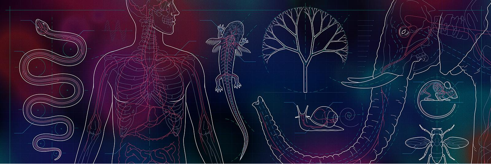
As editor-in-chief and executive editor of Knowable Magazine from Annual Reviews, we’re grateful for the invitation to write a post here at The Node about a special report on developmental biology — “Building Bodies” — that Knowable just published. We hope that the articles, written in accessible language, will intrigue and be of use to many of you.
Both of us started in research before taking a sideways step into journalism, and one of us (Rosie) became hooked on developmental biology early on: As a postdoc in the lab of UCLA and HHMI investigator Larry Zipursky in the late 1980s, she watched as the team there tracked down a key gene, bride-of-sevenless, involved in development of the Drosophila retina. (Her interest only grew after learning she had just one kidney, a developmental error that occurs in about one in 2,000 births.) It was a rare treat to fashion a package of dev bio articles all these years later. There were endless topics we could have chosen, and in the end we plumped for four in-depth feature articles focusing on body architecture topics:
As another part of the report, we wanted to touch on some older, key experiments in developmental biology. We considered presenting them in just that manner (“five seminal experiments,” or somesuch). But in the end, we decided to pose five questions of the kind that experimenters often had in mind when they did their work, and ones that a curious child might ask. Why is my heart more on the left? Why does my arm come out at my shoulders and not down at my waist? We’re grateful for the time and thoughts of Stanford developmental biologist Dominique Bergmann as we decided which questions to pick (there are zillions, and we had to limit them to five!) and made sure that a couple touched on newer focuses, such as the nascent field of systems developmental biology and the growing interest in timing during development.
Credit: James Provost
Anyone can republish these stories, either individually or as a package, if they follow some straightforward guidelines. We are proud of what we do, and the more eyes on our content, the happier that makes us! (Our current republishing partners include the Washington Post, Atlantic, Smithsonian and Scientific American.)
In addition, we very much hope that these stories might also prove useful as teaching aids, and with that in mind, we are preparing a PDF collection of them that are similarly free to obtain and use. Please contact Katie Fleeman (kfleeman@annualreviews.org) if you are interested.
Our “Building Bodies” report only begins to touch on the myriad lines of inquiry preoccupying developmental biologists today, and we hope that it offers a taste that will delight those in the know as well as members of the public. That includes people who never knew they were interested in developmental biology before they stumbled upon an article about it. Development is a theme we’ll continue to explore in future articles, comics, Q&As and multimedia content.
Finally, a bit more about KnowableMagazine. Annual Reviews, our nonprofit parent company, is well-known as the publisher of review articles on a broad range of academic topics. Its leaders are passionate about sharing established scholarly knowledge, and Knowable, which was launched in 2017, is one prong of its effort to do so. Our work is made possible by ongoing support from the Gordon and Betty Moore Foundation, as well as initial support from the Alfred P. Sloan Foundation. As with all of Knowable Magazine’s content, these articles are written by seasoned science journalists, many of whom got their start working in science labs as we did. The pieces are carefully fact-checked and copy-edited and are accompanied by attractive graphics — many of which are also free to re-use.
 (No Ratings Yet)
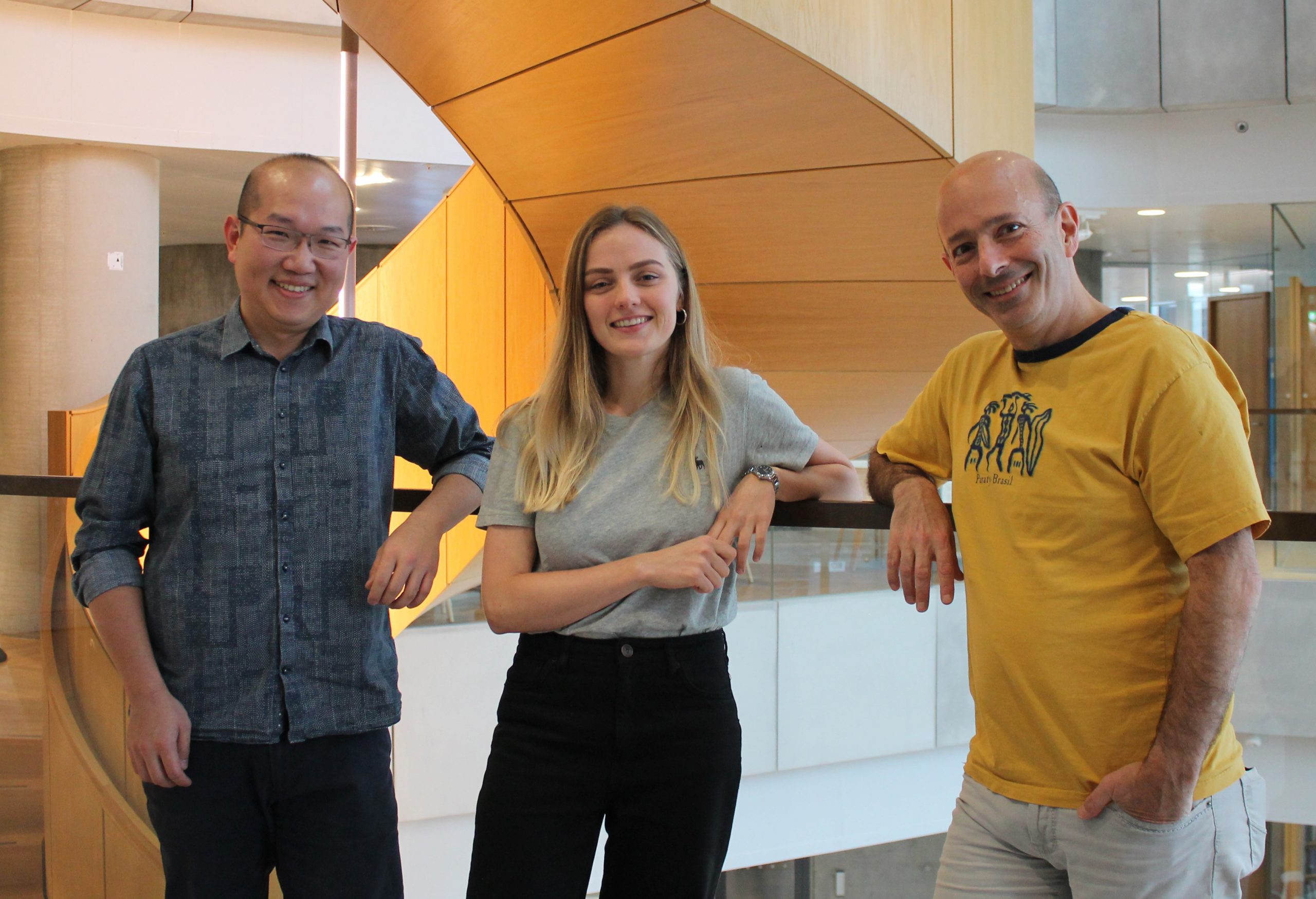


 (3 votes)