This editorial was recently published in Development and written by our editors Benoit Bruneau, Haruhiko Koseki, Susan Strome, Maria-Elena Torres-Padilla. Check out the Special Issue’s full table of contents here.
The development of an organism is regulated by tightly coordinated changes in gene expression. From zygotic gene activation, through to lineage specification and organogenesis, and into postnatal physiology and disease, broad programs of gene activation and repression are deployed in a carefully orchestrated manner. Eventually, the deployment of such developmental programs results in the formation of hundreds of different cell types, all containing the same DNA. Based on this, Conrad Waddington proposed more than half a century ago that development is an example of his epigenetic landscape.
DNA-binding transcription factors (TFs) are the leading drivers of these dynamic and cell type-specific gene regulatory networks, but TFs must function within the topological and physical constraints presented by the dense packing of DNA in chromatin, and within the context of histone modifications, DNA methylation and other aspects of chromatin-mediated regulation. All of these aspects of gene regulation – from TF binding sites, to nucleosomes, to topologically associated domains – are emerging as interlocked layers of developmentally important gene regulation.
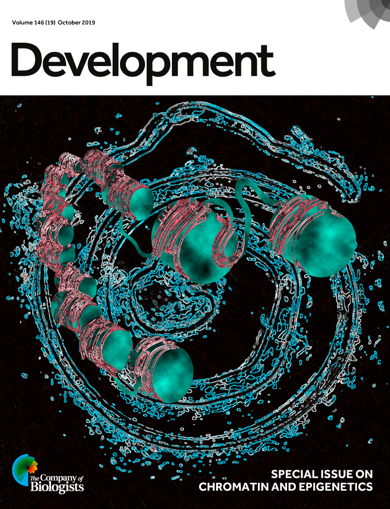
Representation of nucleosomes superimposed on an enhanced image of whole-mount mouse embryonic intestinal tissue (E18.5), illustrating the chromatin dynamics underlying small intestine development. See the article by Lei Chen, Michael Verzi and colleagues
In this Special Issue, we feature the myriad levels of chromatin regulation that impact developmental gene regulation. The Spotlights, Reviews and Research articles herein span topics covering TF interactions with chromatin, histone modifications, chromatin remodelers, DNA methylation, RNA-binding proteins and 3D organization of the genome. All of these levels of gene regulation have clear and broad roles in development, as exemplified by the variety of processes that are featured. These include limb patterning, cardiac differentiation, intestinal development, heart regeneration, zygotic genome activation, control of pluripotency, retrotransposon regulation, cell cycle regulation, dosage compensation, and maintenance of the body plan. Moreover, multiple experimental systems including Caenorhabditis elegans, zebrafish, mice, and human embryonic stem cells are featured, bringing into focus the broad importance of chromatin as an essential component of developmental control.
One Spotlight article features a summary of a discussion that was held at a recent workshop on ‘Chromatin-based regulation of development’ (https://www.biologists.com/workshops/april-2019/) organized by The Company of Biologists. The discussion centered on emerging evidence for the role of 3D genome organization and phase-separated condensates as potentially important regulators of gene expression. The discussion was lively, and the Spotlight captures the essence of the insights and controversies in the field. A second Spotlight highlights a new field of research termed ‘EvoChromo’ that considers the origin and evolution of chromatin, while several timely Reviews synthesize important and sometimes overlooked aspects of gene regulation in development. For example, how do chromatin remodelers find the right loci to act upon to achieve specificity? How do histone modifications participate in developmental disorders? What role does heterochromatin serve in controlling cell identity? What are the optimal means to visualize 3D genome organization?
With the emergence of exciting new technologies and paradigm-shifting insights, we now appreciate how chromatin and epigenetics are complex and important developmental regulators. This Special Issue highlights many of these exciting new aspects of developmental biology, and will hopefully inspire our readers to explore this field further. We would like to thank everyone – authors and reviewers – who contributed to this Special Issue and hope you will consider sending your next manuscript on this topic our way!
These movies from Hiroshi Kimura and colleagues’ Techniques and Resources article in the Special Issue are the latest addition to our YouTube channel
Postdoctoral Fellow positions (can join anytime between 2019 fall to 2020 spring) are available in Lee laboratory at Johns Hopkins University. These positions are not for stem cell experts, rather for who has expertise on non-stem cell fields are preferable.
Here are the focused research areas for this hiring for 3-4 Postdoctoral Fellows.
– General biochemistry
– Modeling neurodegeneration with organoids
– Optogenetics
The Lee lab has been establishing novel methodologies to specify human induced pluripotent cells (hiPSCs) into multiple lineages and to model human diseases, including induced neural crest (Kim et al., Cell Stem Cell, 2014), peripheral neurons (Oh et al., Cell Stem Cell, 2016; Oh et al., Nature Neuroscience, 2017), Schwann cells (Mukherjee-Clavin et al., Nation Biomedical Engineering, 2019) and skeletal muscle cells (Choi et al., Cell Reports, 2016; Choi et al., submitted; Sun et al., submitted) using multiple genetic reporter systems. We continue to study human developmental and degenerative disorders to unravel the underlying cellular/molecular mechanism toward realistic therapeutic approaches.
Compensation is following NIH guideline and JHU is an equal opportunity and affirmative action employer. Applicants can send a CV (with three reference contact info) to the address listed below:
Gabsang Lee, DVM, PhD
Associate Professor, Institute for Cell Engineering, Department of Neurology, Johns Hopkins University School of Medicine, Baltimore, MD 21205, USA
leelabjob@gmail.com
We are seeking to recruit two talented and highly motivated Postdoctoral Research Scientist to investigate the molecular regulation of lineage specification during the development of human preimplantation embryos. One position is for an experimental researcher and one position is for a bioinformatician.
These new posts are part of the exciting Wellcome-funded Human Developmental Biology Initiative (HDBI) that aims to define how cell lineages form during human development and to improve our understanding of fertility, birth defects and regenerative medicine.
The successful applicants with work collaboratively with members of the Reik, Kelsey and Rugg-Gunn groups within the Epigenetics Programme of the Babraham Institute. The job holder will benefit from being part of the HDBI and will work closely with other members of the initiative.
Fixed term contacts of 3-4 years in the first instance, with the expectation of additional funding up to five years.
Full details: https://www.babraham.ac.uk/vacancies-training
Highly motivated postdoctoral candidates are invited to lead several new projects to address fundamental questions in protein and RNA homeostasis related to neurodegenerative diseases in the laboratory of Jiou Wang. Experimental approaches, including biochemistry, genetics, and cell biology, from invertebrate to mammalian systems are employed. New techniques applied in the lab include iPSC neurons, genome editing, single cell analysis, and metabolite studies. Candidates with a strong background in biochemical, molecular, and/or cellular biology are encouraged to apply.
The Johns Hopkins Medical Institutions provide a stimulating and collaborative environment for biomedical research. Our lab is affiliated with the Department of Biochemistry and Molecular Biology at the Bloomberg School of Public Health and the Department of Neuroscience at the School of Medicine. The Baltimore/Washington D.C. area also offers rich professional and living opportunities.
Candidates should have a doctoral degree and strong research background. Please send a statement of research experience and career goals, a copy of Curriculum Vitae, and contact information of at least one reference to Dr. Jiou Wang at jiouw@jhmi.edu.
A complete listing of PubMed-accessible publications can be accessed at the following URL: http://www.ncbi.nlm.nih.gov/pubmed/?term=Jiou+Wang.
More information available at: https://www.jhsph.edu/faculty/directory/profile/2251/jiou-wang.The Johns Hopkins University is an Equal Opportunity Employer.
Cancer Research UK have launched two funding calls to drive progress in our understanding of paediatric cancers. We would like to encourage proposals to investigate one or more of the following concepts-
The basis of tumour initiation and progression
Novel therapeutic approaches
Development of novel biomarkers or methodologies to predict disease progression, to enhance efforts in primary and secondary prevention and intervention
Novel models that would enhance pre-clinical research
Development of more effective and/or less toxic treatments to improve long-term health and quality of life
The Stand up to Cancer-Cancer Research UK Paediatric New Discoveries Challenge focuses on multidisciplinary, multi-institutional, transatlantic teams that want to pursue a step change in our understanding of the drivers of paediatric cancers and the development of novel or repurposed medicines, treatment strategies or technologies
If you have any questions then please don’t hesitate to contact Sheona Scales (Cancer Research UK Lead for Paediatric Research) sheona.scales@cancer.org.uk.
A great opportunity for computationally inclined developmental biologists. Note tight deadline.
-D Parichy
The University of Virginia invites applications for a tenure-track Assistant Professor position with joint appointment in the Department of Biology and the School of Data Science. We seek applicants whose research programs address fundamental questions at the interface of Biology and Data Science. Of particular interest are researchers aiming to develop innovative computational tools to improve biological understanding in areas potentially including but not limited to: genomics and phenotype prediction; cell state and signaling; biological network architecture and information processing; multiscale modelling; cellular, organismal or population dynamics; biological image acquisition and analysis. Applicants are sought whose work will synergize with existing labs in the Department of Biology and elsewhere, with research emphases ranging from molecules to cells and tissues, and organisms to populations and ecosystems, as well as programs in the new School for Data Science in the areas of data acquisition, engineering, analysis, visualization or dissemination. Applicants employing computational methods with or without experimental approaches will be considered.
A successful candidate is expected to establish a vigorous, independent, and externally funded research program as well as provide instruction and scientific training at the undergraduate and graduate levels. Applicants with a respect for diversity and a passion for making a positive impact on the world in a collaborative, open environment are strongly encouraged to apply. The position will begin on August 25, 2020.
Located within the College of Arts and Sciences, the Department of Biology provides an interdisciplinary and collaborative environment for basic research and teaching that spans multiple levels of biological organization. The newly formed School of Data Science, founded with the largest gift in the university’s history, is dedicated to open interdisciplinary research of societal benefit with data science at the core. With the schools of Medicine, Engineering & Applied Sciences, UVA offers a diverse, collegial, interdisciplinary, and collaborative environment.
Applicants must have a Ph.D. in life sciences, computer science, statistics or a related field by the start of their appointment. A successful applicant will also have research accomplishments and plans of outstanding quality and significance at the interface of biology and data science as well as a commitment to excellence in teaching and mentoring. A proven commitment to participate in and further develop a diverse, collegial, interdisciplinary, and collaborative environment needs to be demonstrated.
Please apply online at uva.wd1.myworkdayjobs.com/en-US/UVAJobs/job/Charlottesville-VA/Assistant-Professor-of-Biology-Data-Science_R0010887 and attach a cover letter that succinctly highlights your most significant research accomplishments, experiences, and qualifications; a curriculum vitae; a research statement that describes your vision for your research program at the university (≤ 3 pages); a statement of teaching goals; a diversity statement that describes your past experience working on issues of diversity, equity and inclusion and/or working with diverse populations; and the contact information of three references.
Review of applications will begin November 3, 2019; candidates who apply by then will be given priority consideration, but the position will remain open until filled.
Assistant Professor of Developmental Biology, Washington University in St. Louis, School of Medicine
The Department of Developmental Biology at Washington University School of Medicine invites applications at the level of assistant professor on the tenure track. We are seeking outstanding colleagues with an interest in any area of developmental biology, including the genetic and epigenetic mechanisms of embryogenesis, cell fate specification and reprogramming, regeneration, aging, tissue engineering and quantitative approaches to developmental biology. Faculty in the Department of Developmental Biology employ a broad range of cell culture systems and model organisms including human embryonic stem cells, C. elegans, D. melanogaster, zebrafish and mouse. For more information, please visit our website at http://devbio.wustl.edu/.
Review of applications will start October 15, 2019. Interested applicants are required to submit their cover letter, curriculum vitae, and summary of their research accomplishments and plans through the online application website found at https://facultyopportunities.wustl.edu/Posting/Detail/1010397.
Applicants should also be prepared to provide the names and email contact information for three referees to provide letters of recommendation during the application process.
Washington University seeks an exceptionally qualified and diverse faculty; women, minorities, protected veterans and candidates with disabilities are strongly encouraged to apply.
The apical hook is a transient structure that functions to protect the vulnerable apical meristem from damage when the seedling penetrates the soil. Although some of the molecular players regulating its development have been identified, many aspects have remained opaque, including how an early auxin asymmetry in the hypocotyl is established. A paper in Development now provides a link between hormone signalling and the gravitropic response of the seedling’s growing root in apical hook development. We caught up with co-first authors Qiang Zhu and Marçal Gallemí and their supervisor Eva Benková, Professor at the Institute of Science and Technology Austria in Klosterneuberg, to find out more about the project.
Qiang, Marçal and Eva (L-R)
Eva, can you give us your scientific biography and the questions your lab is trying to answer?
EB I studied Molecular Biology and Genetics at the Masaryk University in the Czech Republic, and then obtained my PhD at the Institute of Biophysics of the Czech Academy of Sciences. As a postdoctoral fellow, I joined the laboratories of Professor Klaus Palme at the Max Planck Institute, Cologne, and Professor Gerd Jurgens at the University of Tübingen. During my PhD studies and later as a postdoctoral fellow I became interested in plant hormones and their exceptional impact on all aspects of plant development. I was fascinated by the simple and (among plant biologists) well-known experiment by Murashige and Skoog in reported 1957: they demonstrated that a modulation of the ratio between two plant hormones, auxin and cytokinin, can re-define the developmental programme of meristematic cells, resulting in either shoot or root formation. Thus, in 2007, when I got the opportunity to lead my independent research as group leader at the Plant Systems Biology department of the VIB in Belgium, I decided to pursue this topic and fully focus on plant hormones and the mechanisms underlying their cross-talk. In 2012, I moved to the Institute of Science and Technology Austria, where I continue this research line. My team is trying to dissect the molecular mechanisms underlying complex hormonal interactions and identify key points at which hormonal signalling pathways converge to control plant growth and development.
Qiang and Marçal, how did you come to work in the lab, and what drives your research today?
QZ As a plant biologist, I am impressed by how plants have developed complicated mechanisms that enable rapid and flexible adaptation of their growth and development to their ever-changing surrounding environment, and I’m curious about the mechanisms behind this. When I was in Ghent University, Eva and my previous lab had some joint projects on development of the apical hook, a very important transient structure for dicots when they grow out of the dark. At that time, I got to know this interesting model, and wanted to know more about it. It’s this interest in the topic and project that mainly drives my research. I am so lucky that I had the chance to join the Benková lab and work on the project. My research is greatly supported by Eva, who is talented, hard-thinking and dedicated. It’s really nice to work with her: we have a lot of discussions about this project, which also drive me to deeper investigation into the secret behind the formation of the apical hook.
MG Working in a lab, and specifically a molecular biology lab, is like playing games every day! We assemble and disassemble pieces of a puzzle trying to get to know what are they for, how they work and how they interact. Curiosity is what mainly drives my research but, with the current status of our planet, including extremely exploited natural resources, pollution and global warming, understanding how plants grow and adapt to the environment is also a key question for humanity.
Working in a lab…is like playing games every day
What was known about apical hook development before your current work, and in particular the role of gravity in the process?
EB The apical hook is developmentally a very important structure, which is transiently formed during germination to protect a delicate apical meristem and cotyledons from damage when seedlings penetrate through the soil. Several studies have reported that apical hook formation is driven by asymmetric distribution of auxin accumulating at the inner side of the hook. However, how this asymmetric pattern of auxin distribution is established, and which mechanisms determine formation of the apical hook during early phases of seedling germination, was largely unknown. Although previously there have been some indications that gravity might play a role in the apical hook development, for example from plants observed in space and from our own research, no systematic studies were reported. Compared with other plant developmental processes, research on the apical hook has lagged behind, probably due to the lack of a suitable technique that would enable observation of this process occurring in darkness. Luckily, with establishment of the dark-imaging system we could start to monitor the whole process of apical hook development in real time. I think this has been the most important technical advance and enabled us to address many interesting questions.
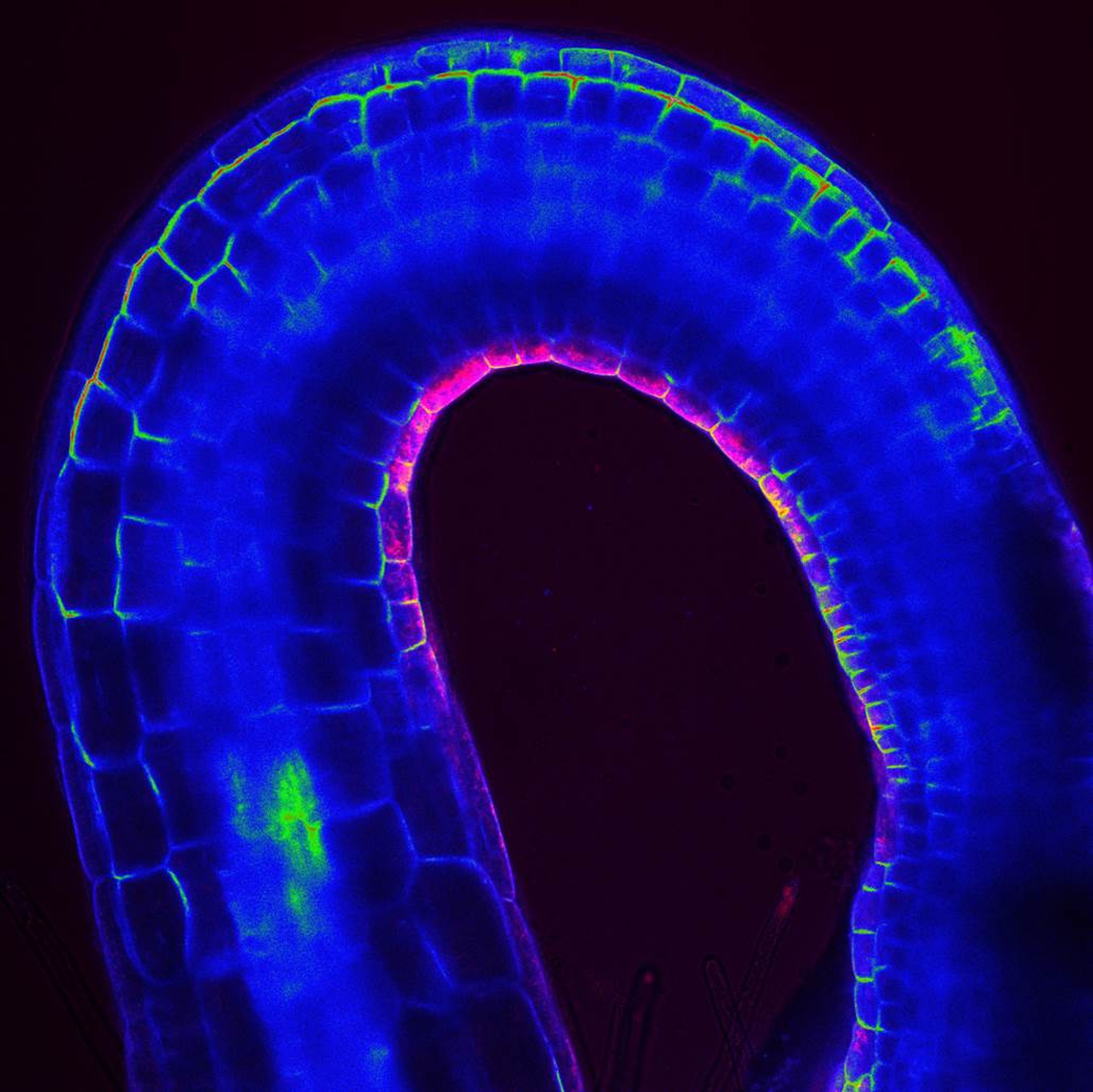
An apical hook with the DR5::RFP auxin-sensitive reporter in magenta and PIN3::PIN3-GFP in green.
Can you give us the key results of the paper in a paragraph?
EB In our work, we showed that during early phases of germination, gravity-stimulated bending of the root acts as the initial cue to coordinate the formation of the apical hook. The core machinery mediating the root’s response to gravity is required for apical hook formation, and PIN2, which so far was considered to be a root-specific auxin efflux carrier, acts as an essential integrator of root-to-hypocotyl communication. We found that during early phases of germination, PIN2 activity is not restricted to the root: it manages transport of auxin to the hypocotyl, thus providing initial asymmetry in auxin distribution, which is further reinforced by the polar auxin transport machinery that is gradually established in the hypocotyl. Hence, two distinct developmental events taking place at opposite poles (root and shoot) of the plant axis are regulated by common regulatory machinery. Finally, we demonstrated that establishment of such inter-organelle transport system is a result of tight interplay between two hormonal pathways, abscisic acid (ABA) and gibberellins (GA), acting antagonistically during embryo maturation and early germination.
How do you think gravity-stimulated, auxin-driven root bending is transmitted to promote development of the apical hook?
QZ Based on our work and published findings, we hypothesise that the initial gravity-stimulated, auxin-driven root bending might promote differences in mechanical forces at opposite sides of hypocotyl, which eventually would lead to an asymmetry in PIN expression. In support of such a scenario, it has been reported that changes in mechanical strains, such as modifications of turgor pressure or the application of external force, have considerable impact on subcellular trafficking and membrane localisation of PINs.
What do you think ABA and GA signalling are doing in the process?
MG Several recent studies including our work have reported that ABA affects the expression and membrane localisation of PIN auxin efflux carriers, key players in the regulation of apical hook formation. Intriguingly, ABA has been found to control stability of Rho guanine nucleotide exchange factors (RopGEFs), well-established regulators of ROPs, a family of small GTPases implicated in various cellular processes including control of PIN polar membrane localisation. This potential mechanistic link would definitely be worth further investigation. Unlike ABA, which reduces levels of PINs, GA has been found to interfere with lytic degradation of PIN proteins and consequently to enhance their accumulation at plasma membrane. So a speculative and plausible scenario is that the dynamic balance between ABA and GA signalling during embryo maturation and the early phases of germination tightly controls the expression and plasma membrane localisation of PIN auxin efflux carriers, thus leading to proper hypocotyl growth and apical hook formation. How these two hormonal pathways mutually interact and which components of their respective pathways are specifically involved in apical hook formation requires further research.
When doing the research, did you have any particular result or eureka moment that has stuck with you?
QZ The most exciting moment for me was when I realised that the root gravitropic response is important for apical hook formation, by carefully observing kinetics of newly forming hooks. And when I found that green embryos are unable to form apical hooks despite a normal root gravity response, I knew this had provided an exciting model to study the mechanisms behind its development and in particular communication between these two organs.
MG My ‘aha!’ moment of this study was when I realised that seedlings germinating from mature embryos on medium supplemented with ABA resembled those grown from green embryos (at certain concentration the root is still growing, whereas cells in the shoot are not able to elongate anymore). This nicely supported our hypothesis that seedlings developing from green embryos might not able to form apical hooks because of their high levels of ABA. Thus, to stimulate formation of the apical hooks in these seedlings we might only need to deplete the excess ABA!
And what about the flipside: any moments of frustration or despair?
QZ For me, the moments of frustration were at the very beginning when I started this project. I had to peel off the Arabidopsis seed coats and isolate the embryos: you know, it’s very difficult to do this even under the microscope. I am so happy that after a long time practicing, my hands do not shake and I can operate freely with the seeds.
MG In line with the previous question, I tried many things to stimulate the growth of green embryos and observe the formation of an apical hook (auxin, GA, Norflurazon, abamine, etc.). It was really frustrating to see no rescue of growth, even in the ABA synthesis mutants or using chemicals that are supposed to inhibit ABA synthesis. It was not until the last trial, in which we combined several treatments (abamine and GA together in the aba2-1 mutant background), that we finally succeeded and could observe growth and apical hook formation in seedlings germinating from green embryos.
So what next for you two after this paper?
QZ I established my own research group in China after I left the Benková lab. Currently, we are studying bamboo as a new research model. Bamboo is probably the fastest growing plant on earth but until now nobody knew why it grows so fast: our group is trying to reveal the molecular mechanisms behind this. The research experiences in Eva’s lab really contributed a lot to my current research, and all the projects are running smoothly. So far, so good.
MG Next paper! We always have the next project in mind, and I am currently finishing experiments for what will be the next manuscript.
Where will this work take the Benková lab?
EB Now we know that if we want to fully understand mechanisms underlying formation of the apical hook, we have to also take into consideration processes occurring well before the bending of the hook itself, such as embryo maturation, outgrowth of the embryonic root and its alignment with the gravity vector. There are also many questions related to the cross-talk between ABA and GA and role of these hormones in coordination of the polar auxin transport required for proper formation of the apical hook. Finally, yet importantly, we still know very little about the pathways downstream of auxin that control the differential cell growth essential for hook development, but also bending of roots, hypocotyls or stems during gravi- or phototropism.
Finally, let’s move outside the lab – what do you like to do in your spare time?
QZ I left Vienna 4 years ago, and this question makes me recall the wonderful time I had in Austria, when my wife, my daughter and I would spend most of our free time traveling Vienna and the small towns around. We really enjoyed the charm of the music capital, and were impressed by the culture and atmosphere there.
MG I enjoy doing sport a lot, and the IST campus is a really good place for that. So I enjoy playing football and volleyball with my colleagues in the yards we have in the campus, and I also enjoy cycling and running in the woods around the campus. I feel privileged to have so many green areas surrounding the campus: there is nothing better for the brain than freshly produced oxygen from the woods!
EB I love gardening, not only because it is nice to have fresh vegetables and fruits, but I very much enjoy observing plants grow, seeing how they change from day to day, getting new leaves, flowers, tasty fruits… I also like travelling with my family, and especially hikes in mountains; a beautiful view into valley after a long tour is always worthwhile.
An exciting opportunity has arisen to join the Childhood Leukaemia Research Group led by Professor Anindita Roy at the University of Oxford. The group’s research explores how childhood leukaemia develops before birth, with a focus on developmental haematopoiesis and treatment resistant infant leukaemia. This post has funding for a total of five years from Wellcome Trust.
In the latest episode we’re getting our hands dirty by delving into the poop-ome – the trillions of bacteria that live inside our guts and make up what’s known as the microbiome. Rather than simply being a bunch of bugs, the microbiome is now believed to play a role in virtually every aspect of health and disease. But what are they up to? How do we even know what species are in there? And can you blame your stinky farts on your gut bacteria?
If you enjoy the show, please do rate and review and spread the word. And you can always send feedback and suggestions for future episodes and guests to podcast@geneticsunzipped.com
 (1 votes)
 (No Ratings Yet)