Mutualistic relationships between bacteria and complex organisms have repeatedly evolved and this has allowed host organisms to exploit new environments and foods. One of the most extreme and fascinating cases of symbiosis in the animal kingdom is observed in annelid worms of the genus Riftia and Osedax. These animals are able to live in particularly extreme environments, including deep sea hydrothermal vents and carcases thanks to bacteria from the environment that they acquire as juveniles. Ultimately, this induces a drastic developmental change where they degenerate their guts and rely entirely on the bacterial symbionts to produce the essential nutrients they require for survival in those hostile environments. Which cellular and genetic mechanisms control this bacterial symbiosis? How did these mechanisms evolve? How did its change contribute to animal evolution?
In this project you would rigorously answer these questions sequencing and comparing the genome of these symbiotic worms with their closest asymbiotic counterparts.
You would have access to a large genomic database, field collections and in-house live organisms to fuel your investigation.
You would gain experience of molecular techniques (nucleic acid extraction, next generation sequencing), bioinformatics (e.g. genome assembly, RNA-seq analyses, gene family evolution), and statistics.
You will be encouraged to develop your own ideas and hypotheses.
The studentship is fully funded and available to EU and UK citizens. It will cover tuition fees as well as provide an annual tax-free maintenance allowance for 3 years at Research Councils UK rates (£17,009 in 2019-20).
Skills preferred
In a multidisciplinary project like this, candidates are unlikely to have a background in all disciplines involved. The most important qualification is motivation, enthusiasm and that the project appeals to you. However, previous computational experience would be a plus. We can envisage strong candidates coming through a variety of routes including:
practical molecular biology
evolutionary theory and phylogenomics
computational biology
To apply, students should have a first class degree or have received a MSc in a relevant field (i.e. marine biology, evolutionary biology, bioinformatics) or are about to finish their MSc.
Cavanaugh, C. M., Gardiner, S. L., Jones, M. L., Jannasch, H. W. & Waterbury, J. B. (1981) Prokaryotic Cells in the Hydrothermal Vent Tube Worm Riftia pachyptilaJones: Possible Chemoautotrophic Symbionts. Science 213, 340-342.
Rouse, G. W., Goffredi, S. K. & Vrijenhoek, R. C. (2004) Osedax: bone-eating marine worms with dwarf males. Science 305, 668-671.
Thornhill, D. J., Fielman, K. T., Santos, S. R. & Halanych, K. M. (2008) Siboglinid-bacteria endosymbiosis: A model system for studying symbiotic mechanisms. Commun Integr Biol 1, 163-166.
Hilario, A. et al.(2011) New perspectives on the ecology and evolution of siboglinid tubeworms. PLoS One 6, e16309
A postdoctoral position is available to study cell signaling mechanism using eye development as a model. Among the current projects, we are studying how extracellular signals induce cytoskeletal dynamics to control cell shape and adhesion. We are also interested in the crosstalk between FGF and other signaling pathways. The aim is to combine mechanistic studies in vitro with mouse genetics in vivo. Potential candidate should be highly motivated with a Ph.D. degree in biomedical science and a strong background in biochemistry and cell biology. Experience in mouse genetics is desirable but not necessary. The successful postdoc candidate will receive training in genetic, molecular and biochemical approaches in vision research, interacting with basic scientists and physicians in a multidisciplinary environment. For details of the research projects, please visit: https://www.pathology.columbia.edu/profile/xin-zhang-phd
Relevant Publications
Li H, Mao Y, Bouaziz M, Yu H, Qu X, Wang F, Feng GS, Shawber C, Zhang X. Lens differentiation is controlled by the balance between PDGF and FGF signaling. 2019. PLoS Biol. 17(2):e3000133.
Collins TN, Mao Y, Li H, Bouaziz M, Hong A, Feng GS, Wang F, Quilliam LA, Chen L, Park T, Curran T, Zhang X. Crk proteins transduce FGF signaling to promote lens fiber cell elongation. eLife. 7:e32586.
Garg A, Hannan A, Wang Q, Collins T, Teng S, Bansal M, Zhong J, Xu K, Zhang X. FGF-induced Pea3 transcription factors program the genetic landscape for cell fate determination. PLoS Genetics. 14(9):e1007660.
Please send CV with names and contact information of three references to: Xin Zhang, Ph.D., Departments of Ophthalmology, Pathology & Cell Biology, Columbia University, New York, NY 10032. Email:xz2369@columbia.edu
The Columbia University Medical Center is located in New York City, which offers world-class museums, performing arts and numerous culture opportunities. With top ranked Ph.D. programs, medical school and affiliated hospitals, it presents a collaborative and stimulating academic environment for research excellence.
Research Group Julien Royet: “Host pathogen interactions in the Drosophila model”
IBDM (UMR CNRS 7288) • Parc Scientifique de Luminy • 13288 Marseille Cedex 9 • France
A PhD position is available at the Institute of Developmental Biology of Marseille (IBDM) for a motivated student to work on a research project investigating the genetic basis of host-bacteria interactions in the Drosophila model. This is a full-time position for 3 years. The candidate must be free to start in September/October 2019.
Background
It is now very well established that gut-associated bacteria can impact the behavior and the physiology of their eukaryotic host. The PhD thesis project is aimed at using the powerful genetic tools available in the Drosophila model and the relative simplicity of its gut microbiota to study, at the molecular level, the molecular dialog between the microbiota and its host. In two recent publications, (Kurz et al, Elife, 2017: Charroux et al, Cell Host Microbe, 2018), the lab has shown that a metabolite produced by gut-associated bacteria, called peptidoglycan, can cross the gut epithelium and reach the insect blood where it interferes with various organs (fat body, ovaries, brain…) and modifies functions (behavior changes, organ wasting…). The PhD student will use the newest genome editing technologies (Crispr…), genetic tools and latest imaging microscopy technics to dissect the precise cellular and molecular mechanisms of the dialog that exist between gut-resident bacteria and some specific cells of the host. Recent results showing that mice deficient in peptidoglycan-sensing proteins exhibit social behavioral alterations suggest that the mechanisms that we study in Drosophila also exist in mammals. The research will be performed in the Institute of Developmental Biology of Marseille, an internationally recognised interdisciplinary research center and a very stimulating scientific environment (http://www.ibdm.univ-mrs.fr/).
Profile of the candidate
We look for an enthusiastic and ambitious student with a strong interest in the genetics of host-bacteria interactions. The candidate is expected to have a background in molecular biology and should hold a Master Degree in Bioscience Engineering, Biotechnology or Biology. The candidate should have a level of proficiency in English which is sufficient to communicate effectively with colleagues.
Applications
Application documents should include a motivation letter, a curriculum vitae and a grade transcript. Additionally, the applicant is expected to arrange for two letters of recommendation to be sent to the address below. The application deadline is May 1th, 2019. Applications should be sent electronically as one single file in pdf format to Julien.royet@univ-amu.fr, leopold.kurz@univ-amu.fr and Olivier.zugasti@univ-amu.fr
Publications
Kurz et al, 2017. Peptidoglycan sensing by octopaminergic neurons modulates Drosophila oviposition. Elife.Mar 7;6. pii: e21937.
Charroux et al, 2018. Local and systemic immune responses to microbiota are respectively controlled by cytosolic and secreted peptidoglycan degrading enzymes in Drosophila. Cell Host and Microbe. Feb 14;23(2):215-228
Jarema Malicki, a pioneer in developmental studies of the vertebrate retina, died on 4th January 2019, shortly after being diagnosed with cancer. Here, I reflect on Jarema’s life and work, with a particular focus on his research interests in zebrafish as a model organism for vertebrate retinogenesis and human ciliopathies.
Jarema was born in Warsaw, Poland, in March 1965. The family visited their small ‘orchard’ property outside of the city at the weekends, and his father often took him into the Tatry Mountains near Zakopane.
These early experiences inculcated a deep life-long affection for the natural world and walking in the mountains; in particular, he loved the Tatry Mountains and the Tramuntana Mountains of Mallorca. He enrolled at Warsaw University as an undergraduate but in 1987, at some personal risk and with great ingenuity, he escaped the political repression and authoritarianism of the Communist regime. He initially headed east, not west: on leaving Poland, he took a train across Siberia, ostensibly as a holiday trip to Japan. He then entered the USA via California, travelling on to Maine by bus with little money or fluency in English, to stay with friends of his parents. He was forever grateful to Bates College, Maine, for accepting him onto their undergraduate study programme, and for their generosity with scholarships and part-time work as he built a new life in the USA. Jarema did not see his parents again until 1991 and did not return to Poland until 1994. He became a US citizen in 2010.
In 1989, after one year at Bates College, he enrolled on the molecular biology PhD programme at Yale University, working in Bill McGinnis’ lab. Bill had made his name by defining the spatial and temporal expression of homeotic genes during Drosophila development. It was an exciting time to enter the Drosophila field: new molecular biology techniques were providing insights into the role of maternal gene expression, and positional cloning of mutants was beginning to define the often unexpected functions of key players. But the extent to which these insights into fly development applied to other vertebrates, the development of which appeared so radically different, was at the time unclear. Studies were just beginning to show that homeotic genes and the homeobox were highly homologous in metazoans, and Jarema’s PhD work took this a stage further by showing that cis-regulatory elements in the mammalian HOX4B gene (now renamed HOXD4) and the homologous fly gene Deformed (Dfd) were functionally equivalent during head development (Malicki et al., 1992). Jarema was very proud that this research was published in Nature just as he was graduating from Yale in 1992.
Around the same time, Wolfgang Driever was pioneering the use of the zebrafish as a genetically tractable vertebrate model organism that was suitable for large-scale mutagenesis screening in his lab at Harvard Medical School (Boston, MA, USA). The Driever lab started the so-called Boston zebrafish mutagenesis screen in 1992, and Jarema joined the lab in 1993. Jarema was very excited to work on this new model organism for embryogenesis and organogenesis, and was enthusiastic to enter a promising new field at such an early point. He spoke warmly about the camaraderie in the Driever lab, the life-long friends he made during this period, and his enjoyment of sailing in Boston Harbor and Cape Cod as well as hiking in the hills of north eastern USA. The research culminated in the publication of the famous zebrafish issue of Development in December 1996 (Driever et al., 1996), in which Jarema co-authored 11 papers and was first author on two that described mutations affecting the retina and ear (Malicki et al., 1996a,b). The positional cloning of these mutants, which Jarema often gave whimsical names using Polish words, was a rich resource for molecular characterization of function in subsequent years, and provided the basis for many projects in Jarema’s labs in Boston and later in Sheffield, UK.
As a faculty member of Harvard Medical School from 1996 and Tufts University (Boston, MA, USA) from 2009, Jarema focused his research on the genetic basis of vertebrate eye and ear development, how it related to the underlying cell biology of polarity and intracellular transport, and how these cellular processes related to morphogenesis and embryonic patterning. Early successes were the characterization of retinal patterning loci derived from the mutagenesis screen. These included oko meduzy (now known as crb2a; translated as ‘jellyfish eye’), which encodes a crumbs gene homologue (Omori and Malicki, 2006), and glass onion (cdh2), which encodes N-cadherin (Malicki et al., 2003). But his seminal contribution during this period was work on nagie oko (mpp5a; translated as ‘naked eye’; Wei and Malicki, 2002), which encodes a large scaffolding protein in the membrane-associated guanylate kinase (MAGUK) family; subsequent work by Ronald Roepman and colleagues identified it as an interaction partner of the Crumbs complex (Kantardzhieva et al., 2005). This work contributed to the realization that MAGUK proteins regulate plasticity and adhesion at tight junctions by stabilizing multi-protein complexes, as exemplified by the Crumbs complex during retinal development. In a satisfying parallel narrative, mutations in several members of the Crumbs complex were found to be a major cause of human inherited retinal dystrophies. In Jarema’s mind, these insights vindicated the medical potential of zebrafish and the power of forward genetic screens in the model.
Another series of mutants from the screen had photoreceptor loss and kidney cysts, phenotypes that are associated with defective cilia formation. In a prescient paper, Jarema’s group demonstrated that oval (ift88) encoded IFT88, a component of the ciliary intraflagellar transport (IFT) system that is essential for cilia maintenance and sensory neuron survival (Tsujikawa and Malicki, 2004). This work presaged a worldwide effort over the next decade to identify human disease genes for human inherited retinal dystrophies and ciliopathies, and a growing recognition that the cilia is a fundamental mediator and regulator of developmental signalling and cellular homeostasis. Jarema’s lab demonstrated that elipsa (traf3ip1) encoded a second IFT protein, TRAF3IP1/IFT54 (Omori et al., 2008), which led to another satisfying link with medical genetics: mutations in TRAF3IP1/IFT54 are now known to cause Senior-Løken syndrome (Bizet et al., 2015), a ciliopathy that is characterized by nephronophthisis and retinal degeneration. Subsequent work on IFT by Jarema’s group revealed molecular mechanisms for the selective transport of specific ciliary cargoes such as opsin within the photoreceptor (Zhao and Malicki, 2011). These insights were the rationale for the recent development of an elegant zebrafish model of conditional in vivo opsin transport, and this remains an area of active interest by Jarema’s trainees and colleagues.
I first met Jarema in person soon after he had moved to the Bateson Centre at the University of Sheffield in 2012. With an established reputation in zebrafish genetics and an interest in inherited retinal dystrophies, he was an excellent recruit to the zebrafish community that was established in Sheffield by Philip Ingham. We hit it off instantly and over numerous visits, talking about ciliary biology and the challenges facing this new field, we eventually collated our conversations into a review article (Malicki and Johnson, 2017) and set up the ‘Northern’ Cilia Club (now part of the UK Cilia Network). Jarema was an enthusiastic and incisive writing partner: his approach was characterized by lucid and insightful scholarship, attention to detail and generous advice. Chatting to his students and trainees, I soon realized that he was equally rigorous, self-disciplined and dedicated in his professional commitments to them. He was tireless in supporting not only his Sheffield students but also those from the Erasmus Programme, as well as younger scientists from Poland. At the same time, he was keen to share his passions from outside of the lab: good food, good wine, good coffee and the great outdoors. These interests often aligned during lab weekends at his hideaway in the Tatry Mountains, or in meals at one of his favourite restaurants. Above all, he was motivated by his love of science: his enthusiasm inspired those around him to work a bit longer and to try a bit harder. Żegnaj ‘Gruba Rybo’.
Acknowledgements
I am grateful to Jarema’s many students, colleagues and collaborators who shared their memories and provided anecdotes of his life and science on which this obituary is based. I extend particular thanks to Frederick Walters, Lilianna Solnica-Krezel, Zhou Zhu, Xiaoming Fang, Pawel Lysyganicz, Pamela Yelick, Dominic Norris, Katarzyna Szymanska, Andrew Furley, Ronald Roepman and Marysia Placzek for their comments and feedback on the text, and to Zhou Zhu for the photograph.
References
Bizet, A. A., Becker-Heck, A., Ryan, R., Weber, K., Filhol, E., Krug, P., Halbritter, J., Delous, M., Lasbennes, M. C., Linghu, B. et al. (2015). Mutations in TRAF3IP1/IFT54 reveal a new role for IFT proteins in microtubule stabilization. Nat. Commun. 6, 8666.
Driever, W., Solnica-Krezel, L., Schier, A. F., Neuhauss, S. C., Malicki, J.,
Stemple, D. L., Stainier, D. Y., Zwartkruis, F., Abdelilah, S., Rangini, Z. et al. (1996). A genetic screen for mutations affecting embryogenesis in zebrafish. Development 123, 37-46.
Kantardzhieva, A., Gosens, I., Alexeeva, S., Punte, I. M., Versteeg, I., Krieger, E., Neefjes-Mol, C. A., den Hollander, A. I., Letteboer, S. J., Klooster, J. et al. (2005). MPP5 recruits MPP4 to the CRB1 complex in photoreceptors. Invest. Opthalmol. Vis. Sci. 46, 2192-2201.
Malicki, J. and Johnson, C. A. (2017). The cilium: cellular antenna and central
processing unit. Trends Cell Biol. 27, 126-140.
Malicki, J., Cianetti, L. C., Peschle, C. and McGinnis, W. (1992). A human HOX4B regulatory element provides head-specific expression in Drosophila embryos. Nature 358, 345-347.
Malicki, J., Neuhauss, S. C., Schier, A. F., Solnica-Krezel, L., Stemple, D. L.,
Stainier, D. Y., Abdelilah, S., Zwartkruis, F., Rangini, Z. and Driever, W. (1996a). Mutations affecting development of the zebrafish retina. Development 123, 263-273.
Malicki, J., Schier, A. F., Solnica-Krezel, L., Stemple, D. L., Neuhauss, S. C., Stainier, D. Y., Abdelilah, S., Rangini, Z., Zwartkruis, F. and Driever, W. (1996b). Mutations affecting development of the zebrafish ear. Development 123, 275-283.
Malicki, J., Jo, H. and Pujic, Z. (2003). Zebrafish N-cadherin, encoded by the glass onion locus, plays an essential role in retinal patterning. Dev. Biol. 95-108.
Omori, Y. and Malicki, J. (2006). oko meduzy and related crumbs genes are
determinants of apical cell features in the vertebrate embryo. Curr. Biol. 16,
945-957.
Omori, Y., Zhao, C., Saras, A., Mukhopadhyay, S., Kim, W., Furukawa, T., Sengupta, P., Veraksa, A. and Malicki, J. (2008). Elipsa is an early determinant of ciliogenesis that links the IFT particle to membrane-associated small GTPase
Rab8. Nat. Cell Biol. 10, 437-444.
Tsujikawa, M. and Malicki, J. (2004). Intraflagellar transport genes are essential for differentiation and survival of vertebrate sensory neurons. Neuron 42, 703-716.
Wei, X. and Malicki, J. (2002). nagie oko, encoding a MAGUK-family protein, is essential for cellular patterning of the retina. Nat. Genet. 31, 150-157.
Zhao, C. and Malicki, J. (2011). Nephrocystins and MKS proteins interact with IFT particle and facilitate transport of selected ciliary cargos. EMBO J. 30, 2532-2544.
IRIBHM is a research institute of the Medical School of the Free University of Brussels (Université Libre de Bruxelles, ULB). The institute offers an internationally prominent research environment in molecular biology and life sciences, that engage different topics that span receptor pharmacology and new therapeutic targets discovery, early embryonic development, neurobiology, stem cells and cancer. The institute has trained over the years a number of talented young scientists both at the graduate and postdoctoral levels. In order to expand internationally and keep rising its level of excellence, the IRIBHM launches an international graduate programme in order to prepare the future leaders in biomedical sciences.
At least 2 PhD scholarships are available. The successful candidates will have the opportunity to work in a warm and stimulating research environment aligned with the highest international standards.
As the administrative center of the European Union, Brussels is a perfect location for an International PhD programme. In addition, the city shows an active cultural life and is hosting most nationalities from all over the world.
Closing date for application is April 28, 2019. The short-listed candidates will be invited for an interview, which will be held in Brussels (between June 3 and 4, 2019). Accepted candidates are expected to start their research project on November 1st, 2019.
To find out more about the IRIBHM International PhD Program, please visit our web site at http://iribhmphd.ulb.be
CamBioScience Limited is a premium global provider of education and educational technology. The Courses & Conferences Department works with world-leading experts to provide intensive training courses and conferences. Courses are in a broad spectrum of emerging and established life science technologies for academic and industry professionals. In-Person courses and conferences take place in various locations around the world including United Kingdom, Austria, South Korea and China.
CamBioScience Ltd. are looking for a highly-motivated, outgoing and ambitious person who will sell in-person courses and conferences to life science researchers. The position will be full- or part- time.
We have courses in the developmental biology field including organoids and gastruloids. You will also have the opportunity to attend these courses to see how they are organised and delivered.
Please contact: michelle@cambioscience.com if you have any questions and for information on how to apply.
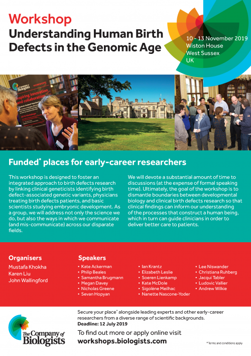
At the beginning of November 2018, thirty researchers congregated at Wiston House to attend a workshop titled ‘Evo-chromo: towards an integrative approach of chromatin dynamics across eukaryotes’. The workshop was organised by Frederic Berger (Gregor Mendel Institute) and Ines Anna Drinnenberg (Institut Curie), and was the 27th workshop hosted by The Company of Biologists since 2010. Its objective was to explore the biology of chromatin – the macromolecular complex of DNA, RNA and protein that makes up chromosomes – in light of evolutionary questions and ideas, particularly those relating to eukaryotic evolution.
Chromatin was first observed in the 19th century, when it was visualised by staining chromatin with basic dyes (the term ‘chromatin’ derives from the Greek word khroma meaning ‘colour’). During the molecular biology revolution of the 20th century, chromatin came to sit at the heart of our understanding of gene regulation, cell differentiation and inheritance. As our discussions at the meeting highlighted, the increasing wealth of phylogenetic data, exciting new technologies, and an increasing number of tractable model systems has produced a series of puzzling and pressing evolutionary questions. However, in the excitement and industry of modern molecular biology, these questions often remain skulking in the background. The focus of this meeting was to clarify those questions and bring them to the foreground.
Alongside the 20 invited speakers, we were lucky enough to attend as one of 10 early-career researchers (PhD students, postdocs, and young PIs). These places are fully funded by the Company of Biologists, and it was a fantastic opportunity for us to present our work and receive feedback. Alexander is a graduate student in the Henderson Group (University of Cambridge) specialising in plant meiotic recombination, and James is postdoc in the Rentzsch Group (Sars Centre) specialising in cnidarian development and evolution. This was an incredibly valuable opportunity for us to meet world-class scientists and expand our academic network. Furthermore, as we are both planning to move deeper into the world of chromatin biology, it was even more valuable in helping us to better orientate in this research area and gain an understanding of the most pertinent questions in the field.
Back row standing: Harmit Malik, Kenneth Wolfe, Peter Andersen, Ulrich Technau, Aidan Maartens, Tobias Wernecke, Simon Elsässer, James Briscoe, Peter Sarkies, Zachary Harvey. Middle row standing: Brandon Gaut, Chema Martin-Duran, James Gahan, Siavash Kurdistani, Frederic Berger, Steven Henikoff, Bob Schmitz, Catherine Peichel, Alexander (Sasha) Blackwell, Douglas Erwin, Bernardo Lemos. Front row seated: Juan Ausio, Joe Thornton, Mia Levine, Kinga Rutowicz, Ines Anna Drinnenberg, Karolin Luger, Marilyn Renfree, Joyce Kao, Wendy Bickmore
The workshop was far from regular. Firstly, we had never attended a meeting that was both so small and so diverse. Talks ranged from genomic imprinting in marsupials, developing biomarkers of human aging, the structure of archaeal nucleosomes, developing quantitative ChIP-seq methods, and the role of genetic conflict in centromere evolution. However, all of these diverse research areas were looked at through the lens of evolutionary biology, with the objective to find commonalities and shared general questions. One consequence of this diversity was that everyone was there to learn and to think, as everybody was to some extent outside of their comfort zone. Secondly, the fact that we were all there to address the issue of chromatin evolution meant that we searched for the common threads and broader significance connecting the talks. This led to a greater examination of the conceptual framework(s) surrounding chromatin biology, and a probing of the limitations and future possibilities. Personally, we found the meeting to be pleasingly free of the ‘look-how-many-RNA-seq-experiments-I-did’ kind of attitude, and the focus was almost exclusively centred on biology and ideas. This was reflected in the large amount of preliminary and unpublished data shown at the meeting. Thirdly, alongside the marvellous attendees, the house itself, located in West Sussex, provided a wonderful context. The 16th century house is situated in the South Downs National Park, and is the location of Wilton Park, a project established in 1946 with the aim of promoting peace and democracy in Europe. It therefore seemed an appropriate setting for the meeting, given the role that international science has played in promoting integration and cooperation in a once fractured world. This couldn’t help but feel more poignant, given the current resurgence of nationalism and the risks this poses to science. Indeed, perhaps the stand out speaker of the meeting was not a scientist but rather a local historian who led us on a captivating historical tour of the house.
The small size of the conference, the absence of phone signal and the remote location all encouraged meaningful social interaction. There was a chance to engage with everybody, with no shortage of time for discussion and questions. The organisers aimed to restrict the number of attendees known by each attendee – this had the benefit of promoting interaction, an unpretentious attitude, and removed the usual hierarchies emerging at most more specialised conferences. This lack of hierarchy was further demonstrated by every speaker having equal time. Interaction was stimulated by having a table plan for all of the dinners, which was rearranged each night. Altogether, the quality of organisation and the thought put into every detail was mindboggling.
It is our belief that this meeting was a milestone in the development of chromatin biology. From our perspective, a consistent problem is that the worlds of ‘mechanism’ and ‘theory’ rarely interact in meaningful ways; the former either disregarding the latter, or else having a problematically naïve understanding of their concepts, whilst the latter remain behind with experimental advances and have a tendency of gross oversimplification. This meeting was at least a step towards redressing this balance, and felt very much like the beginning of a connection between the two. Hopefully, this will be strengthened by continued meetings and interactions of this kind.
As early stage researchers, we left feeling inspired to move deeper into the field of chromatin biology and more than ever convinced that there are huge questions left to be answered. We thoroughly recommend early career researchers to apply to one of these workshops in the future (for a list of upcoming workshops go to http://www.biologists.com/workshops/). If you are interested to read more about the questions discussed and identified during the meeting, please take a look at the forthcoming perspective article – watch this space!
The Node’s Aidan Maartens and The Company of Biologists’ Meetings Organiser Nicky Le Blond also made a film during the event. See what the organisers and participants had to say about this successful Workshop here:
Welcome to our monthly trawl for developmental biology (and related) preprints.
February was notable for the amount of neural development, from retina to cortex and fly to fish. We also found butterfly wings, human segmentation, a plenitude of of plants and sequenced genomes for komodo dragons, parasitic wasps, copepods and one celebrity cat.
The preprints were hosted on bioRxiv, PeerJ, andarXiv. Let us know if we missed anything, and use these links to get to the section you want:
A neurodevelopmental origin of behavioral individuality
Gerit Linneweber, Maheva Andriatsilavo, Suchetana Dutta, Liz Hellbruegge, Guangda Liu, Radoslaw Ejsmont, Lisa Fenk, Andrew Straw, Mathias Wernet, Peter Robin Hiesinger, Bassem Hassan
Dynamic chromatin targeting of BRD4 stimulates cardiac fibroblast activation
Matthew S Stratton, Rushita A Bagchi, Rachel A Hirsch, Andrew S Riching, Marina B Felisbino, Blake Y Enyart, Keith A Koch, Maria A Cavasin, Michael Alexanian, Kunhua Song, Jun Qi, Madeleine E Lemieux, Maggie P.Y. Lam, Saptarsi M Haldar, Charles Y Lin, Timothy McKinsey
Active wetting of epithelial tissues
Carlos Pérez-González, Ricard Alert, Carles Blanch-Mercader, Manuel Gómez-González, Tomasz Kolodziej, Elsa Bazellières, Jaume Casademunt, Xavier Trepat
Dynamics of microRNA expression during mouse prenatal development
Sorena Rahmanian, Rabi Murad, Alessandra Breschi, Weihua Zeng, Brian A Williams, Mark Mackiewicz, Brian Roberts, Sarah Meadows, Dianne Moore, Carrie Davis, Diane Trout, Chris Zaleski, Alexander Dobin, Lei-Hoon Sei, Jorg Drenkow, Alex Scavelli, Thomas R Gingeras, Barbara Wold, Richard M. Myers, Roderic Guigo, Ali Mortazavi
Zebrafish vasculature from Jung, et al.’s preprint
The Tug1 Locus is Essential for Male Fertility
Jordan P. Lewandowski, Gabrijela Dumbović, Audrey R. Watson, Taeyoung Hwang, Emily Jacobs-Palmer, Nydia Chang, Christian Much, Kyle Turner, Christopher Kirby, Jana Felicitas Schulz, Clara-Louisa Müller, Nimrod D. Rubinstein, Abigail F. Groff, Steve C. Liapis, Chiara Gerhardinger, Norbert Hubner, Sebastiaan van Heesch, Hopi E. Hoekstra, Martin Sauvageau, John L. Rinn
Computer models of facial feature diversity in Qiu, et al.’s preprint
Oligogenic effects of 16p11.2 copy number variation on craniofacial development
Yuqi Qiu, Thomas Arbogast, Sandra Martin Lorenzo, Hongying Li, Tang Shih, Richardson Ellen, Oanh Hong, Shawn Cho, Omar Shanta, Pang Timothy, Christina Corsello, Curtis K. Deutsch, Claire Chevalier, Erica E Davis, Lilia M Iakoucheva, Yann Herault, Nicholas Katasanis, Karen Messer, Jonathan Sebat
SURF1 mutations causative of Leigh syndrome impair human neurogenesis
Gizem Inak, Agnieszka Rybak-Wolf, Pawel Lisowski, Rene Juettner, Annika Zink, Barbara Mlody, Petar Glazar, Christopher Secker, Ummi H. Ciptasari, Werner Stenzel, Tobias Hahn, Sebastian Diecke, Josef Priller, Michael Gotthardt, Ralf Kuehn, Erich E. Wanker, Nikolaus Rajewsky, Markus Schuelke, Alessandro Prigione
A newly-sequenced Komodo dragon from Lind, et al.’s preprint
A high-resolution, chromosome-assigned Komodo dragon genome reveals adaptations in the cardiovascular, muscular, and chemosensory systems of monitor lizards
Abigail Lind, Yvonne Y.Y. Lai, Yulia Mostovoy, Alisha K Holloway, Alessio Iannucci, Angel CY Mak, Marco Fondi, Valerio Orlandini, Walter L Eckalbar, Massimo Milan, Michail Rovatsos, Ilya G. Kichigin, Alex I Makunin, Martina Pokorna, Marie Altmanova, Vladimir Trifonov, Elio Schijlen, Lukas Kratochvil, Renato Fani, Tim S Jessop, Tomaso Patarnello, James W Hicks, Oliver A. Ryder, Joseph R. Mendelson III, Claudio Ciofi, Pui-Yan A. Kwok, Katherine S Pollard, Benoit Bruneau
High-density spatial transcriptomics arrays for in situ tissue profiling
Sanja Vickovic, Goekcen Eraslan, Johanna Klughammer, Linnea Stenbeck, Fredrik Salmen, Tarmo Aijo, Richard Bonneau, Ludvig Bergenstraahle, Joshua Gould, Mostafa Ronaghi, Jonas Frisen, Joakim Lundeberg, Aviv Regev, Patrik L Staahl
An Interscholastic Network to Generate LexA Enhancer Trap Lines in Drosophila
Lutz Kockel, Catherine Griffin, Yaseen Ahmed, Lauren Fidelak, Arjun Rajan, Ethan P. Gould, Miles Haigney, Benjamin Ralston, Rex J. Tercek, Lara Galligani, Sagar Rao, Lutfi Huq, Hersh K. Bhargava, Ailis C. Dooner, Emily G. Lemmerman, Ruby F. Malusa, Tran H. Nguyen, Julie S. Chung, Sara M. Gregory, Kiyomasa M. Kuwana, Jonathan T. Regenold, Alexander Wei, Jake Ashton, Patrick Dickinson, Kate Martel, Connie Cai, Carissa Chen, Stephen Price, Jeffrey Qiao, David Shepley, Joanna Zhang, Meghana Chalasani, Khanh Nguyen, August Aalto, ByungJun Kim, Erik Tazawa-Goodchild, Amanda Sherwood, Ahmad Rahman, Sum Ying Celeste Wu, Joel Lotzkar, Serena Michaels, Hillary Aristotle, Antigone Clark, Grace Gasper, Evan Xiang, Frieda Luna Schlor, Melissa Lu, Kate Haering, Julia Friberg, Alyssa Kuwana, Jonathan Lee, Alan Liu, Emma Norton, Leena Hamad, Clara Lee, Dara Okeremi, Harry diTullio, Kat Dumoulin, Sun Yu Gordon Chi, Grayson S. Derossi, Rose E. Horowitch, Elias C. Issa, Dan T. Le, Bryce C. Morales, Ayush Noori, Justin Shao, Sophia Cho, Mai N. Hoang, Ian M. Johnson, Katherine C. Lee, Maria Lee, Elizabeth A. Madamidola, Katrina E. Schmitt, Gabriel Byan, Taeyoung Park, Jonathan Chen, Alexi Monovoukas, Madison J. Kang, Tanner McGowan, Joseph J. Walewski, Brennan Simon, Sophia J. Zu, Gregory P. Miller, Kate B. Fitzpatrick, Nicole Lantz, Elizabeth Fox, Jeanette Collette, Richard Kurtz, Chris Duncan, Ryan Palmer, Cheryl Rotondo, Eric Janicki, Townley Chisholm, Anne Rankin, Sangbin Park, Seung K. Kim
Multi-immersion open-top light-sheet microscope for high-throughput imaging of cleared tissues
Adam K. Glaser, Nicholas P. Reder, Ye Chen, Chengbo Yin, Linpeng Wei, Soyoung Kang, Lindsey A. Barner, Weisi Xie, Erin F. McCarty, Chenyi Mao, Aaron R. Halpern, Caleb R. Stoltzfus, Jonathan S. Daniels, Michael Y. Gerner, Philip R Nicovich, Joshua C. Vaughan, Lawrence D. True, Jonathan T.C. Liu
Perceptions and Prospects in Life Sciences in a Heterogeneous Latin American Population
Leonardo M.R. Ferreira, Giovanni A. Carosso, Bruno Lopez-Videla, Gustavo Vaca Diez, Laura Ines Rivera-Betancourt, Yara Rodriguez, Dalila G. Ordonez, Natalia Montellano Duran, Diana K. Alatriste-Gonzalez, Aldo Vacaflores, Soad Bohorquez, Lilian Gonzalez Auza, Christian Schuetz, Carolina Alexander-Savino, Omar Gandarilla Cuellar, Mohammed Andres Mostajo Radji
Allison Bardin’s Lab Institut Curie, Dept. of Genetics and Developmental Biology Paris, FR In collaboration with
Nicolas Servant’s Group Institut Curie,
Dept. of Bioinformatics, Paris, FR
Maintaining genome integrity of adult stem cells is important to prevent cancer initiation and stem cell functional decline during aging. We have found that a surprising level of genome instability arises during aging in adult intestinal stem cells of Drosophila (Siudeja, Cell Stem Cell, 2015). This model provides an excellent system in which to address important fundamental questions of how stem cell genomes are maintained. The bioinformatic project will further investigate sequencing data to understand stem cell genome instability using Drosophila genetics and whole-genome sequencing approaches.
We are seeking enthusiastic, collaborative, and highly motivated candidates to join us in our genomic analyses. In particular the recruited person will develop new Nanopore-based pipelines and analyses methodologies. In addition, the bioinformatician will analyse genomic variants (structural variants, single nucleotide variants, de novo transposon insertions) and DamID data from Illumina sequencing using pipelines we have already developed in the lab (https://github.com/bardin-lab/.
Candidates with Master’s or PhD degrees may apply. Experience in NGS sequencing analysis is required. General knowledge of biology and expertise in R, Python, Galaxy, and git would be appreciated.
Our team is situated within a new, dynamic, international department with state-of-the-art imaging, sequencing, and proteomics facilities at the Institut Curie in the heart of downtown Paris. To apply, please send your CV, cover letter, and names of two references to allison.bardin@curie.fr.
Website: https://science.institut-curie.org/research/biology-cancer-genetics-and-epigenetics/developmental-biology-and-genetics/team-bardin/
Fixed term contract (available now until 30th September 2019); Full time preferred
Salary: Grade 5 – £25,482/ year
Deadline for applications: 10th March 2019
A Brain Tumour Research-sponsored research assistant position for a motivated scientist is available in the laboratory of Dr. Claudia Barros at the Peninsula School of Medicine of the University of Plymouth, UK. You will be responsible to provide research support to a project focused on the characterization of identified novel target genes potentially involved in brain tumour initiation and growth, and contribute to laboratory maintenance. The work includes molecular biology, cellular and biochemical techniques, such as gene loss and gain of function assays in vivo and in vitro, RT-qPCR, immunochemistry, western-blotting and confocal imaging. It makes use of Drosophila as in vivo model and human brain tumour cell lines and tissues. A relevant 1st class or minimum 2:1 (or equivalent) Bachelor degree is required. A postgraduate Research degree or extended related research experience may be preferred. Experience in as many of the mentioned techniques is desired. Reliability, organisation, ability to multitask and collaborate with team members are essential.
Please include in your application a brief cover letter detailing suitability, experience and interest, an academic CV and ensure that at least one academic reference is received.